Abstract
In comminuted patellar fractures, we performed modified tension band wiring using a FiberWire (Arthrex) instead of the conventional methods. From March 2016 to March 2018, 63 patients with patellar fractures who needed surgical treatment were treated with modified tension band wiring using two Kirschner wires (K-wires) and FiberWire. We inserted two 1.6-mm K-wires perpendicular to the fracture line after accurate reduction. With the knee flexed over 90°, we sutured around the patella using a FiberWire. Visual analog scale score and Levack's score were improved postoperatively. The mean bone union time was 5.6 months. None of the patients had breakage of wires, and nonunion with deformity occurred in one patient. We think that our method can be easier to handle and reduce irritation or breakage of the wires than conventional methods. In addition, early rehabilitation can be allowed. Therefore, we suggest that this method could be a useful method for the treatment of patellar fractures.
The patella is a triangular bone located within the tendon of the quadriceps and is the largest sesamoid bone in the human body. Patellar fractures account for 1% of all fractures, but they are increasing due to recent development of transportation and industry.1) Patellar fractures are comminuted commonly; therefore, restoration and fixation of the articular surface are difficult to achieve. After treatment, limited range of motion (ROM), weakness of extension mechanism, traumatic osteoarthritis, and osteomalacia are commonly observed.12) For the treatment of patellar fractures, conservative methods or partial or total excision of patella can be performed. Recently, however, the importance of patella in knee extension mechanism has been recognized, and efforts to minimize complications with anatomical reduction, firm internal fixation, and early ROM are made.13) As part of this effort, we evaluated the clinical usefulness of tension band fixation, which is one of the surgical treatment methods for patellar fractures, by using a FiberWire (Arthrex, Naples, FL, USA) without using a stainless roll wire.
We conducted this study in compliance with the principles of the Declaration of Helsinki. The protocol of this study was reviewed and approved by the Institutional Review Board of Sun General Hospital (IRB No. DSH-19-04). Written informed consents were obtained. From March 2016 to March 2018, 63 patients with patellar fractures who needed surgical treatment were treated with modified tension band wiring using Kirschner wires (K-wires; 1.6 mm) and FiberWire. The mean age of the patients was 59.6 years (range, 39 to 76 years). The mean follow-up period was 10.4 months (range, 5 to 11 months). Patients with concomitant knee injuries were excluded from the study. All cases were closed fractures, there were in congruency in articular surfaces, with 3 mm or more separation of bone fragments or 2 mm or more displacement. All fractures were intra-articular fractures, most of them were transverse fractures, and 45 cases had comminution.
If the systemic condition, local skin condition, and edema were suitable for operation, internal fixation was performed within 5 days after injury. A longitudinal incision was made. After skin incision, the soft tissues were preserved as much as possible unless identification of the fracture site was disrupted. After identification of the articular surface and bony fragments, reduction of fractures was done under C-arm guidance to confirm the status of reduction and articular congruency. Then, the main fragments were initially fixed by inserting two 1.6-mm K-wires perpendicular to the fracture plane (Fig. 1A and B). Protruded tips of the K-wires were bent and pushed close to both superior and inferior poles of the patella. The knee joint was flexed more than 90° (Fig. 1C), and the soft tissues around the patella were sutured in multi-directions using a FiberWire instead of the stainless roll wire (Fig. 1D). After the procedure, we examined knee ROM several times to test the fixation strength and occurrence of excessive movement of K-wires and loosening of FiberWire. Finally, the reduction status was confirmed with C-arm.
The knee joint was flexed about 10° and a splint was applied. Partial weight-bearing was allowed 1 day after surgery, and passive knee ROM exercises were performed once a day from 1 week after surgery. The sutures were removed at 2 weeks postoperatively and joint movement and weight-bearing were performed with the knee brace in place.
After the operation, according to Levack's method,2) clinical results were assessed with regard to pain, limitation of activity, quadriceps muscle strength, and subjective functional assessment. Visual analog scale (VAS) score was also assessed. In addition, we assessed the degree of displacement and the union time using plain radiographs of the knee. The mean bone union time was 5.6 months (range, 5 to 8 months), breakage of wires was not observed, and nonunion and deformity occurred in one patient. According to the Levack's scoring system,2) 58 patients were rated as excellent and five patients, good. The mean VAS score improved from 7.3 to 1.7 points. Regarding clinical symptoms, intermittent tightening feeling was reported, but there was no limitation in ambulation and knee ROM. Loss of reduction and infectious sign were observed in one patient: antibiotics treatment was performed, but the patient denied reoperation, and nonunion and malunion occurred.
Conservative treatments such as the use of a cylinder splint should be performed for patellar fractures with minimal injury to the extensor mechanism, marginal fractures, less comminution, or transverse fractures with a displacement of less than 2 mm.13) The indications for surgery are as follow: articular in congruency with a displacement of 2 mm or more, bony fragment separation of 3 mm or more, comminuted fractures with displacement of articular surface, osteochondral fractures with displacement into the intra-articular side, and longitudinal or marginal fractures with comminution or displacement.3) Various surgical methods have been introduced, such as stainless roll wire fixation, tension band wiring, percutaneous fixation, and arthroscopic fixation.245) Percutaneous method is relatively simple and has the advantage of preserving the blood flow of bony fragments. However, there is possibility of inaccurate reduction due to interference of the hematoma or tendon fibers, and the ruptured retinaculum cannot be restored. So, it is mainly used for simple fractures without retinacular rupture or nondisplaced fractures.5) The arthroscopic method has the advantage of less postoperative pain, early rehabilitation, and accurate reduction after the confirmation of the articular surface, but it is not suitable for fractures with severe comminution or ruptured extensor mechanism.6)
Levack et al.2) reported that tension band wiring was a useful method for accurate reduction and firm fixation. In another study on transverse fractures using cadavers, Weber et al.7) compared retinacular repair with internal fixation using four different methods. They reported that the group of retinacular repair had more stability of knee joints. In addition, the group of Magnuson wire or modified tension band wire fixation had less displacement of fracture fragment than the group of stainless roll wire or tension band wire fixation; thus, the former group obtained more rigid fixation.7) Lotke and Ecker8) recommended the use of longitudinal anterior bands with cerclage wire technique and retinacular repair in the transverse fractures of the patella. The methods include the advantages of using the stainless roll wire fixation, longitudinal wire fixation, and AO anterior tension band fixation.
Modified tension band wiring is the most widely used method and has demonstrated good clinical results in various studies.124) However, the problem of this procedure is displacement of the K-wire and stainless roll wire, loss of reduction, and metal irritation. In addition, it is not indicated in displaced fractures with severe comminution. Hung et al.4) reported that 10% of the patients required internal fixation due to the irritation of soft tissues by K-wire or stainless roll wire. Berg9) reported that two cannulated screw fixation combined with anterior tension band wiring minimized soft tissue injury. In addition, it is reported that the two screws maximize the fixation force and anterior tension band wiring can prevent anterior angulation. Weber et al.7) reported that the fixation force was strong in the order of modified tension band wiring, tension band wiring, and stainless roll wire fixation in a biomechanical study.
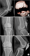
In this study, we used only K-wires and FiberWire instead of stainless roll wire to perform tension band wiring in simple or comminuted transverse patellar fractures. With the procedure, we tried to obtain the stability of reduction while maintaining the tension force as internal fixators. In addition, this method could reduce the irritation of soft tissue caused by the anterior cerclage wire band and enable early ROM exercises and weight bearing. The K-wire alone may had a sufficient fixation force, but we obtained more rigid fixation by adding multidirectional sutures of FiberWire. In the case where the inferior pole of the patella had severe comminution and the K-wire was unable to stabilize the center of bony fragment, FiberWire sutures were performed around the fracture site; with this method, reduction status was maintained and bone union was achieved (Fig. 2).
It is reported that the strength of FiberWire is weaker than the stainless steel wire, but its maximum tensile force is much stronger.10) Therefore, we selected FiberWire, which is easy to manipulate and knot. It appeared that the strength of FiberWire as an internal fixator was weak, but it could convert the compression force to tensile force without loss of reduction during ROM exercises and rehabilitation.
Regarding complications, malunion and reduction loss occurred in a 78-year-old male patient (Fig. 3), who had undergone long-term antibiotics treatment for low-grade infection. So, wound management is important, and if wound healing is satisfactory, ROM exercises should begin as soon as possible.1)
In conclusion, modified tension band wiring using FiberWire is easy to perform and would reduce irritation and breakage of wire. In addition, it could be more useful than anterior cerclage wiring because of the possibility of early rehabilitation and return to daily activities.
Figures and Tables
Fig. 1
Modified tension band wiring using FiberWire. (A) After accurate reduction with reduction forceps, Kirschner wires (K-wires) were inserted from the superior to the inferior pole, perpendicular to the fracture line. (B) In the comminuted fracture of the lower pole, K-wires were passed from the fracture surface of the distal fragment to the inferior pole, and reduction was performed before passage of the K-wires through the superior pole. (C) Then, the knee was flexed over 90°. (D) Sutures were performed around the patella using FiberWire.

Fig. 2
(A) Preoperative radiograph and three-dimensional computed tomographic scan of a 64-year-old male showing comminuted patellar fracture (AO/ASIF classification, 34-C3 type). (B) Postoperative radiographs showing reduction status of the patella after modified tension band wiring using FiberWire. (C) Postoperative radiographs taken 10 weeks after surgery showing bone union.

Fig. 3
Preoperative radiograph (A) and three-dimensional computed tomographic scan (B) of a 78-year-old male showing comminuted patellar fracture (AO/ASIF classification, 34-C3 type). (C, D) Postoperative radiographs showing reduction status of the patella after modified tension band wiring using FiberWire. (E) Postoperative radiograph taken 6 weeks after surgery showing loss of reduction.

References
1. Carpenter JE, Kasman R, Matthews LS. Fractures of the patella. Instr Course Lect. 1994; 43:97–108.

2. Levack B, Flannagan JP, Hobbs S. Results of surgical treatment of patellar fractures. J Bone Joint Surg Br. 1985; 67(3):416–419.

3. Bostrom A. Fracture of the patella: a study of 422 patellar fractures. Acta Orthop Scand Suppl. 1972; 143:1–80.
4. Hung LK, Chan KM, Chow YN, Leung PC. Fractured patella: operative treatment using the tension band principle. Injury. 1985; 16(5):343–347.

5. Leung PC, Mak KH, Lee SY. Percutaneous tension band wiring: a new method of internal fixation for mildly displaced patella fracture. J Trauma. 1983; 23(1):62–64.
6. Tandogan RN, Demirors H, Tuncay CI, Cesur N, Hersekli M. Arthroscopic-assisted percutaneous screw fixation of select patellar fractures. Arthroscopy. 2002; 18(2):156–162.

7. Weber MJ, Janecki CJ, McLeod P, Nelson CL, Thompson JA. Efficacy of various forms of fixation of transverse fractures of the patella. J Bone Joint Surg Am. 1980; 62(2):215–220.

8. Lotke PA, Ecker ML. Transverse fractures of the patella. Clin Orthop Relat Res. 1981; (158):180–184.


 PDF
PDF ePub
ePub Citation
 Print
Print


 XML Download