Abstract
Purpose
To analyze the accuracy of ocular biometry and prediction of postoperative refraction after cataract surgery in acute primary angle-closure glaucoma (ACG) patients treated with laser iridotomy (LI).
Methods
We retrospectively reviewed the medical records of 44 patients who underwent cataract surgery after LI due to ACG (ACG group), and 37 patients who underwent cataract surgery without ocular disease other than cataract (control group) from January 2015 to May 2018. An Acrysof ® single piece (SN60WF) was used as the intraocular lens. We performed preoperative ocular biometry and intraocular lens power calculations using AL-Scan®. The accuracy of the postoperative refractive power prediction was analyzed according to the anterior chamber depth (ACD) and axial length (AL).
Results
The preoperative ACD was 2.29 ± 0.32 mm in the ACG group and 3.15 ± 0.27 mm in the control group (p < 0.01), and the respective AL values were 22.53 ± 0.80 mm and 23.87 ± 1.38 mm (p < 0.01). Using the Haigis formula, patients with an ACD < 2.30 mm in the ACG group (0.52 ± 0.36 diopters [D]) had less accurate results in terms of the mean absolute error than patients with an ACD > 2.31 mm in the ACG group (0.27 ± 0.20 D) and control group (0.27 ± 0.20 D). There was no significant difference in the mean absoluter error between each formula in patients with an AL of < 22.0 mm or > 22.1 mm in the ACG and control groups.
Conclusions
Among patients treated with LI due to ACG, those patients with an ACD > 2.31 mm showed no difference in refractory prediction compared to the control group. However, in patients with an ACD < 2.30 mm, the refractory prediction may be inaccurate when using the Haigis formula, a fourth-generation formula that takes into account the ACD.
Go to : 
References
1. Congdon N, Wang F, Tielsch JM. Issues in the epidemiology and population-based screening of primary abdominal glaucoma. Surv Ophthalmol. 1992; 36:411–23.
2. He M, Foster PJ, Johnson GJ, Khaw PT. Angle-closure glaucoma in East Asian and European people. Different diseases? Eye. 2006; 20:3–12.
4. Hayashi K, Hayashi H, Nakao F, Hayashi F. Effect of cataract abdominal on intraocular pressure control in glaucoma patients. J Cataract Refract Surg. 2001; 27:1779–86.
5. Yoon JY, Hong YJ, Kim CY. Cataract surgery in patients with acute primary abdominal glaucoma. Korean J Ophthalmol. 2003; 17:122–6.
6. Nonaka A, Kondo T, Kikuchi M, et al. Cataract surgery for residual angle closure after peripheral laser iridotomy. Ophthalmology. 2005; 112:974–9.

7. Tarongoy P, Ho CL, Walton DS. Angle-closure glaucoma: the role of the lens in the pathogenesis, prevention, and treatment. Surv Ophthalmol. 2009; 54:211–25.

8. Yang CH, Hung PT. Intraocular lens position and anterior chamber angle changes after cataract extraction in eyes with primary abdominal-closure glaucoma. J Cataract Refract Surg. 1997; 23:1109–13.
9. Hayashi K, Hayashi H, Nakao F, Hayashi F. Changes in anterior chamber angle width and depth after intraocular lens implantation in eyes with glaucoma. Ophthalmology. 2000; 107:698–703.

10. Nonaka A, Kondo T, Kikuchi M, et al. Angle widening and abdominal of ciliary process configuration after cataract surgery for abdominal angle closure. Ophthalmology. 2006; 113:437–41.
11. Kim SA, Kang JH, Park JI, Lee KH. Difference between abdominal refraction and predictive refraction after cataract abdominal in patients with coexisting cataract and primary abdominal glaucoma. J Korean Ophthalmol Soc. 2005; 46:1983–8.
12. Kim KN, Lim HB, Lee JJ, Kim CS. Influence of biometric abdominal on refractive outcomes after cataract surgery in abdominal glaucoma patients. Korean J Ophthalmol. 2016; 30:280–8.
13. Joo J, Whang WJ, Oh TH, et al. Accuracy of intraocular lens power calculation formulas in primary angle closure glaucoma. Korean J Ophthalmol. 2011; 25:375–9.

14. Drexler W, Findl O, Menapace R, et al. Partial coherence abdominal: a novel approach to biometry in cataract surgery. Am J Ophthalmol. 1998; 126:524–34.
15. Lam AK, Chan R, Pang PC. The repeatability and accuracy of axial length and anterior chamber depth measurements from the IOL Master. Ophthalmic Physiol Opt. 2001; 21:477–83.
16. Buckhurst PJ, Wolffsohn JS, Shah S, et al. A new optical low abdominal reflectometry device for ocular biometry in cataract patients. Br J Ophthalmol. 2009; 93:949–53.
17. Shin JY, Lee JB, Seo KY, et al. Comparison of preoperative and postoperative ocular biometry in eyes with phakic intraocular lens implantations. Yonsei Med J. 2013; 54:1259–65.

18. Kwang JY, Choi SH. Comparison of ocular biometry measured by ultrasound and two kinds of partial coherence interferometers. J Korean Ophthalmol Soc. 2011; 52:169–74.

19. Hoffer KJ. The Hoffer Q formula: a comparison of theoretic and abdominal formulas. J Cataract Refract Surg. 1993; 19:700–12.
20. Sanders DR, Retzlaff J, Kraff MC. Comparison of the SRK II abdominal and other second generation formulas. J Cataract Refract Surg. 1988; 14:136–41.
21. Haigis W. Occurrence of erroneous anterior chamber depth in the SRK/T formula. J Cataract Refract Surg. 1993; 19:442–6.

22. Marchini G, Pagliarusco A, Toscano A, et al. Ultrasound abdominal and conventional ultrasonographic study of ocular abdominal in primary abdominal glaucoma. Ophthalmology. 1998; 105:2091–8.
23. Chang SW, Yu CY, Chen DP. Comparison of intraocular lens power calculation by the IOL Master in phakic and eyes with hydrophobic acrylic lenses. Ophthalmology. 2009; 116:1336–42.
24. Kang SY, Hong S, Won JB, et al. Inaccuracy of intraocular lens power prediction for cataract surgery in abdominal glaucoma. Yonsei Med J. 2009; 50:206–10.
Go to : 
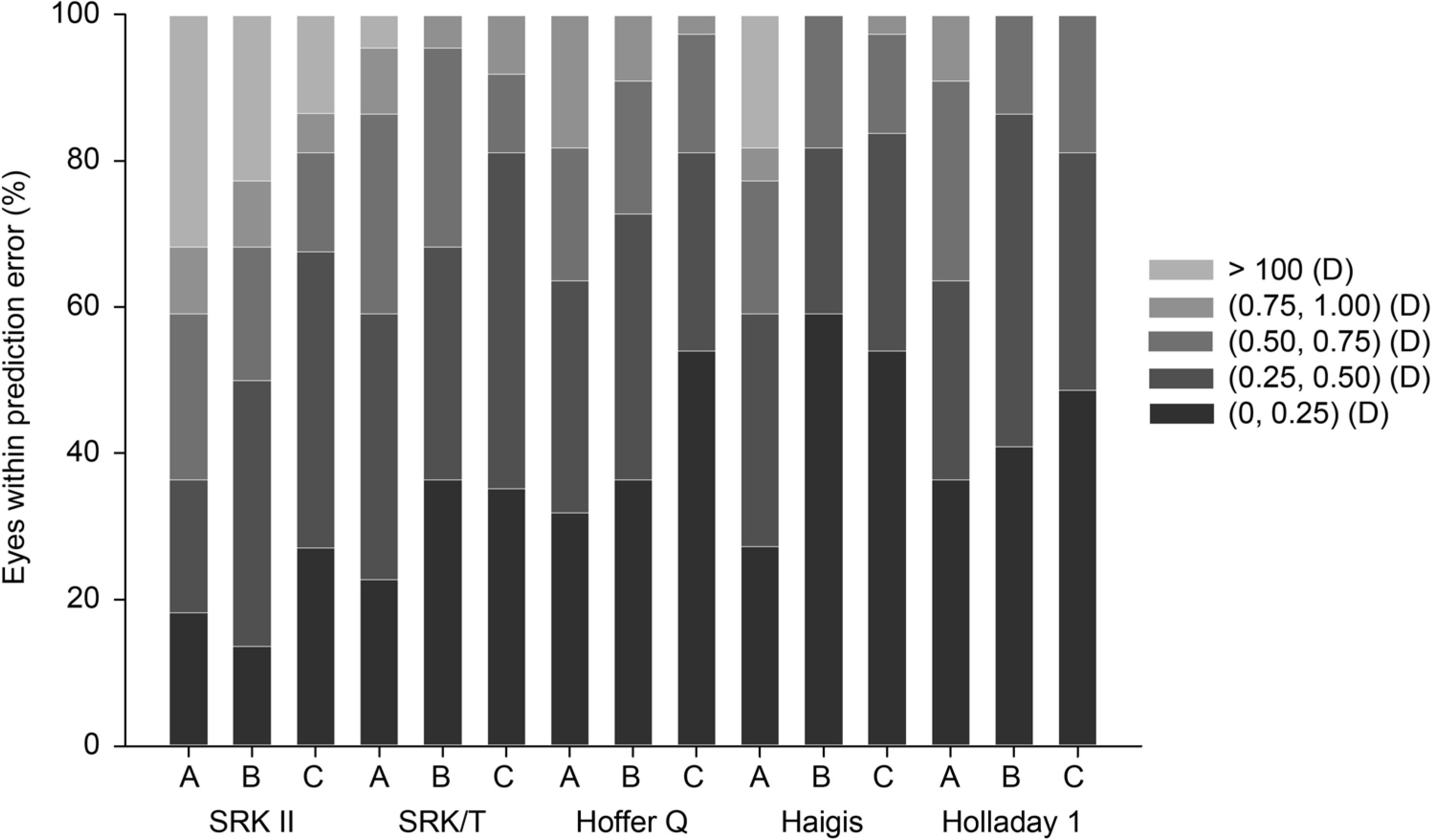
 | Figure 1.Comparison of various IOL power calculation formulas according to anterior chamber depth. Stacked histogram compare the percentage of eyes within a given diopter range of predicted spherical equivalent refraction outcome. A-C indicated group A, group B, and control group individually. IOL = intraocular lens; D=diopter. |
Table 1.
Characteristics of enrolled patients
| All | ACG group | Control group | p-value | |
|---|---|---|---|---|
| Number | 81 | 44 | 37 | |
| Sex (M/F) | 9/35 | 14/23 | 0.137* | |
| Right/left | 16/28 | 15/22 | 0.819* | |
| Age (years) | 69.89 ± 7.52 | 69.20 ± 6.81 | 70.70 ± 8.32 | 0.375† |
| BCVA (logMAR) | 0.42 ± 0.28 | 0.38 ± 0.20 | 0.48 ± 0.35 | 0.117† |
| IOP (mmHg) | 14.67 ± 3.44 | 14.95 ± 4.05 | 14.32 ± 2.56 | 0.415† |
| Spherical equivalent (D) | −0.07 ± 1.80 | 0.14 ± 0.16 | −0.33 ± 1.97 | 0.238† |
| K 3.3 (D) | 44.47 ± 1.65 | 44.86 ± 1.77 | 44.11 ± 1.43 | 0.079† |
| K 2.3 (D) | 44.53 ± 1.65 | 44.78 ± 1.78 | 44.14 ± 1.43 | 0.096† |
| Axial length (mm) | 23.14 ± 1.29 | 22.53 ± 0.80 | 23.87 ± 1.38 | <0.001† |
| ACD (mm) | 2.68 ± 0.53 | 2.29 ± 0.32 | 3.15 ± 0.27 | <0.001† |
| IOL power (D) | 21.59 ± 3.19 | 23.11 ± 1.85 | 19.77 ± 3.51 | <0.001† |
M/F = male/female; BCVA = best-corrected visual acuity; logMAR = logarithm of minimal angle of resolution; IOP = intra ocular pressure; D = diopter; K 3.3 = keratometry calculated at corneal radius 3.3 mm; K 2.3 = keratometry calculated at corneal radius 2.3 mm; ACD = anterior chamber depth; IOL = intraocular lens.
Table 2.
Mean absolute error: comparison of various IOL power calculation formulas according to anterior chamber depth
| Group |
Mean absolute error (diopter) |
p-value* | ||||
|---|---|---|---|---|---|---|
| SRK II | SRK/T | Hoffer Q | Haigis | Holladay 1 | ||
| Total | 0.59 ± 0.44 | 0.38 ± 0.28 | 0.35 ± 0.25 | 0.34 ± 0.27 | 0.31 ± 0.20 | <0.001 |
| (0.01–1.86) | (0.00–1.42) | (0.00–1.00) | (0.01–1.21) | (0.00–0.99) | ||
| Group A | 0.78 ± 0.53 | 0.48 ± 0.32 | 0.43 ± 0.28 | 0.52 ± 0.36 | 0.39 ± 0.26 | 0.067 |
| (0.07–1.86) | (0.00–1.42) | (0.00–0.87) | (0.07–1.21) | (0.00–0.99) | ||
| Group B | 0.60 ± 0.39 | 0.36 ± 0.27 | 0.34 ± 0.26 | 0.27 ± 0.20 | 0.31 ± 0.19 | 0.017 |
| (0.11–1.54) | (0.03–0.84) | (0.00–0.90) | (0.02–0.67) | (0.03–0.75) | ||
| Control | 0.47 ± 0.36 | 0.33 ± 0.24 | 0.31 ± 0.22 | 0.27 ± 0.20 | 0.31 ± 0.20 | 0.139 |
| (0.01–1.86) | (0.00–0.85) | (0.01–1.00) | (0.01–0.79) | (0.01–0.72) | ||
| p-value* | 0.050 | 0.183 | 0.334 | 0.011 | 0.535 | |
Table 3.
Mean error: comparison of various IOL power calculation formulas according to anterior chamber depth
| Group |
Mean absolute error (diopter) |
p-value* | ||||
|---|---|---|---|---|---|---|
| SRK II | SRK/T | Hoffer Q | Haigis | Holladay 1 | ||
| Total | 0.44 ± 0.59 | 0.20 ± 0.42 | −0.11 ± 0.42 | 0.03 ± 0.43 | −0.06 ± 0.39 | <0.001 |
| (−1.46 to 1.86) | (−0.63 to 1.42) | (−0.90 to 1.00) | (−0.64 to 1.21) | (−0.76 to 0.99) | ||
| Group A | 0.76 ± 0.56 | 0.36 ± 0.46 | −0.08 ± 0.51 | 0.35 ± 0.53 | 0.02 ± 0.47 | <0.001 |
| (−0.15 to 1.86) | (−0.47 to 1.42) | (−0.83 to 0.87) | (−0.47 to 1.21) | (−0.76 to 0.99) | ||
| Group B | 0.52 ± 0.50 | 0.16 ± 0.42 | −0.24 ± 0.35 | 0.01 ± 0.34 | −0.14 ± 0.35 | <0.001 |
| (−0.57 to 1.54) | (−0.63 to 0.84) | (−0.90 to 0.54) | (−0.63 to 0.67) | (−0.75 to 0.48) | ||
| Control | 0.20 ± 0.56 | 0.13 ± 0.39 | −0.06 ± 0.38 | −0.08 ± 0.33 | −0.07 ± 0.38 | <0.001 |
| (−1.46 to 1.11) | (−0.42 to 0.85) | (−0.75 to 1.00) | (−0.64 to 0.79) | (−0.69 to 0.72) | ||
| p-value* | 0.002 | 0.172 | 0.231 | 0.006 | 0.526 | |
Table 4.
Refractive results (diopter): comparison of various intraocular lens power calculation formulas according to anterior chamber depth
| Group | Formula | Over 1.0 D myopia* | Over 0.5 D myopia† | Over 0.5 D hyperopia‡ | Over 1.0 D hyperopia§ |
|---|---|---|---|---|---|
| Group A | SRK II | 0.00 | 0.00 | 63.64 | 31.82 |
| SRK/T | 0.00 | 0.00 | 40.91 | 4.55 | |
| Hoffer Q | 0.00 | 27.27 | 9.09 | 0.00 | |
| Haigis | 0.00 | 0.00 | 40.91 | 18.18 | |
| Holladay 1 | 0.00 | 18.18 | 18.18 | 0.00 | |
| Group B | SRK II | 0.00 | 4.55 | 45.45 | 18.18 |
| SRK/T | 0.00 | 4.55 | 27.27 | 0.00 | |
| Hoffer Q | 0.00 | 22.73 | 4.55 | 0.00 | |
| Haigis | 0.00 | 9.09 | 9.09 | 0.00 | |
| Holladay 1 | 0.00 | 13.64 | 0.00 | 0.00 | |
| Control | SRK II | 2.70 | 5.41 | 32.14 | 10.81 |
| SRK/T | 0.00 | 0.00 | 18.92 | 0.00 | |
| Hoffer Q | 0.00 | 8.11 | 10.81 | 0.00 | |
| Haigis | 0.00 | 13.51 | 2.70 | 0.00 | |
| Holladay 1 | 0.00 | 8.11 | 8.11 | 0.00 |
Table 5.
Mean absolute error: comparison of various IOL power calculation formulas according to axial length
| Group |
Mean absolute error (diopter) |
p-value* | ||||
|---|---|---|---|---|---|---|
| SRK II | SRK/T | Hoffer Q | Haigis | Holladay 1 | ||
| Group C | 0.58 ± 0.38 | 0.37 ± 0.26 | 0.41 ± 0.29 | 0.44 ± 0.33 | 0.34 ± 0.20 | 0.487 |
| (0.15–1.37) | (0.06–0.85) | (0.00–0.89) | (0.02–1.21) | (0.08–0.65) | ||
| Group D | 0.74 ± 0.50 | 0.44 ± 0.31 | 0.37 ± 0.27 | 0.37 ± 0.32 | 0.35 ± 0.24 | 0.004 |
| (0.07–1.86) | (0.00–1.42) | (0.00–0.90) | (0.03–1.19) | (0.00–0.99) | ||
| Control | 0.47 ± 0.36 | 0.33 ± 0.24 | 0.31 ± 0.22 | 0.27 ± 0.20 | 0.31 ± 0.20 | 0.139 |
| (0.01–1.86) | (0.00–0.85) | (0.01–1.00) | (0.01–0.79) | (0.01–0.72) | ||
| p-value* | 0.064 | 0.347 | 0.480 | 0.186 | 0.782 | |


 PDF
PDF ePub
ePub Citation
 Print
Print


 XML Download