

 PDF
PDF ePub
ePub Citation
 Print
Print


Abstract
Purpose
To analyze the correlation between clinical features and degree of scleral invasion using anterior segment optical coherence tomography.
Methods
The size, elevation, color, and degree of vascularization were examined in 50 patients (17 males and 33 females) who visited our clinic between February 2017 and April 2018. The thickness and scleral involvement were analyzed by anterior segment optical coherence tomography.
Results
The average horizontal diameter of the pinguecula was 2.37 ± 1.12 mm, which showed significant positive correlations with the thickness and dimension (correlation coefficients: 0.34 and 0.42, respectively). The vertical diameter was 2.21 ± 0.91 mm, which showed significant correlations with thickness, scleral involvement, and dimension (correlation coefficients: 0.22, 0.61, and 0.33, respectively). The correlation coefficient with scleral invasion thickness was as high as 0.61. The average diameter was 2.29 ± 0.97 mm, which was also significantly correlated with thickness and dimension (correlation coefficients: 0.40 and 0.54, respectively). Overall thickness was significantly greater in the elevation group (average 0.66 ± 0.35 mm) than the flat group (0.57 ± 0.23 mm). Thickness was significantly greater in the severe vascularization group (0.70 ± 0.19 mm) than in the less severe vascularization group (0.50 ± 0.13 mm) (p = 0.01).
Figures and Tables
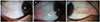
 | Figure 2Anterior segment photographs of pinguecula by elevation. (A) Flat, (B) mild elevation, (C) noticeable elevation.
|
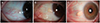
 | Figure 3Anterior segment photographs of pinguecula by vascularization. (A) Mild, (B) moderate, (C) severe.
|
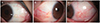
 | Figure 4Anterior segment photography of pinguecula and optical coherence tomography of pinguecula section (yellow line) according to progression. Anterior segment photography and anterior segment optical coherence tomography shows mild (A, B) and moderate (C, D) pinguecula with distinct scleral line (red arrow) without scleral invasion. In severe pinguecula, anterior segment photographs show marked vascularization and elevation (E), anterior segment optical coherence tomography shows thicker and more scleral invasion with distinct scleral line (red arrow) (F).
|
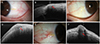
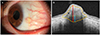 | Figure 5Anterior segment photography and optical coherence tomography of pinguecula with scleral invasion. Anterior segment photograph shows vascularization and elevation of pinguecula (A). Anterior segment optical coherence tomography shows thick (double head red arrow) conjunctival stroma and scleral invasion (double head blue arrow) with large dimension (close yellow line) (B). Scleral invasion refers the distance between normal scleral line (green dotted line) and scleral involvement (double head blue arrow).
|
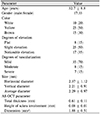
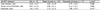
Table 1
Baseline characteristics of the study population to compare the OCT findings and clinical findings of pinguecula
![]()
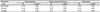
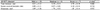
Table 2
Correlation analysis between clinical features and anterior segment optical coherence tomographys of pinguecula
![]()
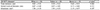
Table 4
Anterior segment optical coherence tomography parameters according to elevation of pinguecula
![]()

Table 6
Post hoc analysis of difference of scleral involved thickness according to elevation of pinguecula

![]()

References
1. Dong N, Li W, Lin H, et al. Abnormal epithelial differentiation and tear film alteration in pinguecula. Invest Ophthalmol Vis Sci. 2009; 50:2710–2715.

2. Bell A. Pinguecula. J Vis Commun Med. 2006; 29:82–83.
3. Jung S, Kwon JW, Hwang HS, Chuck RS. Vascular regression after pinguecula excision and conjunctival autograft using fibrin glue. Eye Contact Lens. 2017; 43:199–202.
4. Blumenthal EZ, Williams JM, Weinreb RN, et al. Reproducibility of nerve fiber layer thickness measurements by use of optical coherence tomography. Ophthalmology. 2000; 107:2278–2282.
5. Chen TC, Cense B, Pierce MC, et al. Spectral domain optical coherence tomography: ultra-high speed, ultra-high resolution ophthalmic imaging. Arch Ophthalmol. 2005; 123:1715–1720.
6. Huang D, Swanson EA, Lin CP, et al. Optical coherence tomography. Science. 1991; 254:1178–1181.
7. Doors M, Tahzib NG, Eggink FA, et al. Use of anterior segment optical coherence tomography to study corneal changes after collagen cross-linking. Am J Ophthalmol. 2009; 148:844–851.e2.
8. Sakata LM, Lavanya R, Friedman DS, et al. Comparison of gonioscopy and anterior segment ocular coherence tomography in detecting angle closure in different quadrants of the anterior chamber angle. Ophthalmology. 2008; 115:769–774.
9. Nanji AA, Sayyad FE, Galor A, et al. High-resolution optical coherence tomography as an adjunctive tool in the diagnosis of corneal and conjunctival pathology. Ocul Surf. 2015; 13:226–235.
10. Soliman W, Mohamed TA. Spectral domain anterior segment optical coherence tomography assessment of pterygium and pinguecula. Acta Ophthalmol. 2012; 90:461–465.
11. Hashemi H, Khabazkhoob M, Yekta A, et al. Prevalence and risk factors for anisometropia in the Tehran eye study, Iran. Ophthalmic Epidemiol. 2011; 18:122–128.
12. Mimura T, Usui T, Obata H, et al. Severity and determinants of pinguecula in a hospital-based population. Eye Contact Lens. 2011; 37:31–35.
13. Ahn SJ, Shin KH, Kim MK, et al. One-year outcome of argon laser photocoagulation of pinguecula. Cornea. 2013; 32:971–975.
14. Hwang JH, Kwon JW. Clinical characteristics of patients with pinguecula between 20 and 39 years of age. J Korean Ophthalmol Soc. 2014; 55:1126–1131.
15. Shin KH, Kwon JW. Clinical features of pinguecula in North-Western Gyeonggi Province. J Korean Ophthalmol Soc. 2013; 54:691–695.
16. Viso E, Gude F, Rodríguez-Ares MT. Prevalence of pinguecula and pterygium in a general population in Spain. Eye (Lond). 2011; 25:350–357.
 XML Download