

 PDF
PDF ePub
ePub Citation
 Print
Print


INTRODUCTION
Mallory-Weiss tears that occur following cardiopulmonary resuscitation in domestic emergency rooms have been reported as a rare complication.1 The complications that may occur after cardiopulmonary resuscitation have appeared in 21% to 60% of patients. The most common complication reported were rib fractures. Other reported complications included sternal fractures, chest wall hematoma, pneumothorax, cardiac contusion, diaphragmatic rupture, liver or spleen rupture, emphysematous cholangitis, and gastrointestinal rupture.2345
Lacerations may occur on the esophagus and stomach junction due to the increasing intragastric pressure which occurs during mouth-to-mouth breathing or Ambu bag breathing and chest compression, which are techniques employed as a part of cardiopulmonary resuscitation.678 A Mallory-Weiss tear, which may occur after cardiopulmonary resuscitation, is rare but can be lifethreatening.
This report shows a case of Mallory-Weiss Syndrome which appeared in a patient who sustained acute myocardial infarctions during exercise, following the cardiopulmonary resuscitation.
CASE
A 79-year-old male patient experienced mental deterioration while swimming and was discovered by a 119 rescue worker who had also been swimming. After chest compression was performed for approximately five minutes and the automated external defibrillator was used once, the patient regained his consciousness and was then transferred to this hospital's emergency room. The patient vomited an estimated 300cc of blood during transport, and complained of mild chest pain and dizziness at the time of his hospital arrival. He had been taking regular prescription medication for hypertension, diabetes, and prostatic hypertrophy. His family medical history showed no exceptional cases of health conditions or disease, and he did not drink or smoke.
At the time of hospitalization, the patient was fully conscious and his vitals were measured as follow. His blood pressure was 144/74mmHg, body temperature was 36℃, pulse rate was 75 BPM, and respiration rate was 20 BRA. There were no specific findings in the cardiac sound auscultation. Minor crackles were auscultated from both lungs and blood was observed around the lips.
According to the blood test results, white blood cells were 11,900/mm3, hemoglobin was 12.1 g/dL, AST was 57 U/L, ALT was 56 U/L, and Troponin was I 1.05 ng/mL. In a separate blood test conducted 19 hours later, hemoglobin decreased to 8.5 g/dL. The ECG findings showed a 2 mm ST segment depression and left ventricular hypertrophy on V4 and V5 lead.
As the patient complained of bloody stool on the first day of hospitalization, a computer tomography (CT) was performed on the chest and abdomen. The CT results suggested a small amount of pleural effusion fluid on the lower portion of both lungs and bloody stool around the rectum.
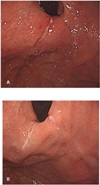
Treatment and Progression: After the patient was diagnosed with upper gastrointestinal tract bleeding and acute myocardial infarction, the fasting, transfusion, and administration of an intravenous proton pump inhibitor were performed. As hemoglobin was maintained at 9.7 g/dL after the 4-pint transfusion for two days during his hospitalization, an upper gastrointestinal endoscopy was performed. As a result, several Mallory-Weiss tear sites were observed at the lower part of esophagus and stomach junction and cardia (Fig. 1A).
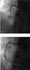
As the patient's condition was stabilized on the seventh day of hospitalization, the cardiac angiography was performed after aspirin and clopidogrel were each administered at a dose of 300mg. Ninety-nine percent stenosis was then observed in the middle of the right coronary artery. The dilatation was performed on the 2.5 mm x 15 mm stricture lesion under the atmospheric pressure of 10 for 30 seconds using the traveler balloon catheter (Fig. 2A). The patient undergoing the procedure did not complain of any symptoms, there was no electrocardiographic change, and, as 37% residual stenosis was observed in the follow-up angiography, a 3 mm x 32 mm Promus primer mesh was implanted (Fig. 2B).
Echocardiography was conducted on the eighth day of hospitalization, and the following were observed: the left ventricular ejection fraction of 42%, the reduced wall motion of the left ventricular lower portion, the side lower portion, and the anterolateral portion.
According to the follow-up upper gastrointestinal tract test conducted on the tenth day of hospitalization, the Mallory-Weiss tear was cured and bleeding was no longer observed (Fig. 1B). As hemoglobin was maintained at 11.2 g/dL on the 11th day of hospitalization, the patient was discharged from the hospital without any showing notable symptoms.
DISCUSSION
Mallory-Weiss tears account for 3% to 10% of upper gastrointestinal tract bleeding, and, in most cases, improve even without special treatment.6
Though rare, Mallory-Weiss tears may occur as a complication even after cardiopulmonary resuscitation is performed. They may cause lacerations at the esophagus, gastric junction, and lesser curvature due to the abruptly increasing intragastric pressure resulting from symptoms such as nausea and vomiting.9
Gastrointestinal damage by cardiopulmonary resuscitation was first detected in reports of gastrointestinal rupture due to chest compression in 1964. The autopsy of deceased patients revealed gastrointestinal laceration and hemorrhage in 5 of 50 cases.10 The major cause of gastrointestinal damage due to cardiopulmonary resuscitation is associated with improper artificial respiration and chest compression.678 Improper artificial respiration and chest compression may increase the pressure of gastrointestinal tract, leading to the rupture of gastric mucosa.678 The factors of gastrointestinal damage occurring after cardiopulmonary resuscitation include the patient's posture, whether to use the cardiac pump, the place where the cardiac arrest occurs, and the rescue worker's cardiopulmonary resuscitation technique.678
In this case, the cardiopulmonary resuscitation, which may have contributed to the patient's Mallory-Weiss tear, was performed by a 119 rescue worker who had received professional training. Although a professional rescuer performed the cardiopulmonary resuscitation, it was presumed that the damages sustained by the patient throughout the ordeal were unavoidable. It was also presumed that his abdominal pressure was elevated due to the inhalation of water into the esophagus resulting from the mental deterioration during swimming. In this case, chest compression alone was performed for five minutes. As the patient gained consciousness relatively quickly, he could be transferred to this hospital's emergency room. Cardiac angiography and mesh implantation were performed when his condition was stabilized after receiving adequate transfusion and an evaluation of upper gastrointestinal tract bleeding. His condition could then be restored.
This case shows the importance of administrating cardiopulmonary resuscitation in a situation where a cardiac arrest occurs in a public place and specialized treatment is required. If the cardiopulmonary resuscitation was performed by a team of trained people rather than by a single rescuer, it may have increased its accuracy and effectiveness. In this regard, it seems necessary to provide even to the general public consistent training on cardiopulmonary resuscitation in preparation for such emergencies.
In this case, the Mallory-Weiss tear occurred as a complication of cardiopulmonary resuscitation. Cardiac angiography and mesh implantation were performed seven days later. The antiplatelet agent was not administered due to the pattern of hemorrhage, and the cardiac angiography was performed when there was no additional hemorrhage after the proton pump inhibitor was administered for seven days. This treatment showed a difference from the treatment in myocardial infarction patients without a pattern of hemorrhage.
Although most patients with Mallory-Weiss Syndrome recover on their own without medical intervention, the disease can be fatal depending on its severity. Patients with acute myocardial infarction with high mortality are at greater risk. This case shows that efficient and accurate administration of cardiopulmonary resuscitation coupled with adequate analysis and treatment for patients with myocardial infarction can save lives.
 XML Download