

 PDF
PDF ePub
ePub Citation
 Print
Print


It is well known that the hepatic artery has numerous variations. The normal pattern of the hepatic artery, as usually represented in textbooks, is only observed in half of cases.1 The common hepatic artery arises from the celiac trunk, dividing into the gastroduodenal artery and proper hepatic artery. However, a common hepatic artery originating from the left gastric artery and the entire hepatic blood supply furnished by the left gastric artery is an extremely rare anomaly. Only a few case of this anomaly has been published in the literature.234
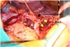
We encountered this anomaly in a 50-year-old man, who underwent subtotal gastrectomy with D2 lymph node dissection for advanced gastric cancer. During dissection from the left edge of the hepatoduodenal ligament towards the esophagus, a thick cord-like structure was found in the lesser omentum. It was the common hepatic artery, originating from the left gastric artery. To our knowledge, intraoperative photographs of this anomaly have never been reported; therefore, surgeons should know this image appearance, which is helpful to recognize this anomaly
A detailed understanding of the vasculature around the stomach and possible anomalies is essential to perform appropriate and safe lymph node dissection during gastric cancer surgery. Without knowledge of this anomaly, given the strategy of extensive lesser sac dissection generally employed during gastric cancer surgery, an inadvertent division of this vessel could result in critical liver damage. We herein report a case of a common hepatic artery originating from left gastric artery and review of the literatures.
CASE
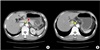
A 55-year-old man was evaluated for dyspepsia and 5 kg weight loss over 3 months. Laboratory testing showed a decreased red blood count (3.9×10⁶/µL), hemoglobin (12.1 g/dL), and hematocrit (37.2 %). Tumor markers including CEA (3.2 ng/ml) and CA 19-9 (26.4 U/ml) revealed no abnormalities. Esophagogastroduodenoscopy demonstrated an ulcerative lesion from the prepylorus to the angle of the stomach. Endoscopic biopsy revealed moderately differentiated adenocarcinoma. Abdominal computed tomography (CT) showed perigastric fat infiltration with lymphadenopathy at the left gastric, common hepatic, and hepatoduodenal ligament lymph node. There is only the splenic artery present along the upper border of pancreas. Common hepatic artery runs toward hepatic hilum along the lower border of left lobe (Fig. 1A, B).
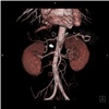
After laparotomy, the greater curvature was freed after ligation, and division of the vessels with lymph node dissection, and then the duodenum was transected. During dissection from the left edge of the hepatoduodenal ligament towards the esophagus, a thick cord-like structure was found in the lesser omentum plane. The common hepatic artery, which runs transversely in parallel with the pancreatic parenchyma, was not found on the upper border of the pancreas. The number 7, 8, and 9 lymph node dissection with vessel preservation was performed carefully. The common hepatic artery had an anomalous origin, the left gastric artery arose from the celiac trunk, two accessary arteries were branched to the lesser curvature, and then it ran toward the hepatic hilum along the lower border of the left lobe (Fig. 2). A distal gastrectomy with Roux-en Y reconstruction was performed. Pathologic findings showed T4aN3M0 and Stage IIIc based on the 7th edition of American Joint Committee on Cancer. The postoperative course of the patient was uneventful and he was discharged on the 9th postoperative day. Multi-dimensional abdominal computed tomography (MDCT) was performed 1 year after the operation, as a routine follow up schedule, and a reconstructed angiographic image showed that the common hepatic artery originated from the left gastric artery (Fig. 3).
DISCUSSION
It is well known that the hepatic artery has numerous variations. Variations of the hepatic artery have been reported on many occasions, including in the reports by Adachi in 1928 and Michels in 1955.56 However, the common hepatic artery originating from the left gastric artery has been rarely reported, and this paper reported the first intraoperative finding.234 In the study by Song, this anomaly was found in eight (0.16%) of 4939 patients.4 In the study by Michels, this anomaly was found in only one (0.5%) of 200 dissected cadaveric specimens.6
Resection of the accessory artery or replacing the left hepatic artery arising from the left gastric artery, during gastrectomy, may cause transient liver dysfunction, usually normalized within 7 days after operation.7 Therefore, a gastric surgeon does not usually consider hepatic artery anatomic variations. However, without knowledge of this anomaly, given the strategy of extensive lesser sac dissection generally employed during gastric cancer surgery, an inadvertent division of this vessel could result in critical liver damage. Thus, the surgeon drew attention to this anatomic variation of the origin of the hepatic artery, ensuring that no damage would occur during gastric surgery. The intraoperative photography of common hepatic artery originating from left gastric artery has never been reported, therefore, this image appearance can be helpful for surgeons during upper gastrointestinal tract and hepatobiliary surgery.
Abdominal computed tomography is routinely performed for patients with gastric cancer to assess local invasion and distant metastasis. With the technological advancement of multi detector computed tomography (MDCT) technology, vascular anatomic reformation can now be easily delineated. Rapid volumetric acquisition of thin-slice high resolution images of the abdominal arteries with the help of MDCT allows reconstructions to be created, providing the surgeon with patient's arterial anatomy.8 In patients with gastric cancer, preoperative reformation vascular anatomy using an MDCT scan may provide useful information to the surgeon, bearing in mind this anatomical variation discovered during gastric surgery.
 XML Download