

 PDF
PDF ePub
ePub Citation
 Print
Print


INTRODUCTION
Aortic dissection can frequently extend into its peripheral territories. Medical literature reports many cases of renal, coronary, intracranial and visceral artery involvement in aortic dissection.1)2) However it is rare for these branches to have dissection in the absence of main aortic trunk involvement.3) Among visceral arteries, superior mesenteric artery (SMA) is the commonest type of dissection when compared with other gastrointestinal arteries such as the splenic, hepatic, celiac and gastric arteries.4) However, isolated SMA dissection is believed to be rare. Due to its rarity, clinical presentation, use of imaging studies, management, and outcome of SMA dissection has not been investigated in detail. The purpose of this systematic review is to identify the burden, provide a classification tool and delineate the diagnostic and management algorithms of isolated SMA dissection.
Watson5) in 1956 for the first time introduced arterial dissection as a condition resulting from blood penetration into arterial wall, causing a split between the vessel layers, with or without a tear of the tunica intima (inner vessel layer). However, the first case of SMA dissection was reported before that by Bauersfeld6) in 1947 as an incidental autopsy finding in a patient who died of multiple vessels aneurysms. Since then, there was a gradual increase in SMA dissection related deaths and 11 more cases were found on autopsy findings up to 1972.
From 1975 to 1999 the number of SMA dissection cases rose to 23, with 71 cases reported from 2000 to 2009. Interestingly, the incidence of SMA dissection related mortality during this period decreased significantly and only one case of SMA dissection related death was reported since 1972.7) Since 2009, fifty two more cases of SMA dissection were reported, with a further decline in mortality, with only one case resulting in death.8) This higher incidence and decreased mortality related to SMA dissection is likely due to the introduction of contrast-enhanced computed tomography (CT) scan for abdominal pain investigation, which results in an earlier diagnosis. Patients can have a self-limited course or the SMA dissection can potentially be fatal depending upon the nature of vessel involvement and the underlying health condition of the patients. It generally can have one of the four courses; cessation of SMA dissection with no long term sequelae, progressive involvement of the whole vessel, dissecting aneurysm joining the true lumen, or rupture of the vessel causing severe bleeding.
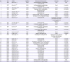
The presumed mechanism of SMA dissection is intimal or vasa vasorum tear leading to hemorrhage in the medial and adventitial layers which can extend over a variable distance.6)9)10)11)12) Segmental arterial mediolysis, congenital connective tissue disorders, arteriosclerosis, cystic medial necrosis, vasculitis and fibromuscular dysplasia have been reported as potential causes of SMA dissection (Table 1).13)14)15) Interestingly, many patients who initially presented with a sudden onset of abdominal pain and had ultrasound and X-ray were diagnosed as having gastroenteritis, gastric or nonspecific pain, and had to return within a week to be correctly diagnosed with having SMA dissection.15)
Table 1
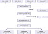
Sakamoto classification of SMA dissection based on CT scan findings and its management

![]()
Sakamoto et al.3) for the first time classified SMA dissection into four types based on the findings on contrast-enhanced CT scanning and described its management as reported in Table 1 and illustrated in Figure 1. However this observation was only based on a study of 12 patients and large-scale studies are required to validate this data.
 | Figure 1Type I: Patent false lumen; Type II: False lumen without re-entry; Type III: thrombosed false lumen with an ulcer-like projection; Type IV: completely thrombosed false lumen with no ulcer like projection.
|
An increasing number of patients with SMA dissection who are hemodynamically stable are treated conservatively. Along with anticoagulation therapy (heparin drip or warfarin), conservative management includes antiplatelets like cilostazol and ticlopidine, bowel rest and control of risk factors like hypertension. Anticoagulation does not revert or halt the progression of dissection but prevents thrombus formation and its distal embolization. It is recommended to have complete bowel rest and administer intravenous heparin until the abdominal pain settles. Oral anticoagulants and antiplatelet medications are continued until resolution of radiological images.16)
Hemodynamically unstable patients having signs and symptoms of ischemia or those with radiological evidence of progression or worsening SMA dissection, such as formation of thrombus, narrowing or saccular aneurysm formation, should have urgent revascularization, as they are at high risk of rupture. The two main revascularization techniques are endovascular or surgical repair. A surgical procedure is inevitable in cases of bowel infarction or SMA rupture.3) The extent and type of surgery depends on the viability of gut, type of dissection and the reversibility of circulation. Sisteron and Viveville17) in 1975 performed the first SMA surgical revascularization using a saphenous vein graft.
Endovascular management includes intralesional thrombolytic therapy, stent placement, embolotherapy and balloon angioplasty. Leung et al.18) first described percutaneous stent placement for SMA dissection. Endovascular technique is a minimally invasive procedure, prevents progression of dissection and provides instant relief from ischemia with shorter hospital stays.
METHODS
Search strategy
A literature search for relevant articles was performed by 2 authors independently on May 25, 2018, using MEDLINE (PubMed, Ovid), Embase, Scopus and Cochrane databases. There was no language or time restriction placed on the search. We were specifically looking for articles on SMA that described the three treatment modalities used for the management of SMA. These included conservative, surgical and endovascular management. The search strategies included various combinations of text-words and medical subject headings (MeSH) to generate two subsets of citations: one for SMA, using the MeSH and terms like “SMA”, “SMA dissection”, “superior mesenteric artery”, “superior mesenteric artery dissection”, “mesenteric artery dissection”, “intestinal artery dissection”, “spontaneous dissection of SMA”, and “spontaneous dissection of superior mesenteric artery” and the other for its management using terms and MeSH like “conservative”, “heparin”, “coumadin”, “antiplatelet”, “graft”, “stent”, “surgery”, “patch”, “resection”. The terms from the 2 subsets were combined in 1:1 combination using the Boolean operators “AND” and “OR”. Results from all the possible combinations were downloaded into an EndNote library. Based on our research question, a third author manually searched the references in all known articles to identify studies that were missed by the initial search.
Selection criteria
The selection criteria for the included studies was all reported case reports, case series and review articles on SMA dissection describing its presentation, diagnosis, management and/or post management follow up. Studies with insufficient data, discussing only the mechanism or histology and conference papers were excluded, as were studies with not enough description of its subjects.
Study selection
The titles and abstracts of the selected articles were reviewed independently by three authors and the articles which met inclusion criteria were reviewed by a fourth author. Full-text articles that were potentially relevant to the study were also reviewed by all the four authors to confirm the eligibility. Disagreements were resolved by mutual consensus and after a detailed group discussion.
RESULTS
Initially, we retrieved a total of 261 articles after removing duplicates, out of which 111 articles were relevant to our case. Interestingly, the timeline of the articles included was very broad, ranging from articles published in 1975 all the way to 2017. It is important to note that the number of published articles on SMA dissection has seen a steep increase in the last couple of decades due to the increasingly widespread use of CT scan and the resultant increase in the number of diagnosed cases.”
SMA dissection occurred in 80.6% males (n=117/145), and 17.2% females (n=25/145). The age of individuals ranged from 33 to 87 years with a mean age of 55.7 years (standard deviation, 9.7; 33–87). Data extraction from the review articles revealed that 41.3% cases (n=60/145) managed SMA dissection by conservative approach using anticoagulants, antiplatelets and antihypertensives. Endovascular approach (stenting, embolotherapy and thrombolysis) was employed in about 29.6% (n=43/145) whereas open surgical repair was done in 28.9% (n=42/145) (Figure 2).
 | Figure 2Prisma flow sheet showing the search strategy on isolated SMA dissection.SMA = superior mesenteric artery.
|
The overall results were satisfactory after treatment as 91.7% (n=133/145) patients survived without complications. Only 5 cases resulted in death. Others developed acute diverticulitis, acute mesenteric ischemia, ligament of Treitz abscess and bilateral lower limb paralysis each. Poor recovery was observed in the case of bilateral lower limb paralysis, whereas the rest fully recovered from their complications.
DISCUSSION
Etiology
The exact etiology of isolated SMA dissection remains unknown, though many conditions have been found to be associated with SMA dissection. In our study, 22.7% (n=33/145) of the patients were known hypertensives or presented with markedly high blood pressures (above 160/100 mmHg). Cigarette smoking was found in 18.6% of the cases (n=27/145).19)20)21) A history of trauma was found to be associated with four or 2.7% (n=4/145). One patient had a seat belt trauma associated SMA dissection due to low-velocity motor vehicle accident, while another had trauma related SMA aneurysm, leading to presumed dissection.22)23) Only a few iatrogenic SMA dissection cases were reported, following translumbar aortography with the use of balloon catheter, or due to mesenteric angioplasty in the treatment of chronic mesenteric ischemia.24)25)26)
Symptoms
The most common presentation in our study was sudden onset of pain, either abdominal 55.8% (n=81/145), epigastric 22.7% (n=33/145), periumbilical 4.8% (n=7/145), back pain 4.8% (n=7/145), or chest 2.0% (n=3/145). Most of these patients presented acutely within 4 weeks of the onset of symptoms possibly due to bowel ischemia and/or infarct.15)27) Only 2.0% patients presented with shock along with abdominal pain (n=2/145) due to rupture of the dissecting SMA and eventually died.7)11) Approximately 2.7% presented with melena (n=4/145). Besides these, patients commonly presented with nausea, vomiting and abdominal distension. We believe that physicians should follow the American Gastroenterological Association guidelines and should consider diagnostic work-up in an appropriate clinical setting for acute mesenteric ischemia in every patient with a history of unexplained abdominal pain for more than 2–3 hours.28)
In our study about twelve patients or 8.2% had chronic symptoms lasting for more than a month, which included nausea, vomiting, diarrhea, melena, postprandial pain, and weight loss.19) This suggests that SMA dissection can have a subacute or chronic course and physicians should have a high index of suspicion in an appropriate setting. Seven patients or 4.8% had SMA dissection discovered either on autopsy or as an incidental finding on CT scan performed for pancreatitis or other reasons.27)29)30)31)
Diagnosis
SMA dissection should be suspected in all patients presenting with intractable abdominal pain, and having one or more risk factors for vascular atherosclerotic disease.21)
Our review showed that contrast enhanced abdominal CT scan and plain CT scan were used almost equally in 35.8% and 36.5% (n=52/145 and n=53/145) of cases. In 42.1% cases (n=61/145) CT angiogram was used, while very few patients had arteriography for the diagnosis of SMA dissection. The accuracy of CT angiogram is almost the same as conventional arteriography with the benefits of decreased morbidity and lower radiation exposure. It also provides a three-dimensional view of luminal borders and extraluminal organs and can be performed more quickly compared to conventional arteriography.12)14)21)32)33)
Eight cases (5.5%, n=8/145) used digital subtraction angiography (DSA) as a diagnostic tool, which has the luxury of doing therapeutic intervention like thrombolysis and stenting if required with very little additional contrast. But we believe it should be reserved for patients with worsening symptoms, who requires endovascular treatment or surgical intervention as it is a very invasive procedure.3)
A number of other imaging and surgical modalities were used to diagnose SMA dissection. Magnetic resonance angiography (MRA) and diagnostic laparoscopy were used in one case each 0.6% (n=1/145) while 2 patients or 1.3% were diagnosed on laparotomy.34)35) Platelet scintigraphy was useful to determine the patency and thrombosis of false lumen (e.g., type IV Sakamoto CT classification).3)29) Ultrasound was successfully used in making the diagnosis of seven patients or 4.8% of the patients, demonstrating the intimal flap while it missed SMA dissection in nine patients or 6.2% of the patients36)37) This flap is sometime hidden behind the thrombus of the false lumen of SMA; contrast-enhanced CT is a better alternative in such cases.38) Doppler ultrasound helped in the diagnosis of only seven patients or 4.8% of the patients but was very useful in the operating room for the assessment of bowel viability. It also helped to decide about the type of vascular intervention and for the post intervention surveillance of patients.39) Of note, there was no role for blood tests or abdominal X-rays in the diagnosis of SMA dissection.
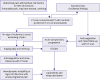
Moreover, this review also showed that the extent and type of diagnostic modality can direct towards appropriate management. Based on the review of the literature we suggest that symptomatology and clinical features of the patient should direct physicians for appropriate diagnostic testing as illustrated in Figure 3.
MANAGEMENT
Conservative
Our systematic review showed that the most common treatment modality utilized was conservative treatment in about 41.3% (n=60/145). These patients were hemodynamically stable and had no clinical or imaging evidence of ruptured SMA dissection.9) They had successful resolution of symptoms during their mean follow up of 16.4 month (0.5–83 month) with no mortality, even in cases of 90% SMA involvement (Table 2). Hence, we deduce that a trial of anticoagulation therapy as a conservative approach is warranted in all cases of uncomplicated SMA dissection.40) This is especially true for Sakamoto type I and IV dissection.
Table 2
Characteristics of conservatively managed isolated SMA dissection
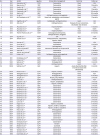
| No. | Year | Author | Age/Sex | Conservative management | Outcome | Follow up |
|---|---|---|---|---|---|---|
| 1 | 2017 | Léonard et al.41) | 57/M | Conservative | Good | N/A |
| 2 | 2016 | Spence et al.42) | 33/F | Conservative | Good | 18 months |
| 3 | 2016 | Hoek et al.43) | 48/M | Conservative | Good | N/A |
| 4 | 2016 | Funahashi et al.44) | 58/M | Conservative | Good | 12 months |
| 5 | 2016 | Funahashi et al.44) | 67/M | Conservative | Good | 60 months |
| 6 | 2016 | Nath et al.45) | 68/F | Conservative | Good | 0.5 months |
| 7 | 2015 | Jia et al.46) | 70/M | Antihypertensives, anticoagulants | Good | 10 months |
| 8 | 2015 | de l'Escalopier et al.47) | 51/M | Bowel rest, antiplatelets, anticholesterol agents | Good | 12 months |
| 9 | 2015 | Daghfous et al.48) | 40/M | Antiplatelets, antihypertensives | Good | N/A |
| 10 | 2015 | Zink et al.49) | 55/M | Antihypertensives | Paralysis of lower extremities, Decreased renal function | N/A |
| 11 | 2015 | Akuzawa et al.8) | 38/M | Heparin/Warfarin | Good | 11 months |
| 12 | 2015 | Akuzawa et al.8) | 62/M | Heparin | Poor | Death |
| 13 | 2015 | Akuzawa et al.8) | 38/M | Heparin/warfarin | Good | 11 months |
| 14 | 2014 | Moreno-Machuca et al.50) | 46/M | Anticoagulant (bemiparine), antihypertensives, analgesics | Good | 24 months |
| 15 | 2014 | Ogul et al.51) | 40/M | N/A | N/A | N/A |
| 16 | 2014 | Zinsser et al.52) | 61/M | Antihypertensives, antiplatelets, heparin, statins | Good | 1 month |
| 17 | 2014 | Ham et al.53) | 56/M | Conservative | Good | N/A |
| 18 | 2014 | Corral et al.54) | 42/M | Antiplatelets | Good | 14 months |
| 19 | 2014 | Corral et al.54) | 85/M | Anticoagulants | Good | N/A |
| 20 | 2013 | Davis and Kendall55) | 46/M | Intravenous labetalol and nitroprusside | N/A | N/A |
| 21 | 2012 | Yoo et al.56) | 56/M | Conservative | Good | 17 months |
| 22 | 2012 | Shimizu and Tokuda57) | 61/M | Conservative | Good | 6 months |
| 23 | 2012 | Kokai et al.58) | 56/M | Antihypertensives, anticoagulants, antiplatelets | Good | 47 months |
| 24 | 2011 | Namikawa et al.59) | 59/M | Conservative | Good | 4 months |
| 25 | 2011 | Kang et al.60) | 46/M | Heparin, steroids | Good | 1 month |
| 26 | 2010 | Saba et al.61) | 49/M | Prostaglandin E1 | Good | 3 months |
| 27 | 2010 | Bair et al.62) | 72/M | Antihypertensives | Good | N/A |
| 28 | 2009 | Subhas et al.63) | 56/F | Heparin | Acute diverticulitis after 2 months (treated with antiobiotics) | 2 months |
| 29 | 2009 | Mousa et al.64) | 57/M | Heparin, warfarin | Good | 18 months |
| 30 | 2009 | Totsugawa et al.65) | 51/M | Prostaglandin E1 | Good | N/A |
| 31 | 2009 | Totsugawa et al.65) | 56/M | Prostaglandin E1 | Good | 10 months |
| 32 | 2009 | Jang et al.66) | 58/M | Proton pump inhibitors | Good | 2 months |
| 33 | 2008 | Tsai et al.21) | 49/M | Antihypertensives | Good | N/A |
| 34 | 2008 | Ghuysen et al.67) | 38/M | Heparin, antiplatelets | Good | 3 months |
| 35 | 2008 | Morris et al.68) | 56/M | Heparin, warfarin | Good | 5 months |
| 36 | 2008 | Morris et al.68) | 62/F | Conservative | Good | 5 months |
| 37 | 2008 | Takayama et al.69) | 58/M | Conservative | Good | 83 months |
| 38 | 2007 | Sakamoto et al.3) | 58/M | Conservative | Good | 7 months |
| 39 | 2007 | Sakamoto et al.3) | 43/M | Conservative | Good | 38 months |
| 40 | 2007 | Sakamoto et al.3) | 60/M | Conservative | Good | 60 months |
| 41 | 2007 | Sakamoto et al.3) | 52/M | Conservative | Good | 72 months |
| 42 | 2007 | Sakamoto et al.3) | 48/M | Conservative | Good | 36 months |
| 43 | 2006 | Chang et al.20) | 49/M | Conservative | Good | 3 months |
| 44 | 2006 | Lee et al.70) | 57/M | Conservative | Good | 24 months |
| 45 | 2004 | Nagai et al.16) | 59/M | Heparin, ticlopidine | Good | 12 months |
| 46 | 2004 | Nagai et al.16) | 56/M | Heparin, warfarin, ticlopidine | Good | 5 months |
| 47 | 2004 | Nagai et al.16) | 49/M | Heparin, warfarin, ticlopidine | Good | 4 months |
| 48 | 2004 | Nozu et al.71) | 55/M | Anticoagulation | Good | 8 months |
| 49 | 2004 | Suzuki et al.39) | 54/F | Anticoagulation | Good | 4 months |
| 50 | 2004 | Suzuki et al.39) | 50/M | Conservative | Good | 4 months |
| 51 | 2004 | Suzuki et al.39) | 60/M | Conservative | Good | 5 months |
| 52 | 2004 | Suzuki et al.39) | 50/M | Conservative | Good | 2 months |
| 53 | 2003 | Sartelet et al.7) | 44/M | Fluid resuscitation | Death | N/A |
| 54 | 2002 | Furukawa et al.33) | 52/M | Conservative | Good | 12 months |
| 55 | 2002 | Takayama et al.72) | 63/M | Warfarin | Good | 6 months |
| 56 | 2001 | Sheldon et al.14) | 41/M | Coumadin | Good | 22 months |
| 57 | 2000 | Matsou et al.73) | 58/M | Conservative | Good | N/A |
| 58 | 1998 | Yasuhara et al.19) | 45/M | Conservative | Good | 24 months |
| 59 | 1998 | Yasuhara et al.19) | 55/M | Conservative | Good | 12 months |
| 60 | 1998 | Dushnitsky et al.74) | 58/M | Conservative | Good | 16 months |
![]()
Surgical revascularization
We found that about 28.9% (n=42/145) patients underwent surgical management and that bypass grafting was the commonest procedure. Bypass grafting was performed in 57.1% (n=24/42) patients, in which a saphenous vein graft was used in 12 cases. Infrarenal aortoiliac bypass, superior aortomesenteric prosthetic bypass, radial artery bypass and right gastroepiploic bypass were used in one case each. Other grafts used in our review included superficial femoral artery, radial artery and prosthetic grafts. SMA was directly anastomosed to the infrarenal artery in a few cases to avoid graft-related complications. Thrombectomy was performed in 16.6% cases (n=7/42) whereas arteriotomy and intimectomy were performed in 9.5% cases each (n=4/42). Ligation of a branch of SMA was carried out in one case (Table 3).
Table 3
Characteristics of surgically managed isolated SMA dissection
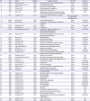
| No. | Year | Author | Age/Sex | Surgical procedure | Outcome | Follow up |
|---|---|---|---|---|---|---|
| 1 | 2016 | Mitsuoka et al.77) | 45/M | Laparotomy, arteriotomy, stenting of SMA | Good | 6 months |
| 2 | 2015 | Dzieciuchowicz et al.78) | 42/F | Thrombendarterectomy | Good | 30 months |
| 3 | 2014 | Wall et al.79) | 65/M | Infrarenal aortoiliac grafting | Good | 6 months |
| 4 | 2011 | Carter et al.80) | 57/F | Great saphenous vein grafting | Good | 6 months |
| 5 | 2011 | Tameo et al.81) | 51/M | Ligation of a branch of SMA | Good | 6 months |
| 6 | 2011 | Mei et al.82) | 58/F | Arteriotomy of the inferior mesenteric artery, thrombectomy, great saphenous vein grafting | Good | N/A |
| 7 | 2010 | Hwang et al.83) | 54/M | Intimectomy, great saphenous vein patch angioplasty | Thrombus formation (resolved with anticoagulation) | 12 months |
| 8 | 2009 | Bruns et al.84) | 47/M | Thrombendarteriectomy | Good | 5 months |
| 9 | 2008 | Morris et al.31) | 39/F | Enterectomy, hemicolectomy, small bowel transplant | Good | 24 months |
| 10 | 2007 | Sakamoto et al.3) | 45/M | Surgery | Good | 40 months |
| 11 | 2006 | Matsushima76) | 51/M | Laparotomy | Good | N/A |
| 2006 | Armstrong and Franklin85) | 64/M | Laparotomy, resection of aneurysm, interposition vein grafting of pancreaticoduodenal artery | Good | 24 months | |
| 12 | 2005 | Picquet et al.27) | 53/F | Saphenofemoral grafting, cholecystectomy, percutaneous jejunostomy | Good | 6 months |
| 13 | 2005 | Kochi et al.86) | 43/M | Bypass grafting | Good | 6 months |
| 14 | 2004 | Tsuji et al.87) | 44/M | Endoaneurysmorraphy | Good | 15 months |
| 15 | 2003 | Javerliat et al.4) | 68/M | Dissection of aneurysm | Good | 6 months |
| 16 | 2003 | Javerliat et al.4) | 61/F | Closure of arteriotomy, thrombus removal | Good | 5 months |
| 17 | 2003 | Javerliat et al.4) | 51/M | Small bowel resection, jejunostomy | Good | 30 months |
| 18 | 2002 | Kugai and Chibana88) | 51/M | Resection, SMA interposition with SV | Good | N/A |
| 19 | 2002 | Hirai et al.89) | 42/M | Radial artery grafting | Good | N/A |
| 20 | 2002 | Yamashiro et al.90) | 67/M | Saphenous vein bypass grafting | Good | 12 months |
| 21 | 2002 | Gouëffic et al.36) | 56/M | Superior aortomesenteric prosthetic bypass grafting, end to end distal anastomosis | Good | 3 months |
| 22 | 2001 | Wadhwani et al.91) | 61/M | Resection of aneurysm | Good | N/A |
| 2000 | Zimmerman-Klima et al.23) | 49/M | Resection of aneurysm, aorto-SMA bypass | Good | N/A | |
| 23 | 2000 | Iha et al.92) | 46/M | Aortomesenteric bypass with Saphenous vein | Good | N/A |
| 24 | 2000 | Sparks et al.93) | 41/M | Resection of aneurysm | Good | 12 months |
| 25 | 1999 | Common et al.94) | 69/M | Laparotomy | Good | 132 months |
| 26 | 1998 | Barmier et al.12) | 48/F | SMA thrombectomy | Good | 0.23 months |
| 27 | 1997 | Nakamura et al.29) | 44/M | Laparotomy, resection of transverse colon | Good | 48 months |
| 28 | 1995 | Ando et al.13) | 47/M | Resection, SMA transposition | Good | 48 months |
| 29 | 1993 | Solis et al.22) | 45/F | SMA thrombectomy, intimectomy, aneurysmorraphy | Good | 6 months |
| 30 | 1992 | Vignati et al.35) | 50/M | Right gastroepiploic artery bypass grafting | Good | 12 months |
| 31 | 1992 | Chaillou et al.1) | 64/F | Bypass grafting | Good | 6 months |
| 32 | 1992 | Suzuki et al.39) | 57/F | Bypass grafting | Good | 9 months |
| 33 | 1992 | Suzuki et al.39) | 78/M | Laparotomy | Good | 3 months |
| 34 | 1992 | Cormier et al.40) | 50/M | Intimectomy, angioplasty | Good | 6 months |
| 35 | 1992 | Cormier et al.40) | 52/M | Bypass grafting | Good | 24 months |
| 36 | 1992 | Cormier et al.40) | 41/M | SMA angioplasty | Good | 36 months |
| 37 | 1992 | Cormier et al.40) | 60/M | Bypass grafting | Good | 48 months |
| 38 | 1989 | Corbetti et al.95) | 62/M | Resection | Good | N/A |
| 39 | 1989 | Corbetti et al.95) | 52/M | Arteriotomy, Fogarty procedure | Good | N/A |
| 40 | 1989 | Koto et al.96) | 53/M | Resection, SV aortomesenteric bypass | Good | N/A |
| 41 | 1988 | Takehara et al.32) | 50/M | Aortomesenteric bypass | Good | N/A |
| 42 | 1985 | Krupski et al.97) | 51/F | SMA thrombectomy, intimectomy, saphenous vein grafting | Good | 48 months |
| 43 | 1976 | Rignault et al.34) | 50/M | SMA transposition | Good | N/A |
| 44 | 1975 | Sisteron et al.17) | N/A | Saphenous vein graft | Good | N/A |
![]()
Some cases among the selected articles were complicated with aneurysms as well. In these cases, aneurysms were resected in 16.6% (n=7/42) and aneurysmorrhaphy was performed in 4.8% cases (n=2/42) along with grafting. In a review of 30 cases by Stanley et al.75) simple ligation of communicating vessels with SMA aneurysm was successful (Table 3).
Only one patient had hemicolectomy and small bowel transplant while another had small bowel resection and jejunostomy due to bowel infarct because of the SMA dissection.31) Cholecystectomy and percutaneous jejunostomy was executed in a solitary case, and in a single patient embolectomy with Fogarty procedure was carried out.
Modified surgical techniques such as endoaneurysmorrhaphy was adopted for extended dissections, as this helped to preserve the patent collateral circulation. Certain limited access procedures like patch angioplasty after intimectomy for small dissection was also beneficial. In some cases laparotomy was performed due to suspected bowel infarction but no intervention was done due to the absence of any ischemia or infarction.76)
These surgical interventions for SMA dissection had successful resolution of symptoms on their mean follow up of 15.8 months where the follow up ranged from 0.23 to 48 months.
Endovascular revascularization
The results of our systematic review showed that 29.6% patients (n=43/145) underwent endovascular repair of SMA dissection (Table 4). SMA stenting was used in 88.3% cases (n= 38/43). Other procedures executed were thrombolysis with urokinase in 18.6% (n=8/43) of patients. However, it was given as an intralesional infusion in only half of the patients (n=4/8), and most of these patients underwent subsequent stenting, while one patient had laparotomy due to ischemic bowel within 4 hours of infusion for pain. Therefore, the utility of urokinase cannot be established by this review and larger scale studies are required. 4.6% (n=2/43) had embolotherapy through a vascular procedure, but regular follow up of such patients is needed to determine its long-term effects. In one case a laparoscopic cholecystectomy was performed alongside stenting highlighting that other intraabdominal pathologies can also be addressed in conjunction with this technique.
Table 4
Characteristics of endovascularly managed isolated SMA dissection
| No. | Year | Author | Age/Sex | Endovascular intervention | Outcome | Follow up |
|---|---|---|---|---|---|---|
| 1 | 2017 | Nishi et al.103) | 49/M | Antihypertensives, antiplatelets, stenting of SMA | N/A | N/A |
| 2 | 2017 | Gao et al.104) | 58/M | Antihypertensives, antiplatelets, stenting of SMA | Good | 6 months |
| 3 | 2016 | Akpınar et al.105) | 53/M | Heparin, thrombolysis, stenting of SMA | Good | 6 months |
| 4 | 2015 | Jia et al.106) | 49/M | Stenting of SMA, antiplatelets, antihypertensives | Good | 24 months |
| 5 | 2015 | Sirignano et al.107) | 45/M | Stenting of SMA | Good | 10 months |
| 6 | 2014 | Chang et al.108) | 56/M | Stenting of SMA | Good | 3 months |
| 7 | 2013 | Saguchi et al.30) | 82/M | Stenting of SMA, heparin, antiplatelets | Death | 24 months |
| 8 | 2013 | Lee et al.109) | 71/F | Stenting of SMA, antiplatelet therapy | Good | 14 months |
| 9 | 2012 | Nakai et al.110) | 73/M | Stenting of SMA | Thrombosis of the SMA pseudoaneurysm after 1 week (treated with warfarin) | 7 months |
| 10 | 2011 | van Uden et al.111) | 52/M | Stenting of SMA | Good | 6 months |
| 11 | 2011 | Lim et al.112) | 46/M | Enoxaparin, aspirin, clopidogrel, stenting of SMA | Good | 13 months |
| 12 | 2011 | Lim et al.112) | 48/M | Enoxaparin, aspirin, clopidogrel, stenting of SMA | Good | 14 months |
| 13 | 2011 | Yang et al.113) | 43/M | Thrombolysis with urokinase, stenting of SMA | Good | 24 months |
| 14 | 2011 | Nomura et al.114) | 70/F | Stenting of SMA | Good | 18 months |
| 15 | 2011 | Carter et al.115) | 45/F | Heparin, stenting of SMA | Good | 6 months |
| 16 | 2010 | Watring et al.15) | 44/F | Clopidogrel, stenting of SMA | Ligament of Treitz abscess (resolved with drainage and antibiotics) | N/A |
| 17 | 2010 | Kwak et al.116) | 52/M | Stenting of SMA | Good | 4 months |
| 18 | 2010 | Patel et al.117) | 75/M | Stenting of SMA | Good | 6 months |
| 19 | 2009 | Wu et al.118) | 53/M | Enoxaparin, clopidogrel, stenting of SMA | Good | 9 months |
| 20 | 2009 | Wu et al.118) | 66/M | Stenting of SMA, aspirin, clopidogrel | Good | 7 months |
| 21 | 2009 | Gobble et al.119) | 43/M | Anticoagulation, stenting of SMA | Good | 19 months |
| 22 | 2009 | Gobble et al.119) | 48/M | Stenting of SMA | Good | 12 months |
| 23 | 2009 | Gobble et al.119) | 78/F | Stenting of SMA | Good | 11 months |
| 24 | 2009 | Baldi et al.120) | 50/M | Heparin, stenting of SMA | Good | 12 months |
| 25 | 2008 | Casella et al.99) | 51/M | Stenting of SMA | Good | 30 months |
| 26 | 2007 | Sakamoto et al.3) | 47/M | Embolotherapy | Good | 50 months |
| 27 | 2007 | Sakamoto et al.3) | 51/M | Embolotherapy | Good | 38 months |
| 28 | 2007 | Sakamoto et al.3) | 61/M | Thrombolysis | Good | 48 months |
| 29 | 2007 | Sakamoto et al.3) | 49/M | Thrombolysis | Good | 36 months |
| 30 | 2007 | Sakamoto et al.3) | 47/M | Thrombolysis | Good | 12 months |
| 31 | 2007 | Sakamoto et al.3) | 44/M | Thrombolysis | Good | 38 months |
| 32 | 2007 | Iwase et al.121) | 57/M | Stenting of SMA, anticoagulation | Good | N/A |
| 33 | 2007 | Kutlu et al.122) | 74/M | Stenting of SMA, heparin, aspirin | Good | 12 months |
![]()
In most cases, stents up to a diameter of 10 mm and lengths of 10 cm were used. There is not enough data on the types of stents to be used but self-expandable stents are popular among the gastroenterologists.98)99) Kim et al.100) in his study described the use of special types of covered stents on two patients due to its high flexibility, stability and minimal shortening. The number of stents varied in different studies ranging from a single up to three stents.15)98)101)102)
In cases of complicated SMA dissection combined approach with endovascular arterial stenting and eventual bowel resection can be considered in unstable patients with a contaminated cavity.99)
Iwase et al.121) and Kutlu et al.122) described the importance of balloon angioplasty in a patient with SMA dissection with complete narrowing of the true SMA lumen. Sakamoto et al.3) used embolotherapy with microcoils in a patient presenting with a large mesenteric hematoma due to SMA rupture. The management protocol is illustrated in Figure 4.
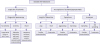 | Figure 4A flow diagram for management approach of isolated SMA dissection.CT = computed tomography; SMA = superior mesenteric artery.
|
The follow up in these patients who underwent endovascular revascularization ranged from 2 to 50 months, with an average follow up of 16 months. Longer follow ups are needed to determine the efficacy of endovascular management.
Follow up
There are no available guidelines for the interval of follow-up and imaging studies for SMA dissection patients. More studies are needed to determine the long-term benefits of each of the different management modalities. In our review, the cumulative follow up for all studies ranged from 1 week to 7.5 years. This longest follow up was observed in a patient who was managed conservatively and there were no further SMA dissection episodes reported. The longest follow-up for endovascular treatment was 4.1 years, and 11 years for surgical procedure in 3 patients.3)93) We believe that repeat CT scans should be performed on follow up in all patients to monitor the progression/resolution of SMA dissection in cases of conservatively managed SMA dissection and to look for the patency of stenting in endovascularly managed cases. Similarly, CT scan, if performed on regular follow up, can give an idea about post-surgical long-term complications in SMA dissection patients. Our review showed that the interval of follow up and hence the duration of post management imaging varied widely among all studies. In a study by Sakamoto et al.,3) a CT scan was performed weekly initially for the first month and then only twice or thrice over the span of years thereafter. We advocate that there should be evidence-based recommendations for regular follow up imaging for each treatment modality of SMA dissection.
CONCLUSION
SMA dissection is strongly associated with hypertension and smoking, presents mostly with intractable acute abdominal pain but can also be picked up as an incidental finding on CT scan or angiography. The conservative and endovascular management approach could be used in most patients, which can reduce costs and surgery-related morbidity and mortality. Surgical management should be reserved for complicated cases where conservative or endovascular management has no role or when there are other compelling indications for surgery like vessel rupture or bowel infarction. Our study furthermore revealed that there was no concordance among the 145 cases when it came to follow up and imaging studies but serial CT scans for monitoring the progress or resolution of SMA dissection is of paramount importance in conservatively managed cases while CT angiography is beneficial as arteriography for monitoring stent patency in endovascularly managed cases and for monitoring post-surgical complications.
 XML Download