

 ePub
ePub Citation
 Print
Print


INTRODUCTION
Atopic dermatitis (AD) is a chronic relapsing inflammatory skin disease. Its pathogenesis is complicated, but skin barrier or immune dysfunctions and genetic susceptibility play an important role in AD occurrence1. Genes encoding the proteins associated with skin barrier functions, including filaggrin (FLG), serine protease inhibitor Kazal-type 5 (SPINK5), and kallikrein 7 (KLK7)234, and genes related to immunity, including β-defensin 1 (DEFB1), kinase insert domain receptor (KDR), tumor necrosis factor alpha56, Fc epsilon receptor 1 alpha, and interleukins (ILs) 4, 5, 9, 10, 12, 13, and 187891011, were reported to be involved in AD development. However, there are considerable differences in mutations between different ethnic and regional groups. It is essential that valuable AD-related genetic variations in patients of the same race or region be clarified for clinical applications.
We recently compared genetic variations related to skin barrier functions and immunity between AD subjects and non-AD controls in Korea using a reverse blot hybridization assay (REBA), which can simultaneously detect multiple genetic mutations. Mutations of KLK7, SPINK5, FLG, DEFB1, KDR, IL-5 receptor alpha (IL5RA), IL9, and IL-12 receptor beta-1 subunit (IL12RB1) genes were significantly frequent in AD patients. Moreover, we found that the larger the number of gene variants, the higher the prevalence of AD12.
We hypothesized that there would be distinguishing characteristics in early-onset AD subjects. We aimed to identify genetic variations and clinical characteristics that could predict early AD development.
MATERIALS AND METHODS
Subjects and comparisons
We defined early-onset AD as AD that occurred at ≤3 years of age and late-onset AD as AD that occurred at >3 years of age. The study population consisted of 28 early-onset AD subjects and 57 non-AD controls from a birth cohort (Cohort for Childhood Origin of Asthma and Allergic Diseases [COCOA], a multi-center prospective birth cohort of a Korean inner-city population13); and 108 early-onset AD subjects, 90 late-onset AD subjects and 189 non-AD controls from a university hospital (Wonju Severance Christian Hospital). The data of the birth cohort were collected from November 2007 to December 2015 and that of the university hospital was collected from December 2008 to July 2017. In the birth cohort, AD subjects and non-AD controls were classified at three years of age by pediatric allergy specialists.
The study was comprised of two main comparisons (Fig. 1). The first main comparison was performed to identify different genetic variations between early-onset AD subjects and non-AD controls. We compared early-onset AD subjects (n=28) with non-AD controls (n=57) in the birth cohort (original comparison). However, there were some limitations in the population of the birth cohort. The number of the subjects was small, and subjects who could develop AD later on were included with the non-AD controls. Therefore, we performed an additional comparison to compensate for these limitations. In the additional comparison, 51 early-onset AD subjects from the university hospital were added to the 28 early-onset AD subjects of the birth cohort (n=79), and they were compared with the non-AD controls from the university hospital (n=189). The 51 early-onset AD university hospital subjects were chosen from a total of 108 early-onset AD subjects because they first visited our clinic at ≤3 years of age. We considered that they would have similar characteristics to the cohort population. The 189 non-AD controls from the university hospital were comprised of subjects, of various ages, without AD, while the non-AD controls of the birth cohort included subjects who could develop AD later on. The second main comparison was performed to identify different genetic variations and clinical characteristics that could predict early AD onset between early- and late-onset AD subjects. The 108 early-onset AD subjects and 90 late-onset AD subjects from the university hospital were included in this analysis.
The Yonsei University Wonju Campus Institutional Review Board approved this study (CR316120). Informed consent was obtained from all patients or the guardians of subjects.
Clinical characteristics
Basic data, including sex, age, the onset age of AD, the duration of AD, and the self-reported personal and family histories of atopic diseases (AD, allergic rhinitis, and asthma), were collected. Total serum immunoglobulin E (IgE) levels and allergen-specific IgE levels for 41 common allergens, including Dermatophagoides farinae (DF) and Dermatophagoides pteronyssinus (DP), were recorded.
Genetic variations
Single-nucleotide polymorphisms (SNPs) in genes associated with skin barrier functions (KLK7, SPINK5, and FLG) and immunity (DEFB1, KDR, IL5RA, IL9, and IL12RB1) were analyzed, because their SNPs were significantly more frequent in AD patients compared with those in non-AD controls in our previous study12.
Specific primer sequences for each gene were obtained from GenBank (http://www.ncbi.nlm.nih.gov/genbank/). Polymerase chain reaction (PCR) was performed, with primers, on genomic DNA extracted from blood samples using the QIAamp DNA Mini Kit (QIAGEN GmbH, Hilden, Germany). The amplification mixture for genes contained 1X primer mix, 2X PCR premix (Genet Bio, Daejeon, Korea), 2 mM of MgCl2, 250 µM of deoxynucleotide triphosphates, and 10 ng of genomic DNA in a final mixture volume of 50 µl. Multiplex PCR was performed using this mixture, followed by the direct sequencing of both strands of PCR products using the ABI PRISM 3100 Genetic Analyzer (Applied Biosystems, Foster City, CA, USA) at Cosmo Genetech Co., Ltd. (Seoul, Korea). Subsequent sequence alignment was performed using multiple sequence alignment programs (http://multalin.toulouse.inra.fr/multalin/).
Reverse blot hybridization assay
Genus-specific oligonucleotide probes of each gene were designed using sequence data from the National Center for Biotechnology Information database, followed by a Basic Local Alignment Search Tool search (https://blast.ncbi.nlm.nih.gov/Blast.cgi) to confirm the sequence homology of probes. A REBA membrane that could detect 13 wild-type (WT) and 13 mutant-type probes was designed. The REBA was performed as follows12: 20 µl of each PCR product was mixed with denaturation solution (0.2 N NaOH and 0.2 mM ethylenediaminetetraacetic acid [EDTA]) and incubated for 5 minutes. Denatured PCR products were diluted with 960 µl of 2X saline–sodium phosphate–EDTA (SSPE)/0.1% sodium dodecyl sulfate (SDS). REBA membrane strips were placed on MiniTrays (Bio-Rad, Hercules, CA, USA) and incubated with 2X SSPE/0.1% SDS for 5 minutes. After removing the residual fluid, slots were filled with denatured single-stranded PCR products. PCR products were incubated at 55℃ for 30 minutes, washed twice with 2X SSPE/0.5% SDS at 62℃ for 10 minutes, and then incubated in 1:2,000 diluted streptavidin-conjugated alkaline phosphatase (Roche Diagnostics GmbH, Mannheim, Germany) with 2X SSPE/0.5% SDS for 30 minutes. Hybridized amplicons were colorimetrically detected by incubating the strips in 1:50 diluted nitro blue tetrazolium chloride/5-bromo-4-chloro-3-indolyl phosphate p-toluidine salt (Roche Diagnostics GmbH) in 67% dimethyl sulfoxide (v/v) with Tris-buffered saline (pH 9.5) for 5 to 10 minutes. The presence of WT and mutant-type probes was confirmed by analyzing the band pattern.
Statistical analysis
Differences in genetic variations were identified by calculating odds ratios (ORs) with a chi-squares or Fisher's exact test, as appropriate. A logistic regression was performed to compare clinical characteristics between subjects in second main comparison. Statistical analyses were performed using IBM SPSS Statistics ver. 23.0 (IBM Co., Armonk, NY, USA). p-values less than 0.05 were considered statistically significant.
RESULTS
Comparisons of genetic variations between early-onset AD subjects and non-AD controls
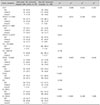
In the original comparison of the birth cohort (Table 1), no differences in the frequency of genetic variations were observed between early-onset AD subjects and non-AD controls.
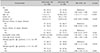
In the additional comparison (Table 2), early-onset AD subjects from the university hospital and birth cohort were compared with non-AD controls from the university hospital. A heterozygous mutant of KLK7 was less prevalent in early-onset AD subjects than its WT (OR, 0.468; 95% confidence interval, 0.265~0.826). Heterozygous mutants of SPINK5 1156 (2.034; 1.167~3.545), DEFB1 (2.498; 1.297~4.813), IL5RA (3.068; 1.778~5.293), IL12RB1a (2.210; 1.281~3.815), and IL12RB1b (2.984; 1.721~5.175) were significantly associated with early-onset AD compared to their WTs.
Comparisons of clinical characteristics and genetic variations between early- and late-onset AD subjects
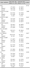
Early-onset AD subjects were younger and had more prolonged disease durations and family histories of atopic diseases than late-onset AD subjects. Late-onset AD subjects had more prevalent allergen-specific IgE positivity (≥3+) for DF and DP (Table 3), than early-onset AD subjects. However, no differences in sex, onset age, the personal history of atopic diseases, eosinophil count, total serum IgE levels, and the number of sensitized allergens were observed between both groups. There were no differences in the frequency of genetic variations between both groups (Table 4).
DISCUSSION
Genetic variations play a significant role in AD occurrence14. Some studies have attempted to identify genetic variations that can predict early AD development and have reported that FLG mutations are associated with an earlier onset21516171819. Dežman et al.20 suggested that polymorphism rs2303067 in SPINK5 is associated with early-onset AD, whereas Heo et al.21 reported that COL6A6 polymorphisms are novel candidate variants in early-onset AD. Additionally, Bergmann et al.22 reported that cord blood IgE levels and parental histories of atopy are predictors of early-onset AD. Paternoster et al.14 showed that AD-related genetic risks and personal or parental histories of atopic diseases are associated with early-onset AD.
We intended to analyze the clinical characteristics and genetic variations associated with early AD development in Koreans. In the comparison between early-onset AD subjects and non-AD controls in the birth cohort, no differences in AD-related genetic variations, even FLG mutations, were observed. Theoretically, this comparison would most likely show the genetic variations associated with early-onset AD occurrence. Considering that genes associated with AD occurrence were analyzed, this negative finding may have resulted from the limited number of subjects and characteristics of the non-AD controls in the birth cohort. AD subjects were classified at three years of age. Therefore, non-AD controls in the birth cohort included subjects who could develop AD later on. To compensate for these drawbacks in the cohort population, an additional comparison was conducted by combining the birth cohort and university hospital's populations. In early-onset AD subjects, although not completely consistent, certain genetic variations in genes associated with AD occurrence tended to be more present than in the non-AD controls.
In our result, there were differences in prevalence of genetic variations of KLK7, SPINK5, DEFB1, IL5RA, IL12RB1a, and IL12RB1b between early-onset AD subjects and non-AD controls. The KLK7 gene encodes stratum corneum chymotryptic enzyme (SCCE), and the SPINK5 gene encodes lymphoepithelial Kazal-type-related inhibitor which affects SCCE activity. They have crucial roles in formation and maintenance of skin barrier. Unlike our previous reports12, the prevalence of mutant of KLK7 was less in early-onset AD subjects compared to non-AD controls. This may be resulted from heterogeneity and limited number of sample. DEFB1 encodes β-defensin 1, one of the antimicrobial peptides (AMPs) that have broad antimicrobial property5. It affects AD pathogenesis in terms of innate immunity. AMPs including defensins are decreased in the skin of AD patients12. IL-5 and IL-12 are cytokines related with adaptive immunity, and dysregulations of their pathways affect the pathogenesis of AD. It is well known that IL-5 activates IL-5 receptor, then their pathway prolongs eosinophil lifespan which is of significance in the AD pathogenesis7. IL-12 is involved in promoting T helper 1 (Th1) immune response and cell mediated immunity. IL-12 receptor is mainly expressed on activated T cells and natural killer cells. Its reduced expression causes increasing Th2 cytokine production and may contributes to occurrence of AD and other allergic diseases23.
Meanwhile, there were no differences in the frequency of genetic variations between early- and late-onset AD subjects, as revealed by the comparisons performed to identify genetic variations that could predict early AD development. It is suggested that the onset age of AD is not decisively determined by AD-related genetic variations.
FLG mutations are associated with earlier AD onset in previous studies151718. Patients with major genetic risks develop symptoms earlier, but in patients whose genetic susceptibility is not prominent, the disease probably initiates later and prolonged environmental exposure is needed to fully develop AD24. Moreover, the frequency of FLG mutations in Asians is much lower than that in Europeans25. This allows us to explain that the difference between our results and those of European studies could have been because (1) mutations of other genes may have a greater effect on AD occurrence than FLG mutations in Koreans and (2) environmental exposures may have a greater effect on AD onset than genetic factors.
Early-onset AD subjects were more likely to have a family history of atopic diseases, which is consistent with the findings of previous studies on predictive factors for early-onset AD1422. In addition, the allergen-specific IgE levels for DF and DP were high in late-onset AD subjects. It is expected that early-onset AD subjects would have higher sensitization rates because allergen exposure through an impaired skin barrier is initiated early. However, our results can be explained by the following: (1) In addition to impaired skin barrier, allergens could be sensitized through the nasal and lung mucosa, (2) late-onset AD subjects were relatively older than early-onset AD subjects, and (3) considering that there were no differences in AD-related genetic variations between both groups, it is suggested that the duration of allergen exposure, or age, are important factors for sensitization. Limitations for this study include the heterogenous nature of the combined birth cohort and university hospital's population and the relatively small sample size.
In conclusion, although AD-related genetic variations can result in AD, the onset age of AD in Koreans cannot be determined. A family history of atopic diseases and environmental exposure are considerable factors that determine AD onset. Neonates with a family history of atopic diseases are likely to develop AD early, and the controlling a person's environmental exposure is important in delaying AD development. Our results may lead to AD prevention and help practitioners provide proper treatment and education to their patients.


 XML Download