

 ePub
ePub Citation
 Print
Print


INTRODUCTION
Skin is an organ that lies between organism and environment. It consists of three layers such as epidermis, dermis and subcutaneous fat. Skin protects organism from external stimuli such as microbes, harmful chemicals and ultraviolet light. Keratinocytes are the major cells in the epidermis that supply the fundamental protective role against harmful insults by making physical barrier. This protective barrier is produced by the sophisticated but well-ordered differentiation program of keratinocytes, in which many genes are spatiotemporally regulated to make solid water-impermeable structure called cornified cell envelope1. In recent years, another important defensive roles of keratinocytes have been recognized. That is, keratinocytes play a role as the immune surveillance cells in innate immunity. Keratinocytes sense the signals from environmental pathogen via the toll-like receptor (TLR) system and initiate the immune responses including the increase of gene expression for many cytokines23. In normal condition, the activation of keratinocyte innate immune system helps to eliminate the external pathogens and to provoke adaptive immune response, thereby contributing to maintenance of homeostasis. However, in pathologic conditions such as psoriasis, keratinocyte activation results in abundant expression of cytokines including interleukin (IL)-1β, tumor necrosis factor (TNF)-α and IL-17, thereby exacerbating the inflammatory condition and then leading to onset of disease456. Considering the importance of these initial innate immune responses, the inhibition of excessive responses by keratinocytes can be a good target of drug development for inflammatory skin diseases such as psoriasis.
We attempted to screen the therapeutic materials for inflammatory skin diseases using an experimental model in which keratinocyte inflammatory reaction was induced using a double-stranded RNA (dsRNA) mimic polyinosinic: polycytidylic acid (poly(I:C))7. We found that salvianolic acid A (SAA) has an inhibitory potential on inflammatory reaction of keratinocytes. Previously, it has been reported that SAA protects against myocardial ischemia/reperfusion injury by reducing platelet activation and inflammation8. SAA also has been reported to alleviate the ischemic brain injury through the inhibition of inflammation and apoptosis in mice9. However, the effects of SAA on skin keratinocytes have never been addressed so far. In this study, we provide evidence that SAA has an inhibitory effects on poly(I:C)-induced inflammatory reaction of skin keratinocytes.
MATERIALS AND METHODS
Ethics statement
Skin tissues were obtained from volunteers under the written informed consent, and the present study was approved from the ethical committee of the Institutional Review Board of Chungnam National University Hospital (IRB no. 1011-135, 2016-07-009).
Cell culture and drug preparations
The immortalized SV40T-transformed human epidermal keratinocytes (SV-HEKs) were cultured using keratinocyte-serum free medium supplemented with bovine pituitary extract (BPE) and recombinant human epidermal growth factor (rhEGF) (Life Technologies Corporation, Grand Island, NY, USA)10. At about 70% confluency, culture medium was changed into MCDB153 (Welgene, Gyeongsan, Korea) supplemented with BPE and rhEGF. After overnight incubation, cells were treated with 1 µg/ml of poly(I:C) (InvivoGene, San Diego, CA, USA). SAA was purchased from Sigma-Aldrich (St. Louis, MO, USA) and dissolved in dimethyl sulfoxide (DMSO).
Cytotoxicity test
For cytotoxicity test, well-established MTT (3-(4,5-dimethyl-2-thiazolyl)-2,5-diphenyl-2H-tetrazolium bromide) assay was employed. After treatment with SAA for 24 hours, cells were incubated with MTT solution (0.5 mg/ml) and cell viability was validated by checking optical density at 570 nm.
Quantitative real-time polymerase chain reaction (qRT-PCR)
Total RNA was purified and reverse transcribed using moloney-murine leukaemia virus reverse transcriptase (MMLV-RTase; Elpis Biotech, Daejeon, Korea). The resulting cDNA was used in real-time PCR. The gene-specific primers were used as below: IL-1β, 5′-TTAAAGCCCGCCTGACAGA and 5′-GCGAATGACAGAGGGTTTCTTAG; IL-6, 5′-CTGCGCAGCTTTAAGGAGTTC and 5′-CCATGCTACATTTGCCGAAGA; IL-8, 5′-CCTTTCCACCCCAAATTTATCA and 5′-TTTCTGTGTTGGCGCAGTGT; CCL20, 5′-CCACCTCTGCGGCGAAT and 5′-TGTGTATCCAAGACAGCAGTCAAA; TNF-α, 5′-CTCCTTCAGACACCCTCAACCT and 5′-CGACCCTAAGCCCCCAATT; GAPDH, 5′-TGCACCACCAACTGCTTAGC and 5′-GGCATGGACTGTGGTCATGAG.
Enzyme-linked immunosorbent assay (ELISA)
The secreted cytokines were quantified by ELISA. The commercial kit for IL-8 was purchased from Life Technologies Corporation. The ELISA kits for IL-1β and TNF-α were obtained from R&D Systems (Minneapolis, MN, USA).
Western blotting
Cellular extracts were prepared using a lysis buffer (Intron, Daejeon, Korea). The cellular proteins were separated by gel electrophoresis and then transferred to nitrocellulose membranes. The membranes were incubated with primary antibody, and then sequentially incubated with peroxidase-conjugated secondary antibodies. The Western bands were obtained by enhanced chemiluminescence (Intron). The following primary antibodies were used: phospho-p65, phospho-IκBα, caspase-1 (Cell Signaling Technology, Beverly, MA, USA); IL-1β (Abcam, Cambridge, MA, USA); NLRP3 and ASC (AdipoGen, San Diego, CA, USA); actin (Sigma-Aldrich).
RESULTS
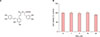
To screen the effective materials that can inhibit poly(I:C)-induced inflammation in skin keratinocytes, we initially used ELISA for IL-8 and single compounds commercially available. Among many compounds tested, SAA showed inhibitory potential on secretion of IL-8, thus we chose SAA for a further study. SAA is a polyphenolic compound (Fig. 1A), which has been originally extracted from Radix Salvia miltiorrhiza11. To determine the cytotoxicity, we treated SV-HEKs with different doses of SAA and performed MTT assay. As a result, SAA did not show the cytotoxic effect up to the dose of 10 µM, although insignificant slight decrease of cell viability was seen at the dose of 20 µM (Fig. 1B). Therefore, we treated keratinocytes with SAA up to 10 µM in the following experiments.
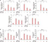
Poly(I:C) is a chemical mimicking the dsRNA of some viruses, which provokes innate immune reaction in a TLR3-dependent fashion in skin keratinocytes12. To examine the effect of SAA, we pre-treated SV-HEKs with SAA then stimulated with poly(I:C). Inflammatory reaction was first determined by qRT-PCR. In the absence of pre-treatment with SAA, poly(I:C) resulted in dramatic increase of mRNAs for inflammatory cytokines including IL-1β, IL-6, IL-8, TNF-α, and CCL20. By contrast, pre-treatment of SAA significantly inhibited poly(I:C)-induced expression of inflammatory cytokines (Fig. 2A). We next checked the secreted cytokines from keratinocytes using ELISA. Consistent with the data obtained from qRT-PCR, poly(I:C) treatment led to dramatic increase of secretion of inflammatory cytokines such as IL-1β, IL-8, and TNF-α from keratinocytes. Pre-treatment with SAA markedly inhibited poly(I:C)-induced secretion of inflammatory cytokines (Fig. 2B).
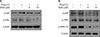
It has been well recognized that nuclear factor-κB (NF-κB) signaling is pivotal in innate immune responses of skin keratinocytes1314. To delineate the potential mechanism, we investigated the effect of SAA on activation of NF-κB signaling. Consistent with previous data, poly(I:C) treatment led to activation of NF-κB signaling that was evidenced by increase of phosphorylation of p65 subunit and IκBα. Pre-treatment of keratinocytes with SAA resulted in significant inhibition of poly(I:C)-induced phosphorylation of p65 and IκBα (Fig. 3A). In addition to NF-κB signaling, mitogen-activated protein kinases (MAPKs) are also implicated in poly(I:C)-induced inflammatory reaction in keratinocytes and other systems151617. Thus, we examined whether SAA can also inhibit MAPK pathway using Western blot. As a result, SAA significantly inhibited poly (I:C)-induced phosphorylation of p38 MAPK, JNK, and ERK1/2 (Fig. 3B).
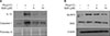
When skin keratinocytes were stimulated by poly(I;C), the large caspase-1-activating complex called inflammasome is assembled and then proteolytic activation of IL-1β is occurred. The active IL-1β is then released and participates in the induction of inflammation, contributing to establishment of inflammatory skin diseases such as psoriasis718. To examine whether SAA affects inflammasome activation, we collected cell culture medium and then checked the released IL-1β and caspase-1. As anticipated, treatment of keratinocytes with poly(I:C) resulted in remarkable increase of IL-1β secretion and inflammasome-activated caspase-1. Pre-treatment with SAA markedly inhibited the secretion of IL-1β and caspase-1 (Fig. 4A). As the inflammasome is a complex structure in which component proteins such as NLRP3 and ASC are involved19, we next examined the protein levels of NLRP3 and ASC. Poly(I:C) increased the protein level of NLRP3, and this poly(I:C)-increased NLRP3 was obviously inhibited by SAA. However, there was no significant effect on ASC level by poly(I:C) treatment (Fig. 4B).
DISCUSSION
Skin keratinocytes play an important role in protection of organism by providing physical and functional defense systems. The one involves barrier structure that is composed of water-insoluble corneocytes and intercellular lipid molecules, the other includes immune modulatory potential in which many cytokines and chemokines are coordinately regulated by pathogen-sensing machinery. Especially for innate immunity, keratinocytes exert their important role as a primary guardian against non-self and/or self antigens through the activation of TLR systems. It has been demonstrated that keratinocytes express a line of functional TLRs including TLR3, 4, 5, 7 and 92021. Among them, TLR3 acts as a receptor for dsRNA that can be found in some viruses. In condition related to skin disease such as psoriasis, keratinocytes are susceptible to viral RNA intermediates, and activation of TLR3 leads to drastic increase of pro-inflammatory cytokines and chemokines including IL-1β, TNF-α, IL-6, IL-8 and CCL2022. The increased inflammatory cytokines in turn contributes to recruit and activate immune cells such as neutrophils and activated T cells23. Thus, the drug screening against inflammatory reaction of keratinocytes is a good approach for the development of therapeutics for skin diseases such as psoriasis. In this study, we provide evidence that SAA inhibits poly(I:C)-induced inflammatory reaction of keratinocytes, suggesting that SAA can be applicable for the treatment of skin disease such as psoriasis.
SAA is a polyphenolic depside that is composed of three of monocyclic aromatic units linked by ester bond (Fig. 1A). It was originally isolated from the dried roots of Salvia miltiorrhiza11. SAA has a variety of biological and pharmacological activities, including anti-tumor, anti-metastatic, anti-oxidant, and anti-inflammatory effects. For example, SAA induces cell apoptosis and suppresses tumor growth in acute myeloid leukemia via the inhibition of PI3K/Akt signaling24. Other evidence indicates that SAA inhibits the migration and invasion of human breast cancer cells by inactivating transgelin225. In addition, SAA inhibits the high-fat diet-induced hepatic inflammation via a NLRP3 inflammasome-dependent way in rat liver26.
In this study, we demonstrated that SAA has inhibitory potential on poly(I:C)-induced inflammatory reaction in skin keratinocytes. The effects of SAA were likely due to its inhibitory potential on NF-κB signaling, because that NF-κB is key player in the inflammatory reaction. As well recognized, NF-κB directly binds to promoter of many inflammatory cytokine genes, thereby provoking consequent relay of inflammatory process. It has been reported that SAA attenuates NF-κB activation in other systems such as mouse macrophages and rat renal tissues2728. Thus, it is plausible that the SAA inhibits the activation of NF-κB in skin keratinocytes thereby affecting downstream cellular events. In this regard, it is likely that the inhibition of inflammasome activation by SAA is also linked to NF-κB signaling. Inflammasome activation is an important process in innate immunity, and it has been also recognized that NF-κB activation plays a role in inflammasome activation. When pathogen-associated molecular patterns or damage-associated molecular patterns are recognized by defense cells, inflammasome component protein NLRP3 associates with the adaptor protein ASC. The assembly of NLRP3 and ASC results in the formation of protein complex that can activate the caspase-1 by proteolytic cleavage of pro-caspase-129. The resultant active caspase-1 is then secreted from keratinocytes, thereby contributing to the pathogenesis of skin disease such as psoriasis30. In our study, SAA markedly inhibited poly(I:C)-induced activation of NF-κB signaling, and also inhibited inflammasome activation in terms of decreasing the NLRP3 level and the IL-1β secretion. Thus, our data provide evidence supporting that SAA can be used for treating innate immunity-related skin diseases.
In summary, we demonstrated that SAA inhibited poly (I:C)-induced inflammatory reaction of epidermal keratinocytes. Our data suggest that SAA would be a candidate for treatment of chronic inflammatory skin disease such as psoriasis.
 XML Download