

 PDF
PDF ePub
ePub Citation
 Print
Print


INTRODUCTION
Crohn's disease (CD) is an inflammatory bowel disease characterized by transmural inflammation that can occur throughout the gastrointestinal tract. However, the terminal ileum is the most frequently involved location. The prevalence of CD among children less than 20 years of age is 43 per 100,000 [1]. In most cases, CD is associated with periods of worsening inflammation and, over time, leads to complications like strictures and fistulas [2]. Intestinal inflammation can typically be managed with medical therapy, however, fibrotic strictures often require surgical or endoscopic treatments like resection or dilation [23]. Therefore, characterization of the bowel wall to determine the extent of inflammation and/or fibrosis is necessary to guide proper clinical management and to assess the response to therapy in these patients.
Ultrasound (US) is a well-established imaging modality that is useful in detecting CD. US parameters such as bowel wall thickness, vascularity, abdominal free fluid, bowel stenosis, and the presence of fistulas or abscesses are considered to be specific markers for CD. However, the ability of conventional grayscale US and Doppler to assess disease activity remains controversial [4]. Contrast-enhanced US (CEUS) allows for qualitative and quantitative assessment of kinetic perfusion parameters in the bowel. Lumason® (Bracco Diagnostics, Inc., Monroe Township, NJ, USA) is an US contrast agent available in the United States and is approved by the Food and Drug Administration for the evaluation of liver and vesicoureteral reflux in the pediatric population. However, off-label uses of US contrast agents in evaluating many conditions in adults and children have been well established in the literature [5]. Some studies have utilized CEUS for distinguishing active and inactive CD, demonstrating a sensitivity of 93.5% and a specificity of 93.7% [6]. Quantitatively, these studies have demonstrated that a shorter time-to-peak intensity and a higher peak enhancement correlates with increased disease activity in these patients [7]. However, other studies have demonstrated no significant association of these same parameters with disease activity [8].
Another useful US technique is elastography, which can be used to map tissue stiffness. Shear wave elastography (SWE) utilizes an acoustic radiation force to excite a region of interest for evaluation. The technology works by a measuring the speed of the shear waves that travel away from this region, which are proportional to tissue elasticity [910]. A few studies have utilized elastography to assess fibrotic from inflammatory strictures in CD. In both, elastography was useful in distinguishing fibrotic from non-fibrotic strictures [1112]. However, because strictures in CD can involve both fibrotic and inflammatory tissue, distinguishing fibrosis from inflammation remains challenging.
Here we present three cases where CEUS and elastography were used to evaluate disease activity in patients with CD. These patients had a spectrum of disease activity including acute inflammation, chronic inflammation with stricture formation, and a post-surgical fibrotic stricture. In all three patient cases, CEUS and elastography were useful in demonstrating the degree of inflammation and presence of fibrosis. We also discuss interpreting CEUS kinetic parameters and elastography values in the evaluation of CD activity.
CASE REPORT
CEUS
This study was approved by the Institutional Review Board of Johns Hopkins Medicine. CEUS scans were performed after the patient or parental off-label consent was obtained. The US exams were performed by a sonographer with over 20 years of experience, with an EPIQ scanner (Philips Healthcare, Bothell, WA, USA) using a broadband 162-element curved array transducer (C5-1, 1–5 MHz, and 55.5 mm). The contrast agent was prepared per the manufacturer's instructions (Lumason®; Bracco Diagnostics, Inc.). A board-certified pediatric radiologist performed the contrast injection. Each patient had two boluses of contrast agent injected (0.03 mL/kg), the first injection to evaluate the terminal ileum and the second injection to evaluate a segment of the proximal jejunum, to serve as the as an internal control, as well as to evaluate the remainder of the bowel loops. The jejunum was utilized as an internal control since solitary jejunal involvement is uncommon in CD, and various studies have demonstrated that jejunal involvement occurs only in 14%–16.7% of patients [1314]. While it may not be appropriate to use the jejunum as a control in every case, in our clinical experience, we have infrequently seen jejunal inflammation in our pediatric patients. In these cases, the scan time was approximately 7 minutes after each bolus, and 1-minute wash-in cine clips of the terminal ileum were obtained along with static images.
A 1 mm2 square region of interest was drawn around the wall of the most narrowed segment of the terminal ileum (or bowel anastomosis) for all three patients. Perfusion kinetics parameters were calculated using the automatic post processing QLAB contrast quantification software on the scanner. The parameters were time to peak, wash-in slope, peak intensity, and area under the curve. A perfusion kinetic curve is generated from the auto fit curve option, which uses a local density random walk model to create a curve and derives quantification parameters from that curve.
SWE
SWE was obtained on the same EPIC scanner (Philips Healthcare) prior to the CEUS evaluation. A 1 cm2 region of interest was placed over the narrowest portion of the terminal ileum to obtain three elastography values, from which the mean elastography value was recorded.
Magnetic resonance enterography (MRE)
MRE was performed on all three patients, using axial and coronal T2-weighted True Fast Imaging with Steady State Precession and T1-weighted, fat saturated, pre- and post-contrast sequences through the abdomen and pelvis. The resulting examination was performed with the patients situated in prone position, using the multichannel phased array abdominal coil. “VoLumen” (biphasic endoluminal contrast) was given by mouth one hour prior to the examination for adequate bowel distension. In this case, intravenous glucagon, corrected for body weight, was given for the suppression of bowel motion during the exam.
Cases
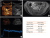
The first patient was a 16-year-old female (height: 1.58 m, weight: 41.7 kg) with a recent diagnosis of CD, who two days prior had received her first infusion of infliximab. She was admitted to the gastroenterology service for severe abdominal pain which began that morning. The patient was unable to tolerate anything by mouth due to severe nausea. At that time, the grayscale US demonstrated a narrowing of the terminal ileum, with a thickened submucosal layer and decreased peristalsis. Contrast US showed avid contrast enhancement of the narrowed terminal ileum. Quantitative evaluation of the terminal ileum, compared to the jejunum as an internal control, demonstrated more prompt wash-in, higher peak intensity, and higher area under the curve. The kinetic perfusion parameters are reported in Fig. 1. Average elastography value of the terminal ileum was 0.64 m/s. MRE demonstrated wall thickening and inflammation in the terminal ileum, resulting in narrowing and proximal dilation in the distal ileal segments. Adjacent to the terminal ileum, there was mesenteric inflammation as evidenced by contrast enhancement (Fig. 1). Given the patient's MRE findings and her clinical improvement with a second infusion of infliximab, her flair was determined to be acute inflammation.
Fig. 1
A case of acute inflammation in a child with Crohn's disease. (A) Contrast-enhanced ultrasound demonstrates avid contrast enhancement in the narrowed terminal ileum. (B) Transverse contrast magnetic resonance imaging of the abdomen demonstrating bowel wall thickening and inflammation in the terminal ileum. (C) Contrast enhancement curve and quantitative parameters generated from the time-intensity curves based on the drawn region of interest in the terminal ileum.
LDRW: local density random walk, WIWO: wash-in, wash-out, AUC: area under the curve.
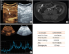
The second patient was a 20-year-old female (height: 1.52 m, weight: 79.8 kg) with a history of ileocolonic CD who had undergone an ileocecal resection five years prior. The patient was receiving adalimumab once every two weeks. The patient was admitted to the gastroenterology service due to abdominal pain of a few weeks' duration. The morning of the admission, the patient had multiple episodes of non-bloody, non-bilious emesis and non-bloody diarrhea. The patient's most recent colonoscopy from six months prior had demonstrated a tight surgical anastomosis through which the scope could not be passed. During her admission, contrast US demonstrated minimal enhancement of the surgical anastomosis and mild enhancement of the more proximal ileum, greater than seen near the anastomosis. The measured mean elastography value was 1.57 m/s at the surgical anastomosis. Kinetic perfusion parameters are reported in Fig. 2. MRE demonstrated mild hyperenhancement of the small bowel loops without definite wall thickening or mesenteric hyperemia. Additionally, there was no high-grade ileal stricture, abscess, or fistula that was demonstrated at that time (Fig. 2).
Fig. 2
A case of a fibrotic stricture in a child with Crohn's disease. (A) Contrast-enhanced ultrasound demonstrates minimal contrast enhancement in the surgical anastomosis. (B) Transverse magnetic resonance imaging of the abdomen demonstrates mild hyperenhancement of the small bowel loops without definite wall thickening or mesenteric inflammation. (C) Contrast enhancement curve and quantitative parameters generated from the time-intensity curves based on the drawn region of interest in the surgical anastomosis.
LDRW: local density random walk, WIWO: wash-in, wash-out, AUC: area under the curve.
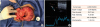
The third patient was a 17-year-old girl (height: 1.66 m, weight: 81.6 kg) with a history of CD who underwent a consultation with a pediatric general surgeon for an ileocolic stricture. A previous colonoscopy demonstrated a stricture at the ileocecal valve with the presence of ulcers in the terminal ileum. CEUS demonstrated a mildly thickened terminal ileum, with decreased peristalsis and minimal contrast uptake. The mean elastography value in the terminal ileum was 1.58 m/s. In this case, MRE demonstrated bowel wall thickening with increased T2 signal and contrast enhancement involving the distal ileum, cecum, and the proximal ascending colon. The patient then underwent an elective ileocecectomy. The surgical findings were of extensive fibrosis and fat creeping of the terminal ileum. The associated pathology demonstrated an ileum with active chronic inflammatory disease (cryptitis, crypt abscess, ulceration, pseudopolyp formation, crypt distortion, stricture formation, transmural chronic inflammation, and noncaseating-granulomas). The intraoperative picture demonstrates the stretched-out bowel stricture (Fig. 3).
Fig. 3
A case of chronic inflammation and stricture formation in a child with Crohn's disease. (A) Intraoperative picture from ileocecectomy demonstrating the stretched-out stricture. The surgical instrument on the top indicates the area of the worst disease activity. (B) Contrast enhancement curve and quantitative parameters generated from the time-intensity curves based on the drawn region of interest in the terminal ileum.
LDRW: local density random walk, WIWO: wash-in, wash-out, AUC: area under the curve.
DISCUSSION
In this case series, we discuss three patients presenting with abdominal pain in the setting of CD, in which CEUS and elastography were useful in detecting the degree of inflammation and fibrosis in the bowel. A few studies have demonstrated the utility of CEUS in evaluating CD activity in the pediatric population. A study by Pallotta et al. [15] demonstrated that CEUS had a sensitivity and specificity for detecting undiagnosed CD of 100% and 100%, compared to a sensitivity and specificity of 75% and 100%, respectively, for conventional transabdominal US alone. However, few studies have used quantification parameters derived from CEUS to evaluate CD activity in the pediatric population. The first study to do so, by Kljucevsek et al. [16], utilized CEUS derived quantification parameters to optimize medical therapy in a 13-year-old boy with CD. The researchers in this study demonstrated that a high intensity of enhancement and a short time-to-peak enhancement were associated with active inflammation of the small bowel, and subsequently optimized the patient's medical therapy, leading to clinical improvement and normalization of the bowel on a subsequent US examination [16]. However, most published studies utilizing CEUS derived kinetic parameters to evaluate CD were performed in adults. Other studies have demonstrated the value of elastography to evaluate children with CD. A study by Fufezan et al. [17] created an elastography scoring system for the assessment of CD inflammation and fibrosis. This classification system demonstrated that elastography could predict increased levels of disease activity markers, like C-reactive protein and erythrocyte sedimentation rate, and could predict disease complications [17]. However, there is a paucity of research that utilizes CEUS and elastography together to assess CD activity in the pediatric population.
The first patient presented with acute inflammation that responded to treatment with infliximab. MRE demonstrated the presence of inflammation, which correlated with US findings. At that time, CEUS demonstrated avid contrast enhancement of the terminal ileum, suggestive of acute inflammation. Quantitatively, compared to the jejunum, the terminal ileum was noted to have more prompt wash-in, increased peak intensity, and increased area under the curve. These findings also favor the presence of active inflammation. The average elastography value in the terminal ileum was 0.64 m/s, suggestive of less stiff and less fibrotic tissue. Our results are consistent with a number of studies that have demonstrated increased contrast enhancement by qualitative and quantitative measures in inflammatory CD [1819]. A number of studies have correlated contrast enhancement and CD activity both histologically and endoscopically in these patients [192021]. A study by Ripollés et al. [20] demonstrated that inflammatory lesions had greater contrast enhancement when compared to fibrotic lesions. Additionally, our patient had a low elastography value, which was consistent with previous studies that demonstrate decreased tissue stiffness associated with acute inflammation when fibrosis is not present [192223]. Together, these findings suggest acute inflammation without the presence of fibrosis. These findings are supported by her clinical improvement with infliximab.
The second patient had minimal inflammation, and her fibrotic surgical anastomosis was most likely the source of her pain. The MRE findings of postsurgical versus mild inflammatory changes without evidence of a high-grade inflammatory stricture correlated with US findings. CEUS demonstrated that the anastomosis had minimal enhancement, suggestive of none to minimal inflammation. Quantitatively, the small values of the peak enhancement and area under the curve also suggested the presence of minimal inflammation. Additionally, the mean elastography value was 1.57 m/s which is suggestive of the presence of a stiffer tissue, containing a more fibrotic component. Taken together, these findings correlate with the fibrotic surgical anastomosis noted on a previous endoscopy.
The third patient underwent an elective bowel resection. The CEUS findings of minimal contrast uptake suggested a lack of acute inflammation. The terminal ileum had an elastography value of 1.58 m/s denoting an increased level of stiffness. These findings were consistent with surgical findings of fibrosis and histological findings of active chronic inflammation with stricture formation in this patient.
In these last two patients, CEUS and elastography were useful in ruling out acute inflammation of the bowel. However, the ability to differentiate fibrotic from inflammatory strictures with CEUS remains controversial. Quaia et al. [24] demonstrated that area under the curve and peak enhancement could be used to differentiate predominantly fibrotic from predominantly inflammatory strictures. However, another study using an animal model demonstrated that quantitative perfusion parameters cannot detect the degree of fibrosis in CD when inflammation is also present. They found that no quantitative parameter could reliably detect fibrosis [23]. Strictures often contain both inflammatory and fibrotic components, and as such, CEUS perfusion parameters are predominantly affected by the degree of inflammation [23]. Our results are consistent with this study, as CEUS effectively identified the presence of acute inflammation, but a lack of contrast enhancement was common to both the surgical fibrotic stricture and the chronic inflammation with stricture formation. Indeed, the quantitative CEUS findings were similar in these two patients. A previous study by Quaia et al. [25] suggested that elastography could complement CEUS in distinguishing the presence of fibrosis, regardless of the degree of inflammation. In our study, elastography demonstrated the presence of a fibrotic stricture in both patients. The elastography value for the patient with active chronic inflammation with stricture formation was comparable to the patient with a surgical fibrotic anastomosis. The increased tissue stiffness in both patients is likely due to underlying fibrosis in the bowel wall, which is present in most ileal strictures, even those with inflammatory infiltrate on histology review [25].
In conclusion, our study demonstrates that CEUS and elastography combined are valuable tools in monitoring disease activity in patients with CD. CEUS is most useful in determining the degree of acute inflammation, and quantitative parameters are not as useful in assessing bowel wall fibrosis. However, elastography can identify bowel wall fibrosis through an evaluation of tissue stiffness. Used together, these techniques allow for a better characterization of the degree of fibrosis and inflammation in bowel strictures in patients with CD. CEUS and elastography offer promise in improving the assessment of CD activity and response to treatment.
 XML Download