

 PDF
PDF ePub
ePub Citation
 Print
Print


INTRODUCTION
Choroid plexus tumors are rare, accounting for only 0.3–0.6% of all brain tumors and 10–20% of those in infants [1]. Choroid plexus tumors include choroid plexus papilloma (CPP), atypical CPP, and choroid plexus carcinoma (CPC). Of these subtypes, CPCs are the most aggressive and malignant at a World Health Organization grade III. CPC primarily occurs in children, and the median age of patients with CPC is 3 years, highlighting the rarity of adult-onset CPC. As CPC is derived from choroid plexus epithelium, it usually presents with cerebrospinal fluid (CSF) obstruction and progresses through CSF metastasis [123]. Mechanical obstruction of the CSF is a common cause of symptoms, including headache, diplopia, and ataxia, and is followed by hydrocephalus [4]. Because of their origin in the choroid plexus epithelium, the majority of CPCs are located within the ventricle. A diffuse border between the tumor and normal brain tissue reflects brain invasion. Maximal surgical resection followed by adjuvant chemotherapy and radiotherapy is the recommended treatment but has yet to be standardized [5].
Here, we report 2 rare cases of adult CPC, which exhibited differing clinical presentation: multiple ventricular seeding and a single juxtaventricular mass. The institutional review board exempt any informed consent from the patients unless the retrospective study reveal personal identifiable information.
CASE REPORT
Case 1
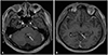
A 40-year-old man who was diagnosed with CPC and leptomeningeal seeding visited our outpatient clinic for a second opinion. The medical history revealed that his chief complaint was dizziness, nausea, and headache, and brain MRI revealed nodular lesions at the 4th ventricle and tectum (Fig. 1). Subtotal removal of 4th ventricular mass was performed, and the pathologic diagnosis was CPC. After the surgery, he complained of sacral area pain. T1-weighted gadolinium enhanced spine MRI showed linear nodular enhancement of whole spinal cord surface, which was compatible of disseminated spinal metastasis (Fig. 2), and CSF cytology was positive for malignant cells. Craniospinal irradiation of 3,600 cGy was delivered in 12 fractions, but the patient progressed to show cauda equina syndrome. Follow-up spine MRI presented aggravated leptomeningeal seeding, and nivolumab was administered as additional salvage chemotherapy. Unfortunately, the patient continued to suffer from worsening symptoms.
Neurological exam at the time of admission to our clinic, 7 months after the brain surgery, showed left side lower extremity weakness of motor grade 4 out of 5 with saddle hypoesthesia, right facial palsy, and facial hypoesthesia. Systemic etoposide and carboplatin (CPT-SIOP-2000) chemotherapies were given [6], and intrathecal methotrexate was administered 4 weeks after the chemotherapy in 2 treatments [7]. However, the patient could get neither symptom improvement nor cytological/radiological response. He deferred any further treatment with slow progression of existing neurological deficiency. Four months after the intrathecal chemotherapy, the patient expired.
Case 2
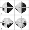
A 49-year-old woman was transferred to our emergency room from another hospital for treatment of an alleged malignant brain tumor. Her chief complaint was visual field disturbance, and she had experienced a generalized tonic-clonic seizure a week prior. She presented right side homonymous hemianopsia upon visual field test (Fig. 3A), and papilledema was observed with fundoscopy.
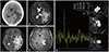
Brain CT revealed a large (diameter of 4 cm) iso-dense intra-axial ovoid mass in the left temporo-occipital area (Fig. 4A). A T2-weighted brain MRI revealed that the mass contained an internal cystic region, abutted the choroid plexus, and had potentially invaded the trigone of the lateral ventricle (Fig. 4B, C). Gadolinium enhancement showed homogenous enhancement of the solid portion, suggesting that the mass was a high-grade glioma or metastatic tumor (Fig. 4D). The radiologist recommended both MR spectroscopy and a metastasis work-up. MR spectroscopy revealed a high choline peak that was consistent with malignant tumors (Fig. 4E). However, the metastasis work-up, including whole-body positron emission tomography, colonoscopy, and esophagogastroduodenoscopy, was negative for systemic primary cancer.
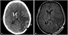
Tumor removal was performed with a transcortical approach via the parieto-occipital junction. The resected tumor was covered with a glistening capsule and was found to be attached to the choroid plexus at the end of the removal. Intraventricular hemorrhage was observed immediately postoperation via a brain CT, and gadolinium enhanced brain MRI confirmed the gross total removal of the tumor 24 hours after the operation (Fig. 5).
Histologically, the tumor was well-encapsulated with a distinct fibrous capsule and was composed of columnar epithelial cells on fibrovascular cores (Fig. 6A, B). The tumor cells had prominent nuclear pleomorphism, suggestive of a carcinoma. Immunohistochemically, tumor cells were focally positive for epithelial membrane antigen and transthyretin (Fig. 6C). Moreover, tumor cells were positive for synaptophysin and negative for other marker including glial fibrillary acidic protein and vimentin. The Ki-67 index was 21.66%. Altogether, these results led to a final diagnosis of CPC. Based on this diagnosis, whole-spine MRI and CSF cytology were performed, and both tests were negative for leptomeningeal seeding.
Right side homonymous hemianopsia gradually improved during follow-up and was largely resolved at postoperative 3 months (Fig. 3B). After consultation with a pediatric oncologist, the patient received 4 cycles of carboplatin-etoposide chemotherapy without any severe side effects. The patient showed neither radiological nor clinical signs of progression at postoperative 10 months.
DISCUSSION
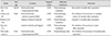
As reviewed by Sun et al. [1], the location of CPCs varies with age. Most lateral ventricular tumors occur in patients younger than 20, whereas 4th ventricular CPCs are evenly distributed amongst all age groups. Presentation with multiple CPCs is relatively rare, occurring in only 5% of CPC patients. The first case in this report presented with 4th ventricular CPC and a tectal mass at 40 years of age. For lateral and 3rd ventricle CPCs, the median age of presentation is 1.5 years, and supratentorial CPCs are extremely rare in adults. In fact, we could only retrieve 4 cases from the literature [891011], which are summarized in Table 1. The second case presented here was diagnosed at age 49, and the location of the tumor was more consistent with ‘extraventricular’ than lateral ventricle. Differential diagnosis based on imaging characteristics are difficult in extraventricular cases of CPC as well as CPP, choroid plexus cyst, ependymoma, primitive neuroectodermal tumor, astrocytoma, germinoma, teratoma, meningioma, metastasis to the choroid plexus, and xanthogranuloma [1]. Differential diagnosis is further complicated by the increased tendency of CPCs to invade the parenchyma.
CPC is associated with poor prognosis, and the 5-year survival rate for patients with CPCs is approximately 40% [12]. Numerous authors have emphasized the importance of gross total removal of CPC as part of the therapeutic strategy [138913]. The extent of tumor removal is associated with significantly superior overall survival (OS); whereas, the effect of adjuvant chemotherapy or radiotherapy on OS remains controversial [14]. Based on meta-analysis of Wolff et al. [13], adjuvant radiation can improve survival even after gross total resection; however, this effect was only statistically significant in older age young patients (3–9 years old) and not in infants. Adjuvant radiation is also used in cases with leptomeningeal dissemination and spinal metastases. Nevertheless, there is no definite established protocol for radiation or chemotherapy in CPC. Current CPC treatment is based on data from pediatric patients who are heavily treated with chemotherapy, and the role of chemotherapy has not been established in adult CPCs [1615]. Evidence from the few reported cases of adult-onset CPC suggests that, even after gross total removal of the lesion with adjuvant chemotherapy or radiotherapy, the outcome of adult CPC varies (Table 1).
In the cases presented here, CPC patients were treated by surgical resection and adjuvant chemotherapy and/or radiotherapy. In case 1, the patient was treated with craniospinal irradiation after craniotomy for spinal metastasis. As spinal metastasis progressed even with radiation, adjuvant chemotherapy was administered for systemic control. The treatment response was poor, and the patient expired within a year. On the other hand, in case 2, the operation was performed with gross total resection of the lesion, and postoperative spinal images and CSF studies showed no evidence of CSF metastasis. Adjuvant chemotherapy was administered for systemic control without any severe side effects, and there was no sign of progression of the disease up to 10 months postoperation. It is evident that further studies are necessary to establish standardized radiation and chemotherapy strategies in adult CPC patients.


 XML Download