Abstract
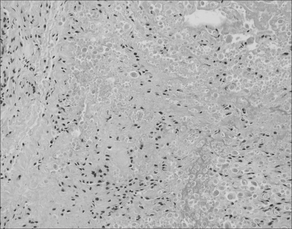
Calcium deposition disease, including calcific tendinitis, rarely affects the knee joint. Only a few cases can be found in the literatures and there is no case report of symptomatic calcific deposition arising from the posterior cruciate ligament in Korea. The authors encountered a case of symptomatic calcific deposition arising from the posterior cruciate ligament, which was excised arthroscopically and confirmed pathologically. This paper reports this case with a review of the relevant literature.
References
1. Chan R, Kim DH, Millett PJ, Weissman BN. Calcifying tendinitis of the rotator cuff with cortical bone erosion. Skeletal Radiol. 2004. 33:596–9.

2. Uhthoff HK, Loehr JW. Calcific tendinopathy of the rotator cuff: pathogenesis, diagnosis, and management. J Am Acad Orthop Surg. 1997. 5:183–91.

3. Holt PD, Keats TE. Calcific tendinitis: a review of the usual and unusual. Skeletal Radiol. 1993. 22:1–9.

4. McKendry RJ, Uhthoff HK, Sarkar K, Hyslop PS. Calcifying tendinitis of the shoulder: prognostic value of clinical, histologic, and radiologic features in 57 surgically treated cases. J Rheumatol. 1982. 9:75–80.
5. Shenoy PM, Kim DH, Wang KH. . Calcific tendinitis of popliteus tendon: arthroscopic excision and biopsy. Orthopedics. 2009. 32:127.

6. Tibrewal SB. Acute calcific tendinitis of the popliteus tendon: an unusual site and clinical syndrome. Ann R Coll Surg Engl. 2002. 84:338–41.
7. Chan W, Chase HE, Cahir JG, Walton NP. Calcific tendinitis of biceps femoris: an unusual site and cause for lateral knee pain. BMJ Case Rep. 2016. 2016. pii: bcr2016215745.

8. Song K, Dong J, Zhang Y. . Arthroscopic management of calcific tendonitis of the medial collateral ligament. Knee. 2013. 20:63–5.

9. Molloy ES, McCarthy GM. Hydroxyapatite deposition disease of the joint. Curr Rheumatol Rep. 2003. 5:215–21.

10. Kim MK, Bae JH, Jeon YS. Conservative and early ar-throscopic treatment of calcific tendinitis. J Korean Ar-throscop Soc. 2009. 13:149–54.
Figure 1.
Plain anteroposterior (A) and lateral (B) radiographs of the left knee demonstrating a calcific lesion just below intercondylar notch (arrows).
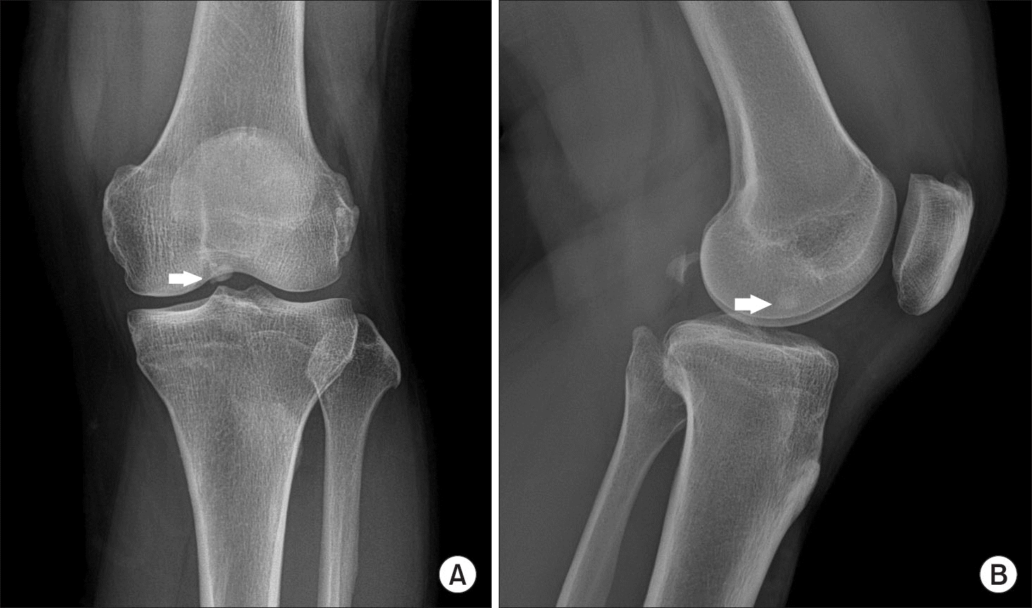
Figure 2.
(A) Sagittal magnetic resonance imaging, T2-weighted of the left knee. A round mass with well circumscribed intermediate signal intensity was observed just inferior to the posterior cruciate ligament (PCL). (B) A low signal intensity mass was noted in the posterior aspect of the PCL in T1-weighted image.
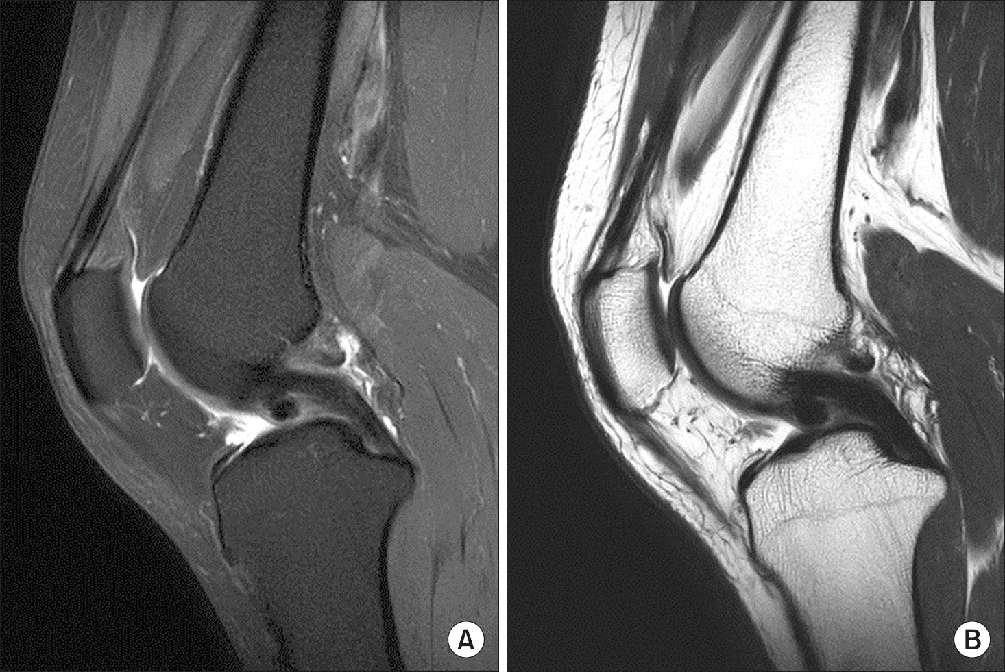
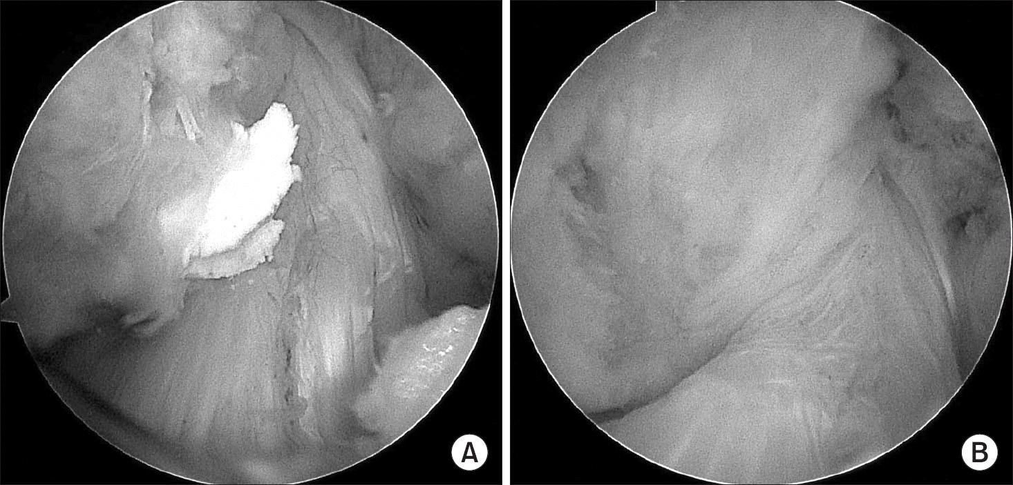
Figure 3.
(A) On the anterolateral portal view, there were calcific deposits with a ‘tooth-paste’ consistency arising from the posterior cruciate ligament. (B) After excising the mass from the anteromedial portal with the shaver, it was removed completely by an arthroscopic technique.


 PDF
PDF ePub
ePub Citation
 Print
Print


 XML Download