

 PDF
PDF ePub
ePub Citation
 Print
Print


Jae-Gu Park, M.D.*, Hyung-Jin Chung, M.D., Ph.D. , Su-Young Bae, M.D., Ph.D., Jung-Hwan Lee, M.D., Hwi-Young Kim, M.D., Jun Seok Lee, M.D.
, Su-Young Bae, M.D., Ph.D., Jung-Hwan Lee, M.D., Hwi-Young Kim, M.D., Jun Seok Lee, M.D.
Abstract
Purpose
This study examined the radiological and clinical outcomes of tibiotalocacalcaneal arthrodesis using retrograde intramedullary nailing in a severe hindfoot deformity and ankle/subtalar arthritis.
Materials and Methods
A total of 22 patients (22 cases) with a severe hindfoot deformity and arthritis underwent tibiotalocalcaneal arthrodesis with retrograde intramedullary nails. The average age was 57.4 years (22–82 years) and the mean follow-up was 29.6 months (12–74 months). The radiological outcomes included an assessment of the preoperative and postoperative coronal ankle alignment, hindfoot alignment, sagittal alignment, and postoperative union time. The clinical outcomes were evaluated using the visual analogue scale (VAS), American Orthopaedic Foot and Ankle Society (AOFAS) score, and postoperative complications. The results were analyzed statistically by dividing the patients into two groups based on a 10° angle of deformity.
Results
Regarding the preoperative coronal ankle alignment, 14 patients had a mean varus deformity of 17.8°±14.5° and six had a mean valgus deformity of 8.1°±6.6°. Postoperatively, a satisfactory postoperative coronal ankle alignment of less than 5° was obtained in all patients. Regarding the preoperative hindfoot alignment, 12 patients showed a mean varus deformity of 15.2°±10.5° and six had a mean valgus deformity of 8.1°±4.2°. In total, 94.4% (17 patients) had satisfactory postoperative hindfoot alignment of less than 5°. Radiological union was achieved in 90.9% at an average of 19.2 weeks (12–32 weeks) and there were 2 cases of nonunion. The clinical outcomes showed improvement in the mean VAS and AOFAS scores (p<0.001, p<0.001, respectively). Even a preoperative severe deformity more than 10° showed a significant deformity correction of coronal ankle alignment and hindfoot alignment, postoperatively (p<0.001, p<0.001, respectively). No significant differences were found between the patients with a preoperative coronal ankle deformity more than 10° and those less than 10° regarding the mean postoperative coronal ankle alignment (p=0.162).
Conclusion
Tibiotalocalcaneal arthrodesis using retrograde intramedullary nailing is an acceptable technique for achieving satisfactory deformity correction, high union rate with minimal complications, and improvement of the clinical outcomes. In addition, tibiotalocalcaneal arthrodesis using retrograde intramedullary nailing is considered an effective treatment option, particularly in severe ankle and hindfoot deformities.
Figures and Tables
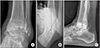 | Figure 1(A) Coronal ankle alignment is defined as the angle between the anatomical axis of the tibia and a line drawn perpendicular to the talar dome in a standing anteroposterior radiograph. (B) Hindfoot alignment is defined as the angle between the anatomical axis of the tibia and the longitudinal axis of the calcaneus in the hindfoot alignment view. (C) Sagittal alignment is defined as the angle between the anatomical axis of the tibia and a line connecting the inferior aspect of the posterior tubercle of the talus to the inferior talar neck in a standing lateral radiograph.
|
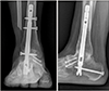 | Figure 2Both anteroposterior and lateral radiographs show the successful fusion trabeculae crossing the fusion site.
|
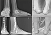 | Figure 3Standing radiographs and photograph of a 54-year-old female with equinocavovarus deformity demonstrates a correction of the deformity and osseous union after tibiotalocalcaneal arthrodesis with intramedullary nail.
|

References
1. Rammelt S, Pyrc J, Agren PH, et al. Tibiotalocalcaneal fusion using the hindfoot arthrodesis nail: a multicenter study. Foot Ankle Int. 2013; 34:1245–1255.

2. Hanson TW, Cracchiolo A 3rd. The use of a 95 degree blade plate and a posterior approach to achieve tibiotalocalcaneal arthrodesis. Foot Ankle Int. 2002; 23:704–710.
3. Mückley T, Ullm S, Petrovitch A, et al. Comparison of two intramedullary nails for tibiotalocalcaneal fusion: anatomic and radiographic considerations. Foot Ankle Int. 2007; 28:605–613.

4. Fang Z, Claaßen L, Windhagen H, Daniilidis K, Stukenborg-Colsman C, Waizy H. Tibiotalocalcaneal arthrodesis using a retrograde intramedullary nail with a valgus curve. Orthop Surg. 2015; 7:125–131.

5. Doets HC, Brand R, Nelissen RG. Total ankle arthroplasty in inflammatory joint disease with use of two mobile-bearing designs. J Bone Joint Surg Am. 2006; 88:1272–1284.
6. Larsen A, Dale K, Eek M. Radiographic evaluation of rheumatoid arthritis and related conditions by standard reference films. Acta Radiol Diagn (Stockh). 1977; 18:481–491.
7. Neri T, Barthelemy R, Tourné Y. Radiologic analysis of hindfoot alignment: comparison of Méary, long axial, and hindfoot alignment views. Orthop Traumatol Surg Res. 2017; 103:1211–1216.
8. Buck P, Morrey BF, Chao EY. The optimum position of arthrodesis of the ankle. A gait study of the knee and ankle. J Bone Joint Surg Am. 1987; 69:1052–1062.
9. Holt ES, Hansen ST, Mayo KA, Sangeorzan BJ. Ankle arthrodesis using internal screw fixation. Clin Orthop Relat Res. 1991; (268):21–28.
10. Morgan CD, Henke JA, Bailey RW, Kaufer H. Long-term results of tibiotalar arthrodesis. J Bone Joint Surg Am. 1985; 67:546–550.
11. Kitaoka HB, Anderson PJ, Morrey BF. Revision of ankle arthrodesis with external fixation for non-union. J Bone Joint Surg Am. 1992; 74:1191–1200.
12. Morrey BF, Wiedeman GP Jr. Complications and long-term results of ankle arthrodeses following trauma. J Bone Joint Surg Am. 1980; 62:777–784.
13. Boer R, Mader K, Pennig D, Verheyen CC. Tibiotalocalcaneal arthrodesis using a reamed retrograde locking nail. Clin Orthop Relat Res. 2007; 463:151–156.
14. Brodsky JW, Verschae G, Tenenbaum S. Surgical correction of severe deformity of the ankle and hindfoot by arthrodesis using a compressing retrograde intramedullary nail. Foot Ankle Int. 2014; 35:360–367.
15. Pelton K, Hofer JK, Thordarson DB. Tibiotalocalcaneal arthrodesis using a dynamically locked retrograde intramedullary nail. Foot Ankle Int. 2006; 27:759–763.
16. Hammett R, Hepple S, Forster B, Winson I. Tibiotalocalcaneal (hindfoot) arthrodesis by retrograde intramedullary nailing using a curved locking nail. The results of 52 procedures. Foot Ankle Int. 2005; 26:810–815.
17. Mader K, Pennig D, Gausepohl T, Patsalis T. Calcaneotalotibial arthrodesis with a retrograde posterior-to-anterior locked nail as a salvage procedure for severe ankle pathology. J Bone Joint Surg Am. 2003; 85:Suppl 4. 123–128.
18. Millett PJ, O'Malley MJ, Tolo ET, Gallina J, Fealy S, Helfet DL. Tibiotalocalcaneal fusion with a retrograde intramedullary nail: clinical and functional outcomes. Am J Orthop (Belle Mead NJ). 2002; 31:531–536.
19. Niinimäki TT, Klemola TM, Leppilahti JI. Tibiotalocalcaneal arthrodesis with a compressive retrograde intramedullary nail: a report of 34 consecutive patients. Foot Ankle Int. 2007; 28:431–434.
20. McGarvey WC, Trevino SG, Baxter DE, Noble PC, Schon LC. Tibiotalocalcaneal arthrodesis: anatomic and technical considerations. Foot Ankle Int. 1998; 19:363–369.
21. Thordarson DB, Chang D. Stress fractures and tibial cortical hypertrophy after tibiotalocalcaneal arthrodesis with an intramedullary nail. Foot Ankle Int. 1999; 20:497–500.
22. Thomas RL, Sathe V, Habib SI. The use of intramedullary nails in tibiotalocalcaneal arthrodesis. J Am Acad Orthop Surg. 2012; 20:1–7.
23. Anderson T, Linder L, Rydholm U, Montgomery F, Besjakov J, Carlsson A. Tibio-talocalcaneal arthrodesis as a primary procedure using a retrograde intramedullary nail: a retrospective study of 26 patients with rheumatoid arthritis. Acta Orthop. 2005; 76:580–587.
24. Goebel M, Gerdesmeyer L, Mückley T, et al. Retrograde intramedullary nailing in tibiotalocalcaneal arthrodesis: a short-term, prospective study. J Foot Ankle Surg. 2006; 45:98–106.
25. Haaker R, Kohja EY, Wojciechowski M, Gruber G. Tibio-talo-calcaneal arthrodesis by a retrograde intramedullary nail. Ortop Traumatol Rehabil. 2010; 12:245–249.
26. Chou LB, Mann RA, Yaszay B, et al. Tibiotalocalcaneal arthrodesis. Foot Ankle Int. 2000; 21:804–808.
27. Kitaoka HB, Patzer GL. Arthrodesis for the treatment of arthrosis of the ankle and osteonecrosis of the talus. J Bone Joint Surg Am. 1998; 80:370–379.
28. Moore TJ, Prince R, Pochatko D, Smith JW, Fleming S. Retrograde intramedullary nailing for ankle arthrodesis. Foot Ankle Int. 1995; 16:433–436.
 XML Download