

 PDF
PDF ePub
ePub Citation
 Print
Print


INTRODUCTION
Foreign body aspiration is the most common respiratory emergency in dental clinics, because dental treatments use small instruments and materials in the oral cavity, which may deteriorate the patient's reflex mechanism [1]. The treatment method depends on whether the foreign body has moved to the gastrointestinal or respiratory tract and whether it could potentially damage the gastrointestinal tract or respiratory mucosa. Aspired foreign bodies are mostly found in the right main bronchus because of its smaller angle and larger diameter compared the left main bronchus. Half the patients with foreign body inhalation and over 90% with foreign body ingestion are asymptomatic and may be unaware that an inhalation or ingestion has occurred [2].
Reports on foreign body aspiration during general anesthesia for dental treatments are rare. Accidental foreign body ingestion has been reported in patients before waking from anesthesia, when the lung function has not fully recovered [3]. No aspiration cases during dental treatments under stable general anesthesia have been reported because these treatments allow neither voluntary movements nor reflex actions. General anesthesia using anesthetic gases and muscle relaxants stops all swallowing and reflex functions, and the patient is subjected to artificial respiration. After complete anesthesia, swallowing is stopped, and aspiration is unlikely because of physical barriers, such as tongue, packing, and endotracheal tube. This is a case report on the fall of a prosthesis in the larynx, which occurs rarely under general anesthesia.
CASE REPORT
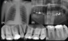
A 36-year-old man visited the dental hospital for treatment of decayed teeth. He had Wilson's disease, which causes impaired copper metabolism. He was able to communicate with people but had mental retardation and extreme fear and anxiety toward dental treatment. He took Altamin and anticonvulsants once a day. His weight was 60 kg, and his general condition was normal. The posteroanterior chest radiograph revealed no abnormal findings (Fig. 1A). Intraoral clinical and radiographic examinations revealed generalized gingivitis and multiple moderate-to-advanced dental caries (Fig. 1B–E). The patient needed multiple operative, periodontal, and prosthodontic treatments. After general examination, two minimal visits of dental treatment using general anesthesia were planned. Before the surgery, he underwent blood tests, chest radiography, and risk assessments. Written informed consent for the planned procedures were obtained from him and his guardian.
Procedures
The patient was instructed to maintain his routine medication. His electrocardiography, heart rate, blood pressure, respiratory rate, end-tidal capnography, body temperature, and entropy levels were monitored during the procedures. An intravenous (IV) line was secured under 4 L/min of O2 and N2O gases (administered via a face mask), and 4% sevoflurane gas and atracurium (IV 25 mg) were also administered. Under an adequate depth of anesthesia, the epiglottis was examined with a bronchoscope and an oral camera, and gentle intubation using a Mac size#3 laryngoscope was performed. The trachea was successfully intubated with a 26-mm endotracheal tube (ETT), without bleeding. Anesthesia was maintained with 2 L/min of O2 and N2O and 2% sevoflurane. The entropy level was recorded every 10 minutes. In both the general anesthesia, response entropy (RE) was maintained in the range of 35–54 and state entropy (SE) of 30–45 during the procedure. No specific events occurred (Table 1).
At the first visit, scaling, composite filling of tooth 12, and preparation and final impression for indirect inlay were performed under general anesthesia, and crown preparation, final impression, and provision of gold crowns for teeth 16 and 37 were achieved. At the second visit, the delivery of five prosthetic teeth (16, 24, 26, 27, and 37), simple extraction of tooth 18, and treatment of some caries were done.
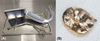
During the try-in step, the gold crown for tooth 36 slid from the surgeon's hand. The prosthesis was found neither on oral examination nor in the operation room. In a subsequent oral examination, when the tongue was tilted, the crown was found near the endotracheal tube balloon. The prosthesis was promptly removed using video-assisted laryngoscopy and curvilinear forceps (Fig. 2). Subsequently, dental floss-tied gauze packing was performed to prevent recurrence of the accident. After 2 h and 35 min from the treatment, the patient recovered upon 100% oxygen administration. During the procedure, the patient exhibited no specific complications. He was discharged 1 hour after stabilization.
DISCUSSION
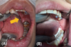
During dental treatment under general anesthesia, various foreign substances (tooth fragments, old restoration pieces, bur, filling material, implant components) are introduced in the oral cavity. If these foreign substances enter the airways, there is a high risk of inhalation in the respiratory tract, causing pneumonia during tube removal after the anesthesia recovery. To prevent such accidents, the rubber dam isolation method should be used for restorative and root canal treatments and oropharyngeal packs (packing gauze) for other surgical and prosthetic treatments [45]. However, packing gauzes could interfere with the dental treatment in patients with an enlarged tongue. Despite gauze packing, gaps may remain because of the tongue position (Fig. 3). The patient in this case had an enlarged tongue. To reduce tongue interference during the occlusal adjustment of the prosthesis, the packing gauze was temporarily removed, which made the prosthesis fall inside the neck.
As the patient's voluntary motions and reflex actions were stopped, the molar prosthesis was expected to be easily visible over the tongue. However, in this case, the prosthesis was not found in the oral cavity, as it entered the retromylohyoid space, which is located below and behind the mylohyoid ridge and represents the S-curve of the mandible, usually covered by the tongue. However, when the tongue was laterally displaced for occlusion, the prosthesis fell in the gap between the tongue and the mandible and was difficult to find. In an attempt to find it, the surgeon moved the tongue from one side to another, causing the prosthesis to fall in the retromylohyoid space, down to the larynx. The missing prosthesis was found caught in the endotracheal tube because of its round and bulky shape. However, if the foreign body is a fractured sharp tip or a small diamond bur, it is difficult for the surgeon to find it. Preventing the foreign body from reaching the larynx is of utmost importance. Therefore, small-size gauze packing must be performed in specific procedures such as try-in of prostheses, and gauze should be bundled with black silk or dental floss for easy removal. In addition, video-assisted laryngoscopy can be used to remove foreign substances more rapidly and accurately in cases of narrow throat and poor visibility [6].
Davis et al. reported on reducing the possibility of aspiration by coating oral cavity with radiopaque material in children undergoing general anesthesia [7]. Studies have shown that blood, saliva, and pus touch the throat tissue even with a well-placed oropharyngeal pack. To prevent this, the wet packing should be frequently replaced during the treatment and suction should be used in the oral cavity.

 XML Download