

 PDF
PDF ePub
ePub Citation
 Print
Print


Introduction
Resin luting cement was developed for the cementation of esthetic materials such as all-ceramic and indirect-composite restorations. In comparison with conventional luting cements, resin luting cements have improved the cementation of base metal alloys due to their lower solubility, better wear resistance, and marginal closure,12 and it is widely used in dentistry.3 The use of self-adhesive resin cement, which combines the advantages of conventional and adhesive luting agents, is increasing since the self-adhesive resin cement is more user-friendly and less technique-sensitive than conventional resin cements.4
However, because of the low chemical affinity of resin cement to metal alloys, surface treatments are recommended to achieve a more durable bonding.567 A variety of surface treatments have been studied in an effort to improve bond strength, including mechanical and chemical bonding as well as combinations of both. The application of several functional monomers is considered as one of the most effective chemical treatments to enhance the physicochemical bonding of resin cements to metal alloy.89
There are various adhesive primers currently used in dentistry, and each primer contains functional monomers such as 4-methacryloyloxyethy trimellitate anhydride (4-META), 10-methacryloyloxydecyl dihydrogen phosphate (10-MDP), 6-methacryloxyhexyl 2-thiouracil-5-carboxylate (MTU-6), and methacryloyloxyalkyl thiophosphate derivatives (MEPS), that increase the retention of resin to a metal surface. However, determining the most effective functional monomer for bonding and the effects of primer application on bond strength remain in debate.5671011
Although macrotests such as tensile and shear bond tests are commonly used in studies to analyze metal-resin bond strength, these studies also have their limitations.12 To overcome some limitations of macrotests, Sano et al.13 used microtensile bond tests, which are considered more appropriate for the evaluation of bond strength since they allow a more uniform distribution of stress, reduce cohesive failure, and provide a more realistic measurement of bond strength at the adhesive interface. Cobalt-Chromium (Co-Cr) metal alloy specimens are difficult to fabricate for microtensile bond tests, and as a result are not widely reported in the literature at this time.
The purpose of this study is to evaluate the effects of various primers on the microtensile bond strength of resin cements to a Co-Cr dental casting alloy.
Materials and methods
In this study, we used Co-Cr alloy, four adhesive primers and two resin cements. Specific information concerning the materials utilized in this study is presented in Table 1. One hundred fifty Co-Cr metal beams (6 mm long, 1 mm wide, and 1 mm thick) were casted. After trimming and polishing the beams, the bonding surfaces were sandblasted with aluminum oxide (125 µm grain) for 5 seconds at 80-psi pressure. The distance from the nozzle to the metal surface was 1.5 cm. The metal beams were cleaned using an ultrasonic cleaner for 1 minute, and were then randomly divided into ten groups according to primer and resin cement; the no primer (NP) group served as the control (n = 15, Table 2). A silicone mold with rectangular cavities (12 mm long, 1 mm wide, and 1 mm thick) was used to fabricate the metal-resin beams (Fig. 1) and the resin cement was applied to the rest part of the mold. All materials were applied and handled according to the manufacturer's instructions. After light-curing, all specimens were allowed to completely set for 24 hours; the metal-resin beams were then stored in distilled water at 37℃ for 24 hours before conducting the microtensile bond test.
Prior to testing, all metal-resin beams were studied under a stereomicroscope (Damisystem, TaeShin BioScience, Namyangju, Korea) at ×30 magnification for flaws, bubbles, or excess resin on the specimen's bonding interface; specimens with defects were excluded. An active gripping method was used by attaching each specimen to the flat grip steel fixture of the microtensile tester (Micro Tensile Tester, BISCO, Schaumburg, IL, USA) using a light-cured adhesive (LIBOND PEN, DFS Diamon, Riedenburg, Germany). The metal-resin beams were loaded under tension at a cross-head speed of 1.0 mm/min using the microtensile tester machine (Fig. 2). Bond strength values were calculated using the formula, σ = L / A, where ‘L’ is the load at failure (N) and ‘A’ is the adhesive area (mm2). The mode of failure of each specimen was determined using a stereomicroscope.
The mean of each group was analyzed via one-way ANOVA with microtensile bond strength (µTBS) as the dependent variable, primer treatment as the independent factor, and Tukey's test as post hoc (α = .05) using SPSS software (SPSS ver. 22.0, IBM, Chicago, IL, USA).
Results
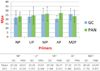
The mean µTBS of all groups was ranged from 20 to 28 MPa (Fig. 3). Panavia F2.0 groups and G-Cem LinkAce groups showed no significant difference in bond strength. In the comparison among groups when primers were used, no statistically significant differences were observed (P > .05).
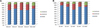
No pre-fabrication failures were found via the fractographic analysis after bonding of the resin cement to the metal alloy in all groups. Mixed failure, which is the combination of adhesive and cohesive failures, is the most prevalent failure mode in both the Panavia F2.0 and G-Cem LinkAce groups (Fig. 4).
Discussion
Traditionally, experimental designs for macrotests were used to compare bond strength to metal alloys in primer systems; however, because larger bonding areas lead to a higher possibility of error and may consequently reduce bond strength,14 microtests were developed.13 Microtensile bond testing is considered more appropriate for the evaluation of bond strength than microshear bond testing, since it allows a more uniform distribution of stress, reduces cohesive failure, and provides a more realistic measurement of the bond strength of the adhesive interface.1516 However, cutting of the specimen for microtensile bond testing is difficult and the vibrations from cutting the specimen using a water-cooled diamond saw during microtensile bond tests may create microcracks on the periphery,17 and these microcracks may result in high levels of pre-testing failures.18 Therefore, most studies on the bonding of metal alloy to resin cement use microshear bond tests, in which specimens can be fabricated without cutting.16 The specimen in this study was fabricated by casting a small-diameter metal beam and bonding it after fabrication, so that a microtensile test could be performed without the cutting.
In this study, we evaluate two adhesive systems: a conventional resin cement, Panavia F2.0, and a self-adhesive resin cement, G-Cem LinkAce. The 10-MDP, contained in Panavia F2.0, is a monomer mainly used as an etching monomer result from the function of dihydrogenphosphate group, and it has a quite hydrophobic property due to a long carbonyl chain render.19 The MDP has a coupling mechanism by: (i) dihydrogen phosphate group which presents great chemical bonding with Co-Cr alloy20 and (ii) the polymerizable methacryloyl group which is essential for copolymerizing the MDP monomer and the resin cement. The 4-META, which is included in the G-Cem LinkAce, is a monomer synthesized in the late 1970s.21 When mixing the 4-META with water, the hydrolysis reaction will occur and change 4-META to 4-MET and subsequently the esterification of 4-MET would promote adhesion.19 The chromium in the Co-Cr alloy produces a thin surface layer of chromic oxide at room temperature that can enhance the chemical bond between the Co-Cr alloy and 4-META.22
However, in this study, the bond strength of the G-Cem group showed no significant difference with the Panavia F2.0 group. The µTBS obtained for all groups were more than 20 MPa, similar to previous studies,23 which is clinically acceptable.24
The effect of a primer on bond strength varies with primer type. However, the use of metal primers for increasing the bond strength of non-precious alloys to resin cement remains controversial. According to Yoshida et al.,5 the bond strength between resin cement and sand-blasted casting alloy was significantly higher when the metal primer was applied due to the affinity of some functional monomers to the oxide layer of base metal alloys. In addition, one recent study reported the tested primer significantly improved the tensile and shear bond strength of the resin cement to metal alloys.25
In contrast, according to Di Francescantonio et al.,10 the use of alloy primer between metal alloy and resin cements did not increase the bond strength for most cementing systems tested. In the present study, although four primers with different functional monomers were applied to Co-Cr alloy, we observed no significant increase in bond strength in any of the four primer groups, which coincides with previous studies.68 It can be assumed that sandblasting increases the surface irregularities of the alloy and improves mechanical bond strength, and when the primer is applied, some of these surface irregularities may be filled. This mechanism can effect total microtensile bond strength, which may explain the results of this study.6
Other factors that may influence the durability of resin bonding and that were not evaluated in this study include pH changes, dynamic fatigue loading, thermocycling, and the various components of the resin cement and primer. Therefore, careful interpretation in the clinical application of these results is suggested and further in vitro research and standardized studies must be conducted to confirm the efficacy of the tested systems.


 XML Download