

 PDF
PDF ePub
ePub Citation
 Print
Print


Seok-Joo Doh , Jin-Hyun Cho
, Jin-Hyun Cho
Abstract
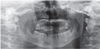
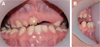
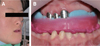
Patients with Down's syndrome have several dental complications such as small teeth caused by underdevelopment of dentin and enamel, periodontitis, agenesis of teeth, prolonged retention of primary teeth and malocclusion due to narrow palate. Removable denture with maxillary double crowns would be a good treatment option to solve the problems of the patient with Down's syndrome. Double crowns compensate the insufficient support and retention of denture and easily solve the cross bite problem. Double crowns also allow easy repair of denture in case of abutment teeth extraction. In this case, 26-year-old female patient with Down's syndrome and dental phobia had small number of teeth with enamel hypoplasia, prolonged retention of primary teeth and dental cross bite. Prosthetic treatment was done using removable denture with double crowns in the maxilla. In the mandible, teeth preparation was done on enamel margin without anesthesia. Anterior laminate and posterior complete zirconia crown restorations were performed. As a result, the cross bite was effectively corrected by denture with double crowns. Pronunciation and appearance were also improved without extraction of teeth and dental anesthesia.
Figures and Tables
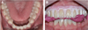
 | Fig. 3Maxillary impression and bite registration. (A)Border molding, (B) Impression, (C) Resulted stone cast, (D) Bite registration.
|
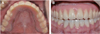
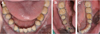 | Fig. 4Mandibular teeth preparation and gingival retraction with cord. (A) Whole occlusal view, (B) Right view, (C) Left view.
|


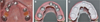
 | Fig. 7Maxillary pick up impression. (A) Inner crown splinting with pattern resin, (B) Pick up impression.
|

References
1. Shajpal A, Siddiqui F. Screening for Down syndrome. Obstet Gynaecol Reprod Med. 2017; 27:333–337.

2. Roizen NJ, Patterson D. Down's syndrome. Lancet. 2003; 361:1281–1289.

3. Crawford D, Dearmun A. Down's syndrome. Nurs Child Young People. 2016; 28:17.
4. van der Linden MS, Vucic S, van Marrewijk DJF, Ongkosuwito EM. Dental development in Down syndrome and healthy children: a comparative study using the Demirjian method. Orthod Craniofac Res. 2017; 20:65–70.
5. Macho V, Palha M, Macedo AP, Ribeiro O, Andrade C. Comparative study between dental caries prevalence of Down syndrome children and their siblings. Spec Care Dentist. 2013; 33:2–7.
6. Severin E, Paun A, Baltag R, Stan A, Funieru C. Common, rare, and individual oro-dental findings in people with Down syndrome. J Int Oral Health. 2016; 8:964–968.
7. Dawson PE. Functional occlusion: from TMJ to smile design. St. Louis: Mosby;2007. p. 114–131.
 XML Download