

 PDF
PDF ePub
ePub Citation
 Print
Print


Kyong-Seop Lee , Young-Jun Lim, Ho-Beom Kwon, Myung-Joo Kim
, Young-Jun Lim, Ho-Beom Kwon, Myung-Joo Kim
Abstract
Excessive wear causes many complications when untreated, so that accurate diagnosis, analysis and predictive treatment plan should be made, and through evaluation of vertical dimension and stepwise treatment, a stable inter-arch relationship can be set. For the long-term success of implant treatment, ideal position and angle of implant is important, and its importance increases especially in multiple implant cases. Therefore, thorough diagnosis and planning, accurate surgery and prosthodontic procedures are significant. In this case, a 68-year-old male patient with a loss of vertical dimension due to multiple tooth loss and overall tooth wear was planned with systematic analyses from the pre-treatment stage to rehabilitate vertical dimension. Full-mouth fixed rehabilitation with computer tomography guided implant surgery was performed to the newly set vertical dimension and attained satisfactory outcomes both functionally and esthetically.
Figures and Tables
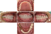
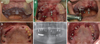
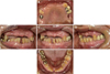 | Fig. 1Preoperative intraoral photographs. (A) Maxillary occlusal view, (B) Right lateral view, (C) Frontal view, (D) Left lateral view, (E) Mandibular occlusal view.
|
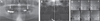 | Fig. 2Radiographic evaluation before treatment. (A) Panoramic radiograph at first visit, (B) TMJ panoramic, (C) Transcranial view.
|
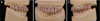
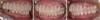
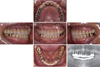
 | Fig. 4Provisional restoration before implant treatment. (A) Maxillary occlusal view, (B) Right lateral view, (C) Frontal view, (D) Left lateral view, (E) Mandibular occlusal view.
|
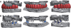
 | Fig. 5Fabrication of implant surgical guide using CAD program. (A) Digital wax up from frontal view, (B) Digital wax up from maxillary occlusal view, (C) Implant planning, (D) Closed sleeve type surgical guide.
|
 | Fig. 6Implant installation with surgical guide. (A, B, C) CT guided implant surgery with surgical guide, (D) After implant 1st surgery, (E) Panoramic radiograph after implant 1st surgery, (F) After implant 2nd surgery.
|
 | Fig. 7Digital wax-up for implant provisional prosthesis and design of titanium customized abutment. (A) Digital wax-up, (B) Titanium customized abutment design.
|
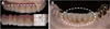
 | Fig. 8Placement of the implant provisional restorations after the connection of titanium customized abutments. The abutments were tightened twice with a torque of 30 Ncm. (A) Right lateral view, (B) Frontal view, (C) Left lateral view.
|
References
1. Lambrechts P, Braem M, Vuylsteke-Wauters M, Vanherle G. Quantitative in vivo wear of human enamel. J Dent Res. 1989; 68:1752–1754.


2. Verrett RG. Analyzing the etiology of an extremely worn dentition. J Prosthodont. 2001; 10:224–233.
3. Turner KA, Missirlian DM. Restoration of the extremely worn dentition. J Prosthet Dent. 1984; 52:467–474.
4. Al-Omiri MK, Lamey PJ, Clifford T. Impact of tooth wear on daily living. Int J Prosthodont. 2006; 19:601–605.
5. Muts EJ, van Pelt H, Edelhoff D, Krejci I, Cune M. Tooth wear: a systematic review of treatment options. J Prosthet Dent. 2014; 112:752–759.
6. Misch CE. Dental implant prosthetics. 2nd ed. St. Louis: Elsevier Mosby;2015. p. 19p. 193–205.
7. BouSerhal C, Jacobs R, Quirynen M, van Steenberghe D. Imaging technique selection for the preoperative planning of oral implants: a review of the literature. Clin Implant Dent Relat Res. 2002; 4:156–172.
8. Orenstein IH. The surgical template: a prescription for implant success. Implant Dent. 1992; 1:182–184.
9. Kola MZ, Shah AH, Khalil HS, Rabah AM, Harby NM, Sabra SA, Raghav D. Surgical templates for dental implant positioning; current knowledge and clinical perspectives. Niger J Surg. 2015; 21:1–5.

10. Oh SC, Jung JH. Morphology and size of clinical crown of permanent upper anterior teeth in Korean adult. J Korean Acad Stomatognathic Funct Occlusion. 2001; 17:38–42.
11. Abduo J, Lyons K. Clinical considerations for increasing occlusal vertical dimension: a review. Aust Dent J. 2012; 57:2–10.
12. Gopi Chander N, Venkat R. An appraisal on increasing the occlusal vertical dimension in full occlusal rehabilitation and its outcome. J Indian Prosthodont Soc. 2011; 11:77–81.
13. Dahl BL, Carlsson GE, Ekfeldt A. Occlusal wear of teeth and restorative materials. A review of classification, etiology, mechanisms of wear, and some aspects of restorative procedures. Acta Odontol Scand. 1993; 51:299–311.
14. Kim Y, Oh TJ, Misch CE, Wang HL. Occlusal considerations in implant therapy: clinical guidelines with biomechanical rationale. Clin Oral Implants Res. 2005; 16:26–35.
15. Raico Gallardo YN, da Silva-Olivio IRT, Mukai E, Morimoto S, Sesma N, Cordaro L. Accuracy comparison of guided surgery for dental implants according to the tissue of support: a systematic review and meta-analysis. Clin Oral Implants Res. 2017; 28:602–612.
 XML Download