

 PDF
PDF ePub
ePub Citation
 Print
Print


Choongkil Kim1 , Wonsup Lee2, Ho-Beom Kwon1
, Wonsup Lee2, Ho-Beom Kwon1
Abstract
Nowadays, dental implant is one of the widely used treatment options for edentulous patients. Recently, a method of improving the accuracy of implant surgery using an implant surgical guide has been introduced and widely used in order to accurately place the implant in a proper position. Full-guided and half-guided implant surgery can be distinguished according to the level of surgical guide application during the implant surgery. It is true that full-guided implant surgery exhibits higher accuracy, but half-guided implant surgery is often performed in a clinical situation due to the factors such as the circumstances of the operation. A partially edentulous patient who lost teeth due to tooth fracture and periodontal disease was treated using implant and fixed prosthesis. Half-guided implant surgery was performed using an implant surgical guide during implant surgery, the accuracy of implant placement was analyzed.
Figures and Tables
Fig. 1
Clinical intraoral photographs at the initial visit. (A) Maxillary occlusal view, (B) Right side lateral view, (C) Frontal view, (D) Left side lateral view, and (E) Mandibular occlusal view.
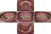
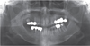
Fig. 3
Intraoral photographs after tooth extraction and old prosthesis removal. (A) Right side lateral view, (B) Frontal view, and (C) Left side lateral view.
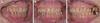
Fig. 4
Pre-op CBCT and STL file image of the maxillary study cast. (A) CBCT image, (B) Maxillary study cast image.

Fig. 5
Implant surgery planning. (A) CBCT and 3D scan data matching, (B) Transverse view, (C) Coronal view, and (D) Sagittal view.

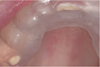
Fig. 7
Guided implant surgery and impression taking. (A) Guided implant surgery on the maxilla, (B) Maxillary area impression with impression copings, (C) Maxillary area digital impression with scanbody, (D) Guided implant surgery on the mandible, (E) Mandibular area impression with impression copings, and (F) Mandibular area digital impression with scanbody.
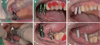
Fig. 8
Custom abutments and provisional prosthesis delivery. (A) Custom abutment delivery, (B) Provisional prosthesis delivery.
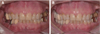
Fig. 9
Facebow transfer and interocclusal records. (A) Maxillary cast mounting procedure, (B) and (C) Eccentric interocclusal records.
Fig. 10
Definitive impression taking and cross mounting. (A) Mandibular occlusal view, (B) Maxillary occlusal view, and (C) Cross mounting.
Fig. 11
Definitive restorations. (A) Right side lateral view, (B) Frontal view, and (C) Left side lateral view.
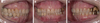
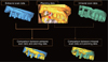
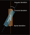
Fig. 14
Coronal deviation in distance between planned and placed implants. Extraoral, result of comparison between planning file and extraoral scanner scan file (left vertical axis); Intraoral, result of comparison between planning file and intraoral scanner scan file (left vertical axis); Difference, The absolute value of the difference between the extraoral value and intraoral value (right vertical axis).
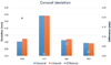
Fig. 15
Apical deviation in distance between planned and placed implants. Extraoral, result of comparison between planning file and extraoral scanner scan file (left vertical axis); Intraoral, result of comparison between planning file and intraoral scanner scan file (left vertical axis); Difference, The absolute value of the difference between the extraoral value and intraoral value (right vertical axis).
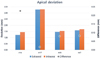
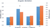
Fig. 16
Angular deviation between planned and placed implants. Extraoral, result of comparison between planning file and extraoral scanner scan file (left vertical axis); Intraoral, result of comparison between planning file and intraoral scanner scan file (left vertical axis); Difference, The absolute value of the difference between the extraoral value and intraoral value (right vertical axis).

References
1. Buser D, Martin W, Belser UC. Optimizing esthetics for implant restorations in the anterior maxilla: anatomic and surgical considerations. Int J Oral Maxillofac Implants. 2004; 19:43–61.

2. González-García R, Monje F. The reliability of cone-beam computed tomography to assess bone density at dental implant recipient sites: a histomorphometric analysis by micro-CT. Clin Oral Implants Res. 2013; 24:871–879.

3. Parsa A, Ibrahim N, Hassan B, van der Stelt P, Wismeijer D. Bone quality evaluation at dental implant site using multislice CT, micro-CT, and cone beam CT. Clin Oral Implants Res. 2015; 26:e1–e7.
4. Bover-Ramos F, Viña-Almunia J, Cervera-Ballester J, Peñarrocha-Diago M, García-Mira B. Accuracy of implant placement with computer-guided surgery: A systematic review and meta-analysis comparing cadaver, clinical, and in vitro studies. Int J Oral Maxillofac Implants. 2018; 33:101–115.
5. Vieira DM, Sotto-Maior BS, Barros CA, Reis ES, Francischone CE. Clinical accuracy of flapless computer-guided surgery for implant placement in edentulous arches. Int J Oral Maxillofac Implants. 2013; 28:1347–1351.
6. Kühl S, Zürcher S, Mahid T, Müller-Gerbl M, Filippi A, Cattin P. Accuracy of full guided vs. half-guided implant surgery. Clin Oral Implants Res. 2013; 24:763–769.
7. Papaspyridakos P, Gallucci GO, Chen CJ, Hanssen S, Naert I, Vandenberghe B. Digital versus conventional implant impressions for edentulous patients: accuracy outcomes. Clin Oral Implants Res. 2016; 27:465–472.
8. Esposito M, Grusovin MG, Willings M, Coulthard P, Worthington HV. The effectiveness of immediate, early, and conventional loading of dental implants: a Cochrane systematic review of randomized controlled clinical trials. Int J Oral Maxillofac Implants. 2007; 22:893–904.
9. Gallucci GO, Benic GI, Eckert SE, Papaspyridakos P, Schimmel M, Schrott A, Weber HP. Consensus statements and clinical recommendations for implant loading protocols. Int J Oral Maxillofac Implants. 2014; 29:287–290.
10. Lee DH, An SY, Hong MH, Jeon KB, Lee KB. Accuracy of a direct drill-guiding system with minimal tolerance of surgical instruments used for implant surgery: a prospective clinical study. J Adv Prosthodont. 2016; 8:207–213.

11. Cassetta M, Stefanelli LV, Giansanti M, Di Mambro A, Calasso S. Accuracy of a computer-aided implant surgical technique. Int J Periodontics Restorative Dent. 2013; 33:317–325.
12. Vasak C, Watzak G, Gahleitner A, Strbac G, Schemper M, Zechner W. Computed tomography-based evaluation of template (NobelGuide™)-guided implant positions: a prospective radiological study. Clin Oral Implants Res. 2011; 22:1157–1163.
13. Vercruyssen M, Cox C, Coucke W, Naert I, Jacobs R, Quirynen M. A randomized clinical trial comparing guided implant surgery (bone- or mucosa-supported) with mental navigation or the use of a pilot-drill template. J Clin Periodontol. 2014; 41:717–723.
14. Arisan V, Karabuda ZC, Ozdemir T. Accuracy of two stereolithographic guide systems for computer-aided implant placement: a computed tomography-based clinical comparative study. J Periodontol. 2010; 81:43–51.
15. Cassetta M, Giansanti M, Di Mambro A, Calasso S, Barbato E. Accuracy of two stereolithographic surgical templates: a retrospective study. Clin Implant Dent Relat Res. 2013; 15:448–459.
16. Valente F, Schiroli G, Sbrenna A. Accuracy of computer-aided oral implant surgery: a clinical and radiographic study. Int J Oral Maxillofac Implants. 2009; 24:234–242.
17. Fluegge T, Att W, Metzger M, Nelson K. A novel method to evaluate precision of optical implant impressions with commercial scan bodies-an experimental approach. J Prosthodont. 2017; 26:34–41.
18. Shimizu S, Shinya A, Kuroda S, Gomi H. The accuracy of the CAD system using intraoral and extraoral scanners for designing of fixed dental prostheses. Dent Mater J. 2017; 36:402–407.
19. Su TS, Sun J. Comparison of repeatability between intraoral digital scanner and extraoral digital scanner: An in-vitro study. J Prosthodont Res. 2015; 59:236–242.
 XML Download