

 PDF
PDF Citation
 Print
Print


INTRODUCTION
Tuberculosis (TB) is a life-threatening infectious disease caused by Mycobacterium tuberculosis (M. tuberculosis). It is estimated that there were approximately 9.6 million new cases of TB in 2014 (1). Furthermore, according to the WHO, in 2016, an estimated 10.4 million people developed active TB (2). M. tuberculosis has a special secretion system called 6-kDa early secretory antigenic target (ESAT6) protein family secretion system-1 (ESX-1), also known as a type VII secretion system, which is encoded in the pathogenic region of difference 1 locus. The virulence proteins ESAT6 and 10-kDa culture filtrate protein (CFP10) of M. tuberculosis are secreted through the ESX-1 secretion system, which are critical for pathogens to resist or evade the host immune system (34).
As a family of representative pattern recognition receptors, TLRs are conserved surface receptors that can sense microbial and endogenous molecules (5). TLR2 and TLR4 are located at the cell surface and recognize lipoprotein and LPS, respectively (6). After sensing, they recruit adaptor molecules, such as myeloid differentiation primary response protein 88 (MyD88) or TIR-domain-containing adapter-inducing interferon-β, and further activate downstream signaling such as NF-κB and MAPKs (7). In addition to lipoprotein and LPS, TLR2 and TLR4 can recognize various bacterial-derived molecules, such as heat-shock proteins and microbial toxins (89). It is known that various proteins derived from M. tuberculosis including ESAT6 require TLR2 or TLR4 to regulate immune responses in host cells (10111213).
Macrophages and dendritic cells (DCs) play important roles in immune responses against M. tuberculosis infection (141516). Of the 2, DCs are the representative antigen-presenting cells that connect innate and adaptive immunities. Although many studies have shown the importance of ESAT6 in the pathogenesis of M. tuberculosis, the role and signaling mechanism of ESAT6 on regulating the activation of immune cells is controversial. Early studies showed that ESAT6 inhibits LPS-induced production of ROS and cytokines such as IL-12p40, IL-6, and TNF-α in macrophages (1718). The ESAT6-mediated inhibition of ROS production leads to decreased DNA-binding activity of NF-κB p65 in LPS-stimulated macrophages (17). ESAT6 also reduces IL-12p40 production induced by MyD88-mediated TLRs stimulation in macrophages, and such inhibition requires TLR2 and AKT activation (18). In contrast, Jung et al. (19) revealed that ESAT6 directly induces IL-6 production in macrophages by activating STAT3. ESAT6 also induces IL-6 and IL-8 production in DCs and enhances IL-23 and IL-1β production induced by LPS and a CD40 ligand (20). It also upregulates the expression of surface molecules such as CD40, CD80, CD86, and MHC-II in DCs (21). However, the underlying mechanism of ESAT6-mediated immune regulation still needs to be elucidated. A recent study of ours revealed that ESAT6 induces IFN-β expression in macrophages via TLR4-dominant signaling pathway (22), although there is evidence showing that ESAT6 mediates immune regulation via TLR2-dependent manner (1318). Therefore, in this study, we sought to clarify the role of TLRs-mediated signaling in regulating ESAT6-induced activation and maturation of DCs.
MATERIALS AND METHODS
Animals
Wild-type (WT), TLR2-, and TLR4-deficient mice with C57BL/6 background were purchased from Jackson Laboratory (Bar Harbor, ME, USA). Animal studies were performed using protocols approved by the Institution Animal Care and the Use Committee of Chonnam National University (approval No. CNU IACUC-YB-2015-32).
Preparation and stimulation of bone marrow derived dendritic cells (BMDCs)
Murine BMDCs were isolated and cultured as described previously (24). Briefly, BMDCs were differentiated with GM-CSF (20 ng/ml; Peprotech, Rocky Hill, NJ, USA) and cultured in RPMI 1640 (Welgene, Gyeongsan, Korea) containing 10% FBS (Corning Costar, Corning, NY, USA), 1% penicillin/streptomycin, 2 mM L-glutamine, and 50 μM 2-mercaptoethanol (Sigma-Aldrich, St. Louis, MO, USA) in a 5% CO2 incubator at 37°C for nine days. Fresh medium was added both three and six days later. In order to measure cytokine levels, BMDCs were seeded in 48-well plates at a density of 2×10⁵ cells/well, incubated overnight, and treated with ESAT6. The culture supernatants were collected 24 h after treatment.
Inhibitor assay
Bay 11-7082 (NF-κB inhibitor) and SP60012 (JNK inhibitor) were obtained from Calbiochem (La Jolla, CA, USA), and SB203580 (p38 inhibitor) and PD98059 (ERK inhibitor) were obtained from Selleck Chemicals (Houston, TX, USA). BMDCs were pretreated with indicated concentrations of each inhibitor 2 h prior to ESAT6 treatment. After 24 h of incubation, the concentration of cytokines in the culture supernatant was measured by ELSIA (R&D Systems, Minneapolis, MN, USA). In order to confirm the endotoxin decontamination, polymyxin B (PMB; Sigma-Aldrich) sulfate was used as LPS inhibitor.
Immunoblotting
For immunoblotting, BMDCs were seeded in six-well plates at a concentration of 2×10⁶ cells/well and incubated overnight. These cells were treated with ESAT6 (50 ng/ml). At the indicated time point after treatment, cells were lysed in a buffer containing 1% Nonidet P-40, 50 mM Tris (pH 7.4), 250 mM NaCl, 5 mM EDTA, 50 mM NaF, 1 mM Na3VO4, and 0.02% NaN3 supplemented with protease inhibitor (complete, Mini, EDTA-free; Roche, Mannheim, Germany), phosphatase inhibitor (Phosphatase Inhibitor Cocktail 2; Sigma-Aldrich), and 2 mM dithiothreitol. In order to detect target protein including MAPKs and NF-κB, lysates were separated by 10% SDS-PAGE and transferred to nitrocellulose membranes. The membranes were then probed with primary antibodies for regular and phosphorylated forms of ERK (Santa Cruz Biotechnology, Santa Cruz, CA, USA), JNK, p38, IκB, and p65 (Cell signaling Technology, Beverly, MA, USA). A primary antibody against β-actin (Santa Cruz Animal Health, Dallas, TX, USA) was used to verify the same amount of protein samples. Following immunoblotting with relevant HRP-conjugated secondary antibodies (Santa Cruz Biotechnology), proteins were detected using Clarity Western ECL Substrate (Bio-Rad, Hercules, CA, USA).
Mixed leukocyte reaction
Mouse naïve CD4+ T-cell was isolated using EasySep™ Mouse Naïve CD4+ T Cell Isolation Kit (STEMCELL Technologies, Vancouver, Canada) from spleens of OT-II mice. BMDCs were prepared from WT, TLR2-, and TLR4-deficient mice. BMDCs (2×10⁵ cells/well) were stimulated with OVA323-339 (OT-II peptide, Sigma-Aldrich) in the presence or absence of ESAT6 for 24 h and co-cultured with naïve CD4+ T-cell (2×10⁶ cells/well) at BMDCs: T cell ratios of 1:10. After five days of co-culturing, the level of IFN-γ and IL-17 in culture supernatant was measured by ELSA (R&D Systems).
Analysis of the expression of surface molecules by flow cytometry
ESAT6-treated BMDCs were resuspended in PBS containing 0.5% BSA (Gibco, Grand Island, NY, USA). After staining with FITC-conjugated anti-CD80, CD86, or I-Ab (BD Biosciences, San Jose, CA, USA) and PE-conjugated anti-CD11c for 20 min at 4°C, cells were washed three times and resuspended in PBS containing 0.5% BSA. The fluorescence was then measured by flow cytometry (MACS Quant Analyzer 10; Miltenyi Biotec, Bergisch Gladbach, Germany), and the data was analyzed using Flowlogic™ software (Miltenyi Biotec).
Statistical analysis
The statistical significance of the differences among the mean values for different groups was tested. Values are expressed as mean ± SD. All statistical calculations were performed using GraphPad Prism version 5.01 (GraphPad Software, San Diego, CA, USA). One-way ANOVA followed by Tukey's and Duncan's post-hoc test was used for multi-group comparisons.
RESULTS AND DISCUSSION
ESAT6 induces cytokine production in BMDCs via TLR4-dependent pathway
It is likely that ESAT6 differently regulates cytokine production in DCs. ESAT6 itself can induce IL-6 and IL-8, but not IL-12p70, IL-23, IL-1β, or TNF-α in human DCs (20). Murine BMDCs can produce TNF-α and IL-1β in response to ESAT6 (21). In addition, TLR4 is essential for ESAT6-induced IFN-β expression in macrophages (22), and TLR4 expression is upregulated in the renal tissues of mice with M. tuberculosis- or ESAT6-induced renal injury (25). It was also reported that TLR2 is important for ESAT6-mediated immune regulation (18). Accordingly, we sought to determine whether TLR2 and TLR4 are required for the ESAT6-induced production of cytokines in BMDCs. ESAT6 treatment led to IL-6, TNF-α, and IL-12p40 production in BMDCs in a dose-dependent manner, and TLR2 deficiency did not affect the cytokine production (Fig. 1A-C). IL-12p70 was not detectable in ESAT6-treated BMDCs (data not shown), which was consistent with previous studies (2021). The cytokine production in response to ESAT6 was abolished in TLR4-deficient BMDCs (Fig. 1D-F). In order to rule out LPS contamination, PMB inhibition assay was performed. The results showed that PMB treatment inhibited LPS-induced production of IL-6, TNF-α, and IL-12p40, whereas the cytokine production by ESAT6 was not affected (Fig. 1G-I). These results indicate that ESAT6 may activate DCs via the TLR4-dependent pathway. There are two major differences compared to previous studies. Unlike our results, Chatterjee et al. (13) showed that ESAT6 can produce IL-6 and TGF-β in WT BMDCs, which was abolished in TLR2-deficient cells. However, Samten's group revealed that TLR2 deficiency does not affect STAT3 phosphorylation and IL-6 production in response to ESAT6 in murine macrophages (19). In our previous study, TLR4 was essential for ESAT6-mediated IFN-β production in murine macrophages, whereas TLR2 was partially involved at only low dose treatment (22). To determine more precise TLRs signaling regulating ESAT6-mediated immune responses, more studies should be accumulated. In addition, in this study, ESAT6 could induce IL-12p40 production in WT BMDCs, which was inconsistent with a previous study showing no production of IL-12p40 in ESAT6-treated BMDCs (13). It seems to be due to differences of preparation of BMDCs and recombinant ESAT6 protein. In our study, BMDCs were prepared at the presence of GM-CSF (without IL-4) for more long time of 9 days according to a previous study by Lutz et al. (24). ESAT6 in our preparation seems to have more strong activity, because 10–100 ng/ml was sufficient to induce cytokines production in BMDCs in this study, whereas up to 5 µg/ml was used in the study by Chatterjee et al. (13).
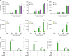 | Figure 1TLR4, but not TLR2, regulates cytokine production by ESAT6 in BMDCs. WT, TLR2-, and TLR4-deficient BMDCs were treated with ESAT6 (10, 50, and 100 ng/ml, respectively) for 24 h. The concentrations of IL-6 (A and D), TNF-α (B and E), and IL-12p40 (C and F) in supernatant were measured by ELISA. Data are presented as mean ± SD.
**p<0.01; ***p<0.001 vs. control cells.
|
TLR4 is required for the activation of NF-κB and MAPKs in response to ESAT6 in BMDCs
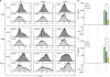
NF-κB and MAPKs are essential signaling cascades for regulating inflammatory responses in host cells (726). ESAT6 upregulates IL-8 gene transcription by increasing NF-κB DNA binding activity, and increases ERK and p38 phosphorylation in lung epithelial cells (27). We investigated whether ESAT6 activates NF-κB and MAPKs in BMDCs, and whether TLR4 is required for such activations. Western blot analysis revealed that ESAT6 induced IκB-α degradation and phosphorylation in WT BMDCs but not in TLR4-deficient cells (Fig. 2A-C). Moreover, phosphorylation of p38, ERK, and JNK was increased in WT BMDCs in response to ESAT6 from 15 min after stimulation, which was slightly reduced in TLR4-deficient cells (Fig. 2A and D-F). ESAT6 is also known to activate p38 MAPK in human lymphocytes (28). Therefore, it is likely that ESAT6 increases the activation of NF-κB or MAPKs in various host cells, and that TLR4 signaling may be essential.
 | Figure 2Activation of MAPK and NF-κB by ESAT6 are involved in TLR4-dependent manner in BMDCs. WT and TLR4-deficient BMDCs were treated with ESAT6 (50 ng/ml) for the indicated time points, and cellular proteins were extracted. The phosphorylation of ERK, JNK, p38, and p65 and IκB-α degradation were analyzed through Western blotting (A-F). Antibody against β-actin was used to confirm the loading doses.
|
NF-κB and MAPKs differently contribute to cytokine production by BMDCs in response to ESAT6
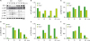
In order to examine the involvement of NF-κB and MAPKs on ESAT6-mediated cytokine production in DCs, an assay using pharmacological inhibitors was performed. A NF-κB inhibitor, Bay 11-7082, dose-dependently reduced the IL-6, TNF-α, and IL-12p40 production induced by ESAT6 in BMDCs (Fig. 3A). At a high concentration of 10 µM, IL-6 and IL-12p40 production were absolutely abolished by BAY 11-7082, whereas the TNF-α level was partially reduced (Fig. 3A). p38 inhibition by SB203580 increased ESAT6-induced production of IL-6 and IL-12p40 in a dose-dependent manner, although it did not influence TNF-α levels (Fig. 3B). However, at the much higher concentration of 40 µM, TNF-α level was also increased by SB203580 (data not shown). These results are consistent with a previous study by Ma et al. (29), showing that the inhibition of p38 MAPK by SB203580 enhances ESAT6-medicated MCP-1/CCL2 production in macrophages. p38 seems to differently regulate cytokine or chemokine production induced by ESAT6 in host cells, as p38 inhibition downregulates IL-8 mRNA and protein expression in lung epithelial cells in response to ESAT6 (27). SP600125 slightly reduced the production of IL-6, but not of TNF-α or IL-12p40, in ESAT6-treated BMDCs (Fig. 3C). In addition, PD98059 partially inhibited TNF-α production in response to ESAT6, but not that of IL-6 or IL-12p40 (Fig. 3D). These findings suggest that ERK and JNK may have modest effects on ESAT6-induced cytokine production in DCs, although these effects are activated by ESAT6. However, in lung epithelial cells, ERK inhibition by PD98059 significantly downregulates IL-8 gene and protein expression in response to ESAT6, whereas JNK inhibition increases IL-8 protein level (27). It needs to be clarified whether these different responses of MAPKs on the ESAT6-mediated production of cytokines and chemokines are specific to certain cell types.
 | Figure 3NF-κB, JNK, and EKR, but not p38, are involved in cytokine production in response to ESAT6 in BMDCs. WT BMDCs were pretreated with the indicated concentrations of each inhibitor, such as Bay 11-7082 (NF-κB inhibitor, A), SB203580 (p38 inhibitor, B), SP600125 (JNK inhibitor, C), and PD98059 (ERK inhibitor, D) for 2 h, and then treated with ESAT6 (50 ng/ml) for 24 h. The concentrations of IL-6, TNF-α, and IL-12p40 in supernatant were measured by ELISA. Values with different letters are significantly different by ANOVA with Duncan's multiple range test at p<0.05.
|
ESAT6 leads to maturation of BMDCs via TLR4-dependent signaling, which is essential for production of IFN-γ and IL-17 by CD4+ T cell
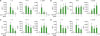
ESAT6 is considered to be a prominent target for cell-mediated immunity against TB, and there have been many trials aimed at developing an efficient TB vaccine to target ESAT6. A recent study revealed that ESAT6 itself enhanced the surface expression of CD40, CD80, CD86, and MHC-II in murine BMDCs (21), which are all markers for DC maturation. Accordingly, in this study, we sought to determine whether TLR2 and TLR4 are required for ESAT6-induced DCs maturation. Flow cytometry analysis showed that ESAT6 enhanced the surface expression of CD80, CD86, and MHC-II in WT BMDCs (Fig. 4A). TLR2 deficiency did not influence the expression of the surface molecules which was increased by ESAT6 (Fig. 4A). In contrast, the surface expression levels of CD80, CD86, and MHC-II were reduced in TLR4-deficient BMDCs as compared to WT cells (Fig. 4A). These findings indicate that TLR4, but not TLR2, is essential for ESAT6-induced DCs maturation.
 | Figure 4TLR4 in BMDCs increases the expression of marker of DCs maturation and activated by ESAT6 play an important role in production of IFN-γ and IL-17 in CD4+ T cell. WT, TLR2-, and TLR4-deficient BMDCs were treated with ESAT6 (50 ng/ml) for 24 h. The expression of CD80, CD86, and MHC class II was analyzed by flow cytometry (A). WT, TLR4-, and TLR2-deficient cells were pretreated with ESAT6 (50 ng/ml) with OVA323-339 (OT-II peptide) for 24 h, then co-cultured with naïve CD4+ T cells for five days. The concentration of IFN-γ and IL-17 in supernatant was measured by ELISA (B and C). Data are presented as mean ± SD.
**p<0.01; ***p<0.001 vs. control cells.
|
The activation and maturation of DCs drives T cell differentiation. Therefore, we investigated whether ESAT6 regulates the differentiation of T cells and whether TLR4 is involved in the process. BMDCs from WT, TLR2-, and TLR4-deficient mice were co-cultured in the presence of ESAT6 and OVA323-339 (an OT-II peptide). Following five days of co-culturing, the IFN-γ and IL-17 level in culture supernatant was measured by ELSA. In the presence of OVA323-339, ESAT6 induced production of IFN-γ and IL-17 in the co-culture of WT BMDCs and naïve CD4+ T cells from transgenic OT-II mice (Fig. 4B and C). TLR2 deficiency in BMDCs did not affect the production of IFN-γ and IL-17 by CD4+ T cells (Fig. 4B and C). However, both IFN-γ and IL-17 level was significantly reduced when co-cultured with TLR4-deficient BMDCs (Fig. 4B and C). It is thought that ESAT6 induces DCs maturation in a TLR4-dependent manner, which may contribute to T cell differentiation.
In conclusion, our study revealed that ESAT6 can induce activation and maturation of DCs via TLR4-dependent signaling, which is required for optimal T cell differentiation. Although there are still controversies regarding the ESAT6-mediated immune regulations, recent studies have revealed that TLR4 signaling is important for ESAT6-induced IFN-β production in macrophages and the development of renal injury in vivo (2225). Further studies are recommended to accumulate experimental evidence to clarify the role of ESAT6 in immune regulation and pathogenesis in TB.
 XML Download