Journal List > J Periodontal Implant Sci > v.49(2) > 1121536

Oh, Chung, and Han: Periodontal regenerative therapy in endo-periodontal lesions: a retrospective study over 5 years

Abstract

Purpose

The aim of this study was to evaluate clinical and radiographic changes and the survival rate after periodontal surgery using deproteinized bovine bone mineral (DBBM) with 10% collagen or DBBM with a collagen membrane in endo-periodontal lesions.

Methods

A total of 52 cases (41 patients) with at least 5 years of follow-up were included in this study. After scaling and root planing with or without endodontic treatment, periodontal regenerative procedures with DBBM with 10% collagen alone or DBBM with a collagen membrane were performed, yielding the DBBM + 10% collagen and DBBM + collagen membrane groups, respectively. Changes in clinical parameters including the plaque index, bleeding on probing, probing pocket depth, gingival recession, relative clinical attachment level, mobility, and radiographic bone gains were evaluated immediately before periodontal surgical procedures and at a 12-month follow-up.

Results

At the 12-month follow-up after regenerative procedures, improvements in clinical parameters and radiographic bone gains were observed in both treatment groups. The DBBM + 10% collagen group showed greater probing pocket depth reduction (4.52±1.06 mm) than the DBBM + collagen membrane group (4.04±0.82 mm). However, there were no significant differences between the groups. Additionally, the radiographic bone gain in the DBBM + 10% collagen group (5.15±1.54 mm) was comparable to that of the DBBM + collagen membrane group (5.35±1.84 mm). The 5-year survival rate of the teeth with endo-periodontal lesions after periodontal regenerative procedures was 92.31%.

Conclusions

This study showed that regenerative procedures using DBBM with 10% collagen alone improved the clinical attachment level and radiographic bone level in endo-periodontal lesions. Successful maintenance of the results after regenerative procedures in endo-periodontal lesions can be obtained by repeated oral hygiene education within strict supportive periodontal treatment.

Graphical Abstract

jpis-49-90-abf001

INTRODUCTION

The proper diagnosis and treatment of endo-periodontal lesions are challenging for clinicians. An endo-periodontal lesion is defined as a pathological communication between the endodontic and periodontal tissues of a given tooth. Most endodontic and periodontal problems are caused by bacteria [123]. There are two types of connections between pulp and periodontal tissues in which bacteria and their byproducts are found [2]. Anatomic connections are associations with the apical foramen, lateral canal, accessory foramina, and dentinal tubules [24]. Non-physiologic connections originate from root canal or pulp chamber perforation and vertical root fracture or cracking [5].
More specifically, endo-periodontal lesions involve an open wound area that requires periodontal regenerative procedures. When patients with total apico-marginal buccal bone loss underwent periapical surgery, the success rate was reported to be 27%–37% [67]. According to Kim et al. [8], the success rate of apico-marginal defects after periapical surgery with a periodontal regenerative procedure was approximately 73.7% over a 5-year follow-up, whereas lesions of only endodontic origin had a success rate of 95.2%.
Periodontal regenerative procedures with many different types of bone grafts and/or substitutes, root surface demineralization, guided tissue regeneration (GTR), growth and differentiation factors, enamel matrix proteins, or various combinations thereof have been shown to yield significant probing pocket depth reduction, clinical attachment gain, and hard tissue fill compared with access flaps alone [91011].
Several histologic studies have shown that deproteinized bovine bone mineral (DBBM) with 10% collagen has the capacity to induce periodontal regeneration [121314]. However, conflicting evidence exists regarding the efficacy of simultaneous GTR and bone replacement grafting in enhancing new bone formation [151617]. Brkovic et al. [16] reported that the amount of new bone formation was not significantly different between bone grafts without a membrane and those with a membrane. Conversely, Lee et al. [17] reported that the combined use of DBBM and a collagen membrane was followed by more new bone formation than was observed with DBBM only. The drawbacks of membrane use include an increased risk of infection, gingival recession, and the need for a second surgical procedure to remove the non-resorbable membrane [1819]. In particular, bone grafting combined with a barrier membrane in a narrow interdental space or thin biotype may cause gingival recession, which exposes the membrane and impairs wound healing [20].
The aim of this study was to evaluate the clinical and radiographic changes, as well as the survival rate, after periodontal regenerative procedures in teeth with endo-periodontal lesions using DBBM with 10% collagen alone or DBBM with collagen membranes.

MATERIALS AND METHODS

Study population

Human subject approval was obtained from the Institutional Review Board of Hanyang University Hospital (No. 2017-07-002). Patients visiting the Department of Periodontology, Hanyang University Hospital from 2007 to 2017 who met the inclusion criteria were screened for enrollment in this study. We included adult patients who had 1) received no active or maintenance periodontal therapy during the preceding 3 months; 2) one or more periodontal sites with a probing depth ≥5 mm; 3) radiographic evidence of bone loss from marginal bone to root apex; 4) a wide, deep periodontal pocket in 1 tooth surface (grade 2) or deep periodontal pockets in more than 1 tooth surface (grade 3) (for periodontitis patients) [5]; 5) at least 2 mm of keratinized tissue; and 6) been complying with a maintenance program. We excluded patients with diabetes, who were smokers, who had regularly used non-steroidal anti-inflammatory drugs, who had used antibiotics within the previous 3 months, who required antibiotic prophylaxis for therapy, and/or who were pregnant. We also excluded patients who had teeth with horizontal or vertical fractures and iatrogenic root perforation. Finally, 41 patients with a total of 52 diseased sites requiring periodontal regenerative procedures were included.

Clinical measurements

The following clinical parameters were collected at baseline (before periodontal surgery) and at 12 months after the surgical procedure: plaque index (PI), bleeding on probing (BOP), probing pocket depth (PPD), gingival recession (GR), relative clinical attachment level (RAL), and tooth mobility. The PPD was measured at 6 sites per tooth (mesiobuccal, mid-buccal, distobuccal, mesiolingual, mid-lingual, and distolingual) as the distance in millimeters from the free gingival margin to the base of the probable pocket using a hand-held periodontal probe (PCP-UNC 15; Hu-Friedy Mfg. Co., Chicago, IL, USA). The GR was defined as the distance from either the cemento-enamel junction (CEJ) or a restorative margin to the gingival margin. The RAL was calculated as the sum of the PPD and GR measurements. Additionally, the PPD measurement for the deepest site was recorded (PPDD), and the corresponding RAL (RALD) was recorded. The PI was recorded according to the criteria described by Silness and Löe [21]. BOP was determined as being present or absent (+/−) within 30 seconds after probing of the aforementioned 6 sites for the designated study teeth. Then, the percent ratio of the number of BOP+ sites relative to the total of 6 sites was calculated. The amount of tooth mobility was evaluated as the Periotest value (PTV) using Periotest Classic (Medizintechnik Gulden e. K., Modautal, Germany).

Treatment protocol

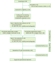
An overview of the study design is presented in Figure 1. Scaling and root planing (SRP) was performed, followed by oral hygiene instruction. Pulp status and the quality of the coronal restoration were verified. The vitality of the diseased teeth was assessed with an electric pulp tester (Gentle PulseTM Analog Pulp Vitality Tester, Parkell, Inc., Edgewood, NY, USA), and root canal treatment was performed if the tooth did not respond to the vitality test. The root canals were cleaned and shaped using nickel-titanium rotary instruments, and irrigated with 5.25% sodium hypochlorite. The root canals were obturated with gutta-percha and AH Plus sealer (Dentsply Maillefer, Tulsa, OK, USA). Thereafter, teeth that showed mobility greater than degree 1 (PTV ≥10) were splinted to adjacent teeth using composite resin and stainless-steel wire. Splinting was used to stabilize mobile teeth during the surgical phase and the maintenance phase.
Figure 1

Flow chart of the participants in this study.

PI: plaque index, BOP: bleeding on probing, PPD: probing pocket depth, RAL: relative clinical attachment level, DBBM: deproteinized bovine bone mineral.
jpis-49-90-g001
Periodontal regenerative surgery was performed by 1 periodontist (H. J.) 3 months after root canal treatment. After local anesthesia, flaps were reflected with a modified or simplified papilla preservation technique according to the width of the interdental space [2223]. Meticulous degranulation and debridement were performed using ultrasonic and hand instruments. Of the 52 operated sites, 23 sites were packed with DBBM (Bio-Oss®, Geistlich Pharma, Wolhusen, Switzerland) and covered by bioresorbable collagen membranes (BioGide®, Geistlich Pharma, Wolhusen, Switzerland). The remaining 29 sites were packed with DBBM containing 10% collagen (Bio-Oss® Collagen, Geistlich Pharma, Wolhusen, Switzerland) without a covering barrier membrane. Flaps were repositioned to their original location and sutured using interrupted suture and horizontal mattress suture techniques with 5-0 Nylon (Prolene®, Ethicon Inc., Skillman, NJ, USA). Patients received general postoperative instructions for periodontal surgical procedures. Oral analgesics (ibuprofen, 600 mg, every 8 hours as necessary) and antibiotics (amoxicillin/clavulanic acid, 375 mg, 3 times daily) were prescribed for 7 days, and patients were instructed to rinse with 0.12% chlorhexidine gluconate 4 times daily for 3 weeks. Patients were seen at 2 weeks after surgery for suture removal.
Supportive periodontal treatment (SPT) of the treated teeth was performed every 3 months for 2 years and every 3 to 6 months thereafter to assess clinical and radiographic signs of healing. At the maintenance appointment, patients again received individualized oral hygiene instruction to remove supragingival plaque.

Radiographic measurements

A digital radiography system (CS9300 Select, Carestream Dental LLC., Atlanta, GA, USA) was used to obtain radiographic images (panoramic and periapical radiographs) of each site at baseline (before periodontal surgery) and at 12 months after the surgical procedure. Periapical radiographs were taken using the parallel cone technique and were imported into the Analysis Toolkit (Adobe Photoshop CS6, Adobe Systems Inc., San Jose, CA, USA). The anatomic landmarks of the intrabony defect, including the CEJ and the bottom of the defect (BD), were identified on the imported radiographic images based on the criteria established by Bjorn et al. [24] (Figure 2). If restorations were present, the apical margin of the restoration was used to replace the CEJ as a fixed reference point [25]. The BD was defined as the most apical extension of the intrabony destruction where the periodontal ligament space retained its normal width. For assessment of the extent of radiographic bone gain, the distance from the CEJ to the BD (CEJ-BD) was measured. Radiographic bone gain was calculated by subtracting the baseline measurement from that in the 12-month follow-up radiograph. As the baseline and 12-month follow-up radiographs were not identical, the vertical distortion between baseline and the 12-month radiographs was estimated as previously described [2526], by measuring an anatomically non-variable distance such as the root length (distance from the CEJ to the root apex [RA]; CEJ-RA) on both imported images, and a correction factor was calculated as follows:
CEJRA(baseline)CEJRA(12monthradiograph)=Correctionfactorjpis-49-90-e001
Figure 2

Pre- and post-operative radiographs and clinical photographs of endo-periodontal lesion. (A) A periapical radiograph before the periodontal regenerative procedure. (B) After reflecting the flap, the bony defect adjacent to the apex was shown. (C) The bone defect was grafted with DBBM and (D) covered with a collagen membrane. (E) Three-dimensional CBCT image before surgery. (F) Three-dimensional CBCT image at the 12-month follow-up showed the bone defect filled with hard tissue. (G) Sagittal CBCT image before surgery showed bone destruction extending to the apex. (H) Sagittal CBCT view at 12 months after surgery. The bone defect was filled with hard tissue. (I) Coronal CBCT image before surgery showed the buccal bone defect extending to the apex. (J) At the 12-month follow-up, the bone defect was filled with hard tissue. (K) Axial CBCT image showing the resorption of the buccal cortical plate. (L) The buccal bone defect was filled with hard tissue at the 12-month follow-up after the periodontal regenerative procedure.

CEJ: cemento-enamel junction, BD: bottom of the defect, RA: root apex, DBBM: deproteinized bovine bone mineral, CBCT: cone-beam computed tomography.
jpis-49-90-g002
The radiographic bone gain at 12 months of follow-up was calculated after applying the correction factor as follows:
Radiographic bone gain = [CEJ-BD(baseline)]−[CEJ-BD(12 months)×correction factor]
All radiographic measurements were performed by a single examiner who was blinded to the procedure [17]. The measurements were repeated after an interval of at least 2 weeks. To evaluate the reproducibility of the 2 measurements, Bland-Altman plots and intraclass correlation coefficients were used. Additionally, cone-beam computed tomography (CBCT) of the teeth with large bone defects was performed at baseline and 12 months after the surgical procedure.

Statistical analysis

The Kolmogorov-Smirnov test and the Shapiro-Wilk test were performed to assess the normality of the data, and the data did not show a normal distribution. The Wilcoxon signed-rank test was used to compare all clinical parameters and radiographic bone gain between baseline and 12 months post-surgery. Comparisons of changes in clinical parameters (PI, BOP, PPD, PPDD, GR, RAL, RALD, and mobility) and radiographic bone gain according to treatment modality were evaluated using the Mann-Whitney U test. The correlation between radiographic bone gain and type of bone defect was assessed using Spearman correlation coefficients. The correlations among clinical parameters were also evaluated using Spearman correlation coefficients. A commercially available software program (SPSS version 21.0, IBM Corp, Armonk, NY, USA) was used to analyze the data. The level of significance chosen for statistical tests was P<0.05.

RESULTS

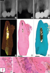
A total of 52 cases (41 patients) were evaluated at baseline and the 12-month follow-up. The mean follow-up period was 70.08±17.21 months (range, 59–122 months). Table 1 shows the demographic information of the patients. The survival rate of the teeth with endo-periodontal lesions was 92.31% at 5 years after the periodontal regenerative procedures. Figure 3 shows the histological findings of the teeth extracted for strategic reasons.
Table 1

Demographic characteristics of patients

jpis-49-90-i001
Variables No.
Sex
Male 20
Female 32
Sites
Mx. anterior area 4
Mx. premolar area 4
Mx. posterior area 24
Mn. anterior area 5
Mn. premolar area 3
Mn. posterior area 12
Defect type
1-wall 34
2-wall 18
3-wall 0
Endodontic treatment
Yes 45
No 7
Guided tissue regeneration
Yes 23
No 29
New prosthesis
Yes 40
No 12
Survived teeth 46
Extracted teeth 6
Due to fracture 3
Due to improper oral hygiene 1
Due to endodontic failure 1
Due to strategic reasons 1
Follow-up period (mean ± SD, mon) 70.08 ± 17.21
Survival period (mean ± SD, mon) 66.77 ± 19.12
Mx.: maxilla, Mn.: mandible, SD: standard deviation.
Figure 3

Radiographic and histologic evaluation of endo-periodontal lesion. (A) A periapical radiograph before surgery showed a bone defect around the apex of the left maxillary central incisor. (B) An endo-periodontal lesion was filled with hard tissue. (C) At 11 years after surgery, the root was fused with the adjacent hard tissue, similar to ankylosis. (D) Reconstructed micro-computed tomography image of the extracted tooth. (E) A histologic evaluation showed that the bone-like mass was directly adhered to the root surface (H&E stain, original magnification, ×1.2, scale bar=2,000 μm). (F) The root was directly in contact with the adjacent bone surrounding the DBBM particles (Masson-Goldner trichrome stain, original magnification, ×1.2, scale bar=2,000 μm) (G) The DBBM particles were surrounded with mature bone (H&E stain, original magnification, ×10, scale bar=500 μm). (H) Higher magnification of (G). The cementum was directly in contact with the adjacent mature bone (H&E stain, original magnification, ×40, scale bar=50 μm).

D: dentin, C: cementum, MB: mature bone, P: DBBM particles, H&E: haemotoxylin and eosin.
jpis-49-90-g003
The clinical parameters obtained at baseline and the 12-month follow-up are presented in Table 2. PI, BOP, and mobility significantly decreased (P<0.001), whereas GR significantly increased (P<0.001) from baseline to the 12-month follow-up. PPD and RAL also significantly improved by 12 months after the periodontal regenerative procedure. The correction factor for the radiographic evaluation was 1.00±0.02. Bland-Altman plots for the radiographic measurements showed good agreement. The intraclass correlation coefficients for the radiographic measurements were 0.927 and 0.982 at baseline and the 12-month follow-up, respectively.
Table 2

Comparison of clinical parameters and radiographic bone gain between baseline and the 12-month follow-up

jpis-49-90-i002
Variables Baseline 12-month follow-up Difference P value
PI 1.06±0.29 0.74±0.22 0.32±0.27 <0.001
BOP (%) 48.08±9.71 13.78±6.37 34.29±10.13 <0.001
PPD (mm) 5.14±0.83 2.96±0.31 2.19±0.71 <0.001
PPDD (mm) 7.59±0.89 3.35±0.48 4.31±0.98 <0.001
GR (mm) 1.43±0.58 2.20±0.77 0.77±0.34 <0.001
RAL (mm) 6.58±0.99 5.16±0.77 1.42±0.76 <0.001
RALD (mm) 9.03±1.01 5.49±1.05 3.54±1.03 <0.001
Mobility (PTV) 26.42±2.28 10.27±2.04 16.15±2.46 <0.001
CEJ-BD (mm) 9.82±1.44 4.58±1.06 5.24±1.67 <0.001
PI: plaque index, BOP: bleeding on probing, PPD: probing pocket depth, PPDD: probing pocket depth at the deepest point, GR: gingival recession, RAL: relative clinical attachment level, RALD: relative clinical attachment level at the deepest point, PTV: the Periotest value, CEJ-BD: radiographic distance from the cemento-enamel junction (or a restorative margin) to the most apical extension of bone destruction.
The changes in clinical parameters and radiographic bone gain according to treatment modality are shown in Table 3 and Figure 4. Among the treatment-related variables, PPDD reduction was greater in the bone-grafted teeth without membranes (4.52±1.06 mm) than in the bone-grafted teeth in which membranes were used (4.04±0.82 mm) at 12 months after periodontal regenerative procedures. However, there was no significant difference between the bone-grafted teeth with membranes and the bone-grafted teeth without membranes. Furthermore, the change in CEJ-BD at the 12-month follow-up was similar between the bone-grafted teeth with membranes (5.35±1.84 mm) and the bone-grafted teeth without membranes (5.15±1.54 mm).
Table 3

Comparison of clinical parameters and radiographic bone gain according to treatment modality

jpis-49-90-i003
Variables No. ΔPPD ΔPPDD ΔRAL ΔRALD ΔMobility ΔCEJ-BD
Membrane
Yes 23 2.12±0.75 4.04±0.82 1.41±0.84 3.33±0.89 16.48±2.86 5.35±1.84
No 29 2.24±0.69 4.52±1.06 1.43±0.70 3.71±0.12 15.89±2.11 5.15±1.54
P value 0.607 0.071 0.264 0.36 0.232 0.416
Endodontic treatment
Yes 45 2.19±0.72 4.44±0.97 1.43±0.79 3.68±1.02 16.36±2.27 5.30±1.73
No 7 2.17±0.68 3.43±0.53 1.38±0.56 2.64±0.56 14.86±3.39 4.83±1.19
P value 0.958 0.009a) 0.916 0.007a) 0.356 0.581
New prosthesis
Yes 40 2.30±0.72 4.38±1.01 1.58±0.76 3.65±1.07 16.28±2.31 5.48±1.72
No 12 1.82±0.56 4.08±0.90 0.90±0.51 3.17±0.83 15.75±2.99 4.45±1.25
P value 0.046b) 0.339 0.005a) 0.14 0.361 0.043b)
Total 52 2.19±0.71 4.31±0.98 1.42±0.76 3.54±1.03 1.94±0.46 5.24±1.67
ΔPPD: difference of probing pocket depth between baseline and the 12-month follow-up, ΔPPDD: difference in the deepest probing pocket depth between baseline and the 12-month follow-up, ΔRAL: difference in the relative attachment level between baseline and the 12-month follow-up, ΔRALD: difference in the relative attachment level in the deepest point between baseline and the 12-month follow-up, ΔMobility: difference in the Periotest value between baseline and the 12-month follow-up, ΔCEJ-BD: difference in the radiographic distance from the cemento-enamel junction (or a restorative margin) to the most apical extension of bone destruction between baseline and the 12-month follow-up.
a)Statistically significant difference (P<0.01); b)Statistically significant difference (P<0.05).
Figure 4

Comparison of clinical parameters and radiographic bone gain according to (A) the use of a membrane, (B) endodontic treatment, and (C) new prosthesis delivery.

ΔPPD: difference in the probing pocket depth between baseline and the 12-month follow-up, ΔPPDD: difference in the deepest probing pocket depth between baseline and the 12-month follow-up, ΔRAL: difference in the relative attachment level between baseline and the 12-month follow-up, ΔRALD: difference in the relative attachment level at the deepest point between baseline and the 12-month follow-up, ΔCEJ-BD: difference in the radiographic distance from the cemento-enamel junction (or a restorative margin) to the most apical extension of bone destruction between baseline and the 12-month follow-up.
a)Statistically significant difference (P<0.01); b)Statistically significant difference (P<0.05).
jpis-49-90-g004
The correlations among clinical parameters were also evaluated (Table 4). The difference in the PI between baseline and 12-month follow-up was significantly related to PPDD reduction (Spearman correlation coefficient=0.422, P=0.002). In addition, RALD gain and PI reduction between baseline and 12-month follow-up showed a significant correlation (Spearman correlation coefficient=0.398, P=0.003). The CEJ-BD reduction was significantly correlated with bone defect type (Spearman correlation coefficient=0.334, P=0.016).
Table 4

Correlations between clinical parameters and radiographic bone gain

jpis-49-90-i004
Variables Spearman correlation coefficients
ΔPI Defect type
PPDD reduction 0.422a) 0.292b)
RALD gain 0.398a) 0.289b)
CEJ-BD reduction 0.209 0.334b)
ΔPI: difference of Silness and Löe plaque index between baseline and the 12-month follow-up, PPDD: probing pocket depth at the deepest point, RAL: relative attachment level at the deepest point, CEJ-BD: radiographic distance from the cemento-enamel junction (or a restorative margin) to the most apical extension of bone destruction.
a)Statistically significant difference (P<0.01); b)Statistically significant difference (P<0.05).

DISCUSSION

The present study showed that periodontal regenerative treatment using DBBM with 10% collagen without a collagen membrane resulted in clinical attachment level and radiographic bone gain in endo-periodontal lesions. In this study, all clinical parameters, including PPDD, RALD, mobility, BOP, and CEJ-BD, were significantly improved.
The aims of periodontal regenerative procedures are to increase the periodontal attachment and amount of bone of a severely compromised tooth, to decrease PPD, and to minimize the increase in GR [10]. For predictable periodontal regeneration, various types of bone grafts and/or substitutes, root surface demineralization, GTR, growth and differentiation factors, enamel matrix proteins, and various combinations thereof have been used since the early 1970s [11]. We used DBBM with 10% collagen without a membrane or DBBM with a collagen membrane in this study. We applied a collagen membrane when the width of the interdental space was >2 mm with a modified papilla preservation flap. If the width of the interdental space was <2 mm, we used only DBBM with 10% collagen without a membrane. Contamination following membrane exposure is a major complication associated with GTR [19]. If we had used bone grafts with membranes in teeth with a narrow interdental papilla (<2 mm), especially in cases where inflammation may have been prolonged and severe or in cases of a thin biotype [20], GR would have interfered with healing by primary intention. GR may cause membrane exposure, which can impair periodontal regeneration. Several studies have presented histologic results after using DBBM with or without collagen membranes [1214272829]. Camelo et al. [27] suggested that DBBM acted as an osteoconductive matrix. They also showed new attachment formation consisting of collagen fibers inserting into new cementum adjacent to the graft after using DBBM with or without collagen membranes. Additionally, another report showed that the use of DBBM with 10% collagen demonstrated histologic evidence of regeneration [12]. The researchers showed the efficacy of DBBM with 10% collagen alone or with a collagen membrane for periodontal regeneration. Hartman et al. [14] demonstrated that DBBM with a collagen membrane had no additive benefit for periodontal regeneration. Similarly, in the present study, there were no statistically significant differences in the clinical attachment level or radiographic bone gain between the groups with and without membranes. A recent systematic review reported an interesting observation, in which a long junctional epithelium was not necessarily formed in cases of nonguided tissue regeneration [11]. New connective tissue attachment might not primarily depend on epithelial downgrowth exclusion (e.g., the GTR principle). This observation suggests that wound stability and space provision are very important for unfolding the innate regenerative potential of the periodontium.
This study showed a tooth survival rate of 92.31% at 5 years after a periodontal regenerative procedure in endo-periodontal lesions. This result is similar to a recent systematic review, which reported that the survival rate of teeth with endo-periodontal lesions was 72.1% to 100% [30]. In the present study, 3 of the 6 failed teeth were extracted due to fracture at 13-month, 31-month, and 42-month follow-up visits. A mandibular second molar without endodontic treatment, with progressive apical radiolucency and increased mobility, was also extracted at a 49-month follow-up visit. A maxillary first molar with an endodontic problem, in which the origin of infection was a fourth canal with incomplete root canal treatment, was extracted. We propose that the prolonged periodontal infection and the defensive action of the pulp had obstructed the root canal and resulted in recurrence of an endo-periodontal lesion at 6 years after the periodontal regenerative procedure. A maxillary central incisor that showed excellent radiographic bone healing was extracted for strategic reasons at 11 years of follow-up. Surprisingly, we observed bone attached to the root after extraction (Figure 3). This tooth showed healing-like ankylosis with excellent bone formation adjacent to DBBM particles. We suggest that the healing-like ankylosis was due to aggressive root planing and degranulation of infected tissue during the surgery or splinting of the treated teeth during the 12-month healing period. We splinted teeth that showed mobility of greater than degree 1 (PTV≥10) with the adjacent teeth, using resin and stainless-steel wire, before periodontal regenerative procedures. At the 12-month follow-up, we re-evaluated the teeth and removed the resin-wire splint. Even though the influence of tooth mobility on regenerative therapy remains controversial, several studies have shown that splinting mobile teeth before a regenerative procedure aided in wound healing [313233].
Several factors influence the results of periodontal regenerative therapies, including defect size and morphology, the patient's smoking habits, and compliance with oral hygiene [181934]. Supportive periodontal maintenance care given after the periodontal regenerative procedure is critical for the long-term success of periodontal therapy. Among patient-related factors, oral hygiene compliance and smoking habits are major determinants [34]. Previous clinical studies showed that professionally delivered supportive periodontal care programs and good oral hygiene provided long-term maintenance of the regenerated attachment [353637]. In the present study, all included patients did not smoke, had good compliance with periodontal maintenance programs, and had a noncontributory medical history. In this study, bone graft procedures using DBBM were performed in 1-wall defects, as well as in 2- and 3-wall intrabony defects. Radiographic bone gain was significantly correlated with the defect type. Additionally, improvement in PI was significantly related with PPDD reduction and RALD gain. Even though the periodontal regenerative procedures were successfully performed, the ultimate success of periodontal healing mainly depends on patients' oral hygiene status and compliance with SPT.
With the increased success rate of implants, it is very difficult for periodontists to decide when to extract severely periodontally involved teeth with endodontic problems. Even though teeth with a severe intrabony defect showed a 96% survival rate for a period of up to 15 years [1038], survival is possible with continuous oral hygiene education and professional tooth cleaning within a stringent recall program. Therefore, we re-evaluated the teeth at 12 months after the periodontal regenerative procedure to decide whether to restore the teeth with a new prosthesis. We also informed the patients that the survival of the treated tooth mainly depends on their compliance with oral hygiene. It is very crucial to consider other factors such as patient cooperation, restorability, and the patient's economic circumstances, all of which influence treatment decisions [2]. In some cases, new prostheses facilitated patients’ oral hygiene due to modulation of the embrasure space and the cervical area of teeth, which were very difficult to clean properly before the procedure. A new prosthesis could also improve the stability of the treated tooth if it was splinted with the adjacent tooth.
This study has several limitations. First, retrospective studies have inherent bias. Second, when assessing the effectiveness of membrane use, endodontic treatment, and new prosthetic treatment, we had to consider economic aspects and patients' preferences. However, the study had several strengths. First, all periodontal regenerative procedures were performed by 1 periodontist. Second, we instructed patients to perform proper oral hygiene before and after the periodontal regenerative procedures. Oral hygiene status was evaluated in all patients with disclosing solution at every visit before and immediately after surgery and during SPT. Therefore, the sources of local and general infections could be minimized. Third, 40 teeth were restored with new prostheses after periodontal regenerative procedures. Fourth, we observed the survival rate and reasons of failure in severely compromised teeth with endo-periodontal problems. Finally, this study presented the 11-year histological follow-up results of DBBM with 10% collagen in endo-periodontal lesions.
This study showed that periodontal regenerative procedures using DBBM with 10% collagen alone improved the clinical attachment level and radiographic bone level in endo-periodontal lesions. We suggest that endodontic treatment before the definitive periodontal regenerative procedure supports periodontal healing in cases of apical involvement. For successful maintenance of the results of periodontal regenerative procedures in endo-periodontal lesions, repeated oral hygiene education within a strict SPT program is of the utmost importance.

ACKNOWLEDGEMENTS

We would like to thank Prof. En-Woo Nam of the Biostatistical Consulting and Research Lab, Hanyang University for assistance with statistical analysis and Seong-Jin Ahn of Hanyang Medical Center for radiographic assistance.

Notes

Author Contributions

  • Conceptualization: Ji-Young Han.

  • Formal analysis: Shin Hye Chung.

  • Investigation: Shin Hye Chung.

  • Methodology: Ji-Young Han, Soram Oh.

  • Project administration: Ji-Young Han.

  • Writing - original draft: Soram Oh, Ji-Young Han.

  • Writing - review & editing: Ji-Young Han.

Conflict of Interest No potential conflict of interest relevant to this article was reported.

References

1. Löe H, Theilade E, Jensen SB. Experimental gingivitis in man. J Periodontol. 1965; 36:177–187.
crossref pmid
2. Zehnder M, Gold SI, Hasselgren G. Pathologic interactions in pulpal and periodontal tissues. J Clin Periodontol. 2002; 29:663–671.
crossref pmid
3. Fabricius L, Dahlén G, Sundqvist G, Happonen RP, Möller AJ. Influence of residual bacteria on periapical tissue healing after chemomechanical treatment and root filling of experimentally infected monkey teeth. Eur J Oral Sci. 2006; 114:278–285.
crossref pmid
4. Simon JH, Glick DH, Frank AL. The relationship of endodontic-periodontic lesions. J Periodontol. 1972; 43:202–208.
crossref pmid
5. Herrera D, Retamal-Valdes B, Alonso B, Feres M. Acute periodontal lesions (periodontal abscesses and necrotizing periodontal diseases) and endo-periodontal lesions. J Periodontol. 2018; 89:Suppl 1. S85–S102.
crossref
6. Hirsch JM, Ahlström U, Henrikson PA, Heyden G, Peterson LE. Periapical surgery. Int J Oral Surg. 1979; 8:173–185.
crossref pmid
7. Skoglund A, Persson G. A follow-up study of apicoectomized teeth with total loss of the buccal bone plate. Oral Surg Oral Med Oral Pathol. 1985; 59:78–81.
crossref pmid
8. Kim E, Song JS, Jung IY, Lee SJ, Kim S. Prospective clinical study evaluating endodontic microsurgery outcomes for cases with lesions of endodontic origin compared with cases with lesions of combined periodontal-endodontic origin. J Endod. 2008; 34:546–551.
crossref pmid
9. Kao RT, Nares S, Reynolds MA. Periodontal regeneration-intrabony defects: a systematic review from the AAP Regeneration Workshop. J Periodontol. 2015; 86:Suppl. S77–104.
10. Cortellini P, Tonetti MS. Clinical concepts for regenerative therapy in intrabony defects. Periodontol 2000. 2015; 68:282–307.
crossref pmid
11. Sculean A, Nikolidakis D, Nikou G, Ivanovic A, Chapple IL, Stavropoulos A. Biomaterials for promoting periodontal regeneration in human intrabony defects: a systematic review. Periodontol 2000. 2015; 68:182–216.
crossref pmid
12. Nevins ML, Camelo M, Lynch SE, Schenk RK, Nevins M. Evaluation of periodontal regeneration following grafting intrabony defects with bio-oss collagen: a human histologic report. Int J Periodontics Restorative Dent. 2003; 23:9–17.
pmid
13. Sculean A, Stavropoulos A, Windisch P, Keglevich T, Karring T, Gera I. Healing of human intrabony defects following regenerative periodontal therapy with a bovine-derived xenograft and guided tissue regeneration. Clin Oral Investig. 2004; 8:70–74.
crossref pmid
14. Hartman GA, Arnold RM, Mills MP, Cochran DL, Mellonig JT. Clinical and histologic evaluation of anorganic bovine bone collagen with or without a collagen barrier. Int J Periodontics Restorative Dent. 2004; 24:127–135.
pmid
15. Guillemin MR, Mellonig JT, Brunsvold MA. Healing in periodontal defects treated by decalcified freeze-dried bone allografts in combination with ePTFE membranes (I). Clinical and scanning electron microscope analysis. J Clin Periodontol. 1993; 20:528–536.
crossref pmid
16. Brkovic BM, Prasad HS, Rohrer MD, Konandreas G, Agrogiannis G, Antunovic D, et al. Beta-tricalcium phosphate/type I collagen cones with or without a barrier membrane in human extraction socket healing: clinical, histologic, histomorphometric, and immunohistochemical evaluation. Clin Oral Investig. 2012; 16:581–590.
crossref pmid
17. Lee JS, Jung JS, Im GI, Kim BS, Cho KS, Kim CS. Ridge regeneration of damaged extraction sockets using rhBMP-2: an experimental study in canine. J Clin Periodontol. 2015; 42:678–687.
crossref pmid
18. Becker W, Becker BE. Clinical applications of guided tissue regeneration: surgical considerations. Periodontol 2000. 1993; 1:46–53.
crossref
19. Villar CC, Cochran DL. Regeneration of periodontal tissues: guided tissue regeneration. Dent Clin North Am. 2010; 54:73–92.
crossref
20. Anderegg CR, Metzler DG, Nicoll BK. Gingiva thickness in guided tissue regeneration and associated recession at facial furcation defects. J Periodontol. 1995; 66:397–402.
crossref pmid
21. Silness J, Löe H. Periodontal disease in pregnancy. II. Correlation between oral hygiene and periodontal condition. Acta Odontol Scand. 1964; 22:121–135.
crossref pmid
22. Cortellini P, Prato GP, Tonetti MS. The modified papilla preservation technique. A new surgical approach for interproximal regenerative procedures. J Periodontol. 1995; 66:261–266.
crossref pmid
23. Cortellini P, Prato GP, Tonetti MS. The simplified papilla preservation flap. A novel surgical approach for the management of soft tissues in regenerative procedures. Int J Periodontics Restorative Dent. 1999; 19:589–599.
pmid
24. Björn H, Halling A, Thyberg H. Radiographic assessment of marginal bone loss. Odontol Revy. 1969; 20:165–179.
pmid
25. Liñares A, Cortellini P, Lang NP, Suvan J, Tonetti MS. European Research Group on Periodontology (ErgoPerio). Guided tissue regeneration/deproteinized bovine bone mineral or papilla preservation flaps alone for treatment of intrabony defects. II: radiographic predictors and outcomes. J Clin Periodontol. 2006; 33:351–358.
crossref pmid
26. Tonetti MS, Pini Prato G, Williams RC, Cortellini P. Periodontal regeneration of human infrabony defects. III. Diagnostic strategies to detect bone gain. J Periodontol. 1993; 64:269–277.
crossref pmid
27. Camelo M, Nevins ML, Schenk RK, Simion M, Rasperini G, Lynch SE, et al. Clinical, radiographic, and histologic evaluation of human periodontal defects treated with Bio-Oss and Bio-Gide. Int J Periodontics Restorative Dent. 1998; 18:321–331.
pmid
28. Paolantonio M, Scarano A, Di Placido G, Tumini V, D'Archivio D, Piattelli A. Periodontal healing in humans using anorganic bovine bone and bovine peritoneum-derived collagen membrane: a clinical and histologic case report. Int J Periodontics Restorative Dent. 2001; 21:505–515.
pmid
29. Camelo M, Nevins ML, Lynch SE, Schenk RK, Simion M, Nevins M. Periodontal regeneration with an autogenous bone-Bio-Oss composite graft and a Bio-Gide membrane. Int J Periodontics Restorative Dent. 2001; 21:109–119.
pmid
30. Schmidt JC, Walter C, Amato M, Weiger R. Treatment of periodontal-endodontic lesions--a systematic review. J Clin Periodontol. 2014; 41:779–790.
crossref pmid
31. Schulz A, Hilgers RD, Niedermeier W. The effect of splinting of teeth in combination with reconstructive periodontal surgery in humans. Clin Oral Investig. 2000; 4:98–105.
crossref pmid
32. Cortellini P, Stalpers G, Mollo A, Tonetti MS. Periodontal regeneration versus extraction and prosthetic replacement of teeth severely compromised by attachment loss to the apex: 5-year results of an ongoing randomized clinical trial. J Clin Periodontol. 2011; 38:915–924.
crossref pmid
33. Siciliano VI, Andreuccetti G, Siciliano AI, Blasi A, Sculean A, Salvi GE. Clinical outcomes after treatment of non-contained intrabony defects with enamel matrix derivative or guided tissue regeneration: a 12-month randomized controlled clinical trial. J Periodontol. 2011; 82:62–71.
crossref
34. Cortellini P, Paolo G, Prato P, Tonetti MS. Long-term stability of clinical attachment following guided tissue regeneration and conventional therapy. J Clin Periodontol. 1996; 23:106–111.
crossref pmid
35. Eickholz P, Krigar DM, Kim TS, Reitmeir P, Rawlinson A. Stability of clinical and radiographic results after guided tissue regeneration in infrabony defects. J Periodontol. 2007; 78:37–46.
crossref pmid
36. Nickles K, Ratka-Krüger P, Neukranz E, Raetzke P, Eickholz P. Open flap debridement and guided tissue regeneration after 10 years in infrabony defects. J Clin Periodontol. 2009; 36:976–983.
crossref pmid
37. Nygaard-Østby P, Bakke V, Nesdal O, Susin C, Wikesjö UM. Periodontal healing following reconstructive surgery: effect of guided tissue regeneration using a bioresorbable barrier device when combined with autogenous bone grafting. A randomized-controlled trial 10-year follow-up. J Clin Periodontol. 2010; 37:366–373.
crossref pmid
38. Cortellini P, Tonetti MS. Long-term tooth survival following regenerative treatment of intrabony defects. J Periodontol. 2004; 75:672–678.
crossref pmid
TOOLS
ORCID iDs

Soram Oh
https://orcid.org/0000-0002-8843-2144

Shin Hye Chung
https://orcid.org/0000-0002-9037-1950

Ji-Young Han
https://orcid.org/0000-0002-2364-8366

Similar articles