

 PDF
PDF ePub
ePub Citation
 Print
Print


Taehoon Kim, Yeon Namgung, Sun Young Jeong1, Sun-Jin Boo
Figures and Tables
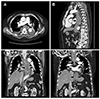 | Fig. 1Initial chest CT. CT scans with axial (A), sagittal (B) and coronal (C, D) images show diffuse wall thickening with intramural low density (arrows) from esophagus to gastric cardia. CT, computed tomography.
|
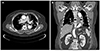 | Fig. 2Follow-up chest CT (3 weeks after medical therapy). CT scans with axial (A) and coronal (B) images reveal the improvement of diffuse wall thickening (arrows) from esophagus to gastric cardia. CT, computed tomography.
|
References
1. Huang YC, Cheng CY, Liao CY, Hsueh C, Tyan YS, Ho SY. A rare case of acute phlegmonous esophagogastritis complicated with hypopharyngeal abscess and esophageal perforation. Am J Case Rep. 2017; 18:125–130.


2. Kim GY, Ward J, Henessey B, et al. Phlegmonous gastritis: case report and review. Gastrointest Endosc. 2005; 61:168–174.
3. Hsu CY, Liu JS, Chen DF, Shih CC. Acute diffuse phlegmonous esophagogastritis: report of a survived case. Hepatogastroenterology. 1996; 43:1347–1352.
4. Jung C, Choi YW, Jeon SC, Chung WS. Acute diffuse phlegmonous esophagogastritis: radiologic diagnosis. AJR Am J Roentgenol. 2003; 180:862–863.
5. Kim HS, Hwang JH, Hong SS, et al. Acute diffuse phlegmonous esophagogastritis: a case report. J Korean Med Sci. 2010; 25:1532–1535.
6. Yun CH, Cheng SM, Sheu CI, Huang JK. Acute phlegmonous esophagitis: an unusual case (2005: 8b). Eur Radiol. 2005; 15:2380–2381.
7. Woo WG, Do YW, Lee GD, Lee SS. Phlegmonous esophagitis treated with internal drainage and feeding jejunostomy. Korean J Thorac Cardiovasc Surg. 2017; 50:453–455.
8. Hu DC, McGrath KM, Jowell PS, Killenberg PG. Phlegmonous gastritis: successful treatment with antibiotics and resolution documented by EUS. Gastrointest Endosc. 2000; 52:793–795.
9. Kim NY, Park JS, Lee KJ, Yun HK, Kim JS. A case of acute phlegmonous gastritis causing gastroparesis and cured with medical treatment alone. Korean J Gastroenterol. 2011; 57:309–314.
10. Chang PC, Wang WL, Hwang TZ, Cheng YJ. Intramural dissection with mucosal rupture alleviating phlegmonous esophagitis. Eur J Cardiothorac Surg. 2012; 41:442–444.
 XML Download