

 PDF
PDF ePub
ePub Citation
 Print
Print


Abstract
Clinical evaluations, nerve conduction studies, and electromyography play major complementary roles in electrophysiologic diagnoses. Electromyography can be used to assess pathologic changes and localize lesions occurring in locations ranging from motor units to anterior-horn cells. Successfully performing electromyography requires knowledge of the anatomy, physiology, and pathology of the peripheral nervous system as well as sufficient skill and interpretation ability. Electromyography techniques include acquiring data from visual/auditory signals and performing needle positioning, semiquantitation, and interpretation. Here we introduce the basic concepts of electromyography to guide clinicians in performing electromyography appropriately.
References
1. AANEM. Proper performance and interpretation of electrodiagnostic studies. Muscle Nerve. 2015; 51:468–471.
2. Preston DC, Shapiro BE. Electromyography and neuromuscular disorders: clinical-electrophysiologic correlations. 3rd Ed.London: Elsevier Saunders;2013. p. 125–266.
3. Rubin DI. Technical issues and potential complications of nerve conduction studies and needle electromyography. Neurol Clin. 2012; 30:685–710.

4. Daube JR, Rubin DI. Needle electromyography. Muscle Nerve. 2009; 39:244–270.
5. Rubin DI. Needle electromyography: basic concepts and patterns of abnormalities. Neurol Clin. 2012; 30:429–456.
6. Mills KR. The basics of electromyography. J Neurol Neurosurg Psychiatry. 2005; 76(Suppl 2):ii32–ii35.
7. Dumitru D, Amato AA, Zwarts M. Electrodiagnostic medicine. 2nd Ed.Philadelphia: Hanley & Belfus, Inc.;2002. p. 257–258.
8. Katirji B, Kaminski HJ, Ruff RL. Neuromuscular disorders in clinical practice. 2nd Ed.New York: Springer;2013. p. 89–152.
9. Kimura J. Electrodiagnosis in diseases of nerve and muscle: principles and practice. 1st Ed.Oxford: Oxford University Press;2001. p. 333–360.
10. Strommen JA, Daube JR. Determinants of pain in needle electromyography. Clin Neurophysiol. 2001; 112:1414–1418.
11. Paganoni S, Amato A. Electrodiagnostic evaluation of myopathies. Phys Med Rehabil Clin N Am. 2013; 24:193–207.
12. Jablecki CK, Busis NA, Brandstater MA, Krivickas LS, Miller RG, Robinton JE, et al. Reporting the results of needle EMG and nerve conduction studies: an educational report. Muscle Nerve. 2005; 32:682–685.
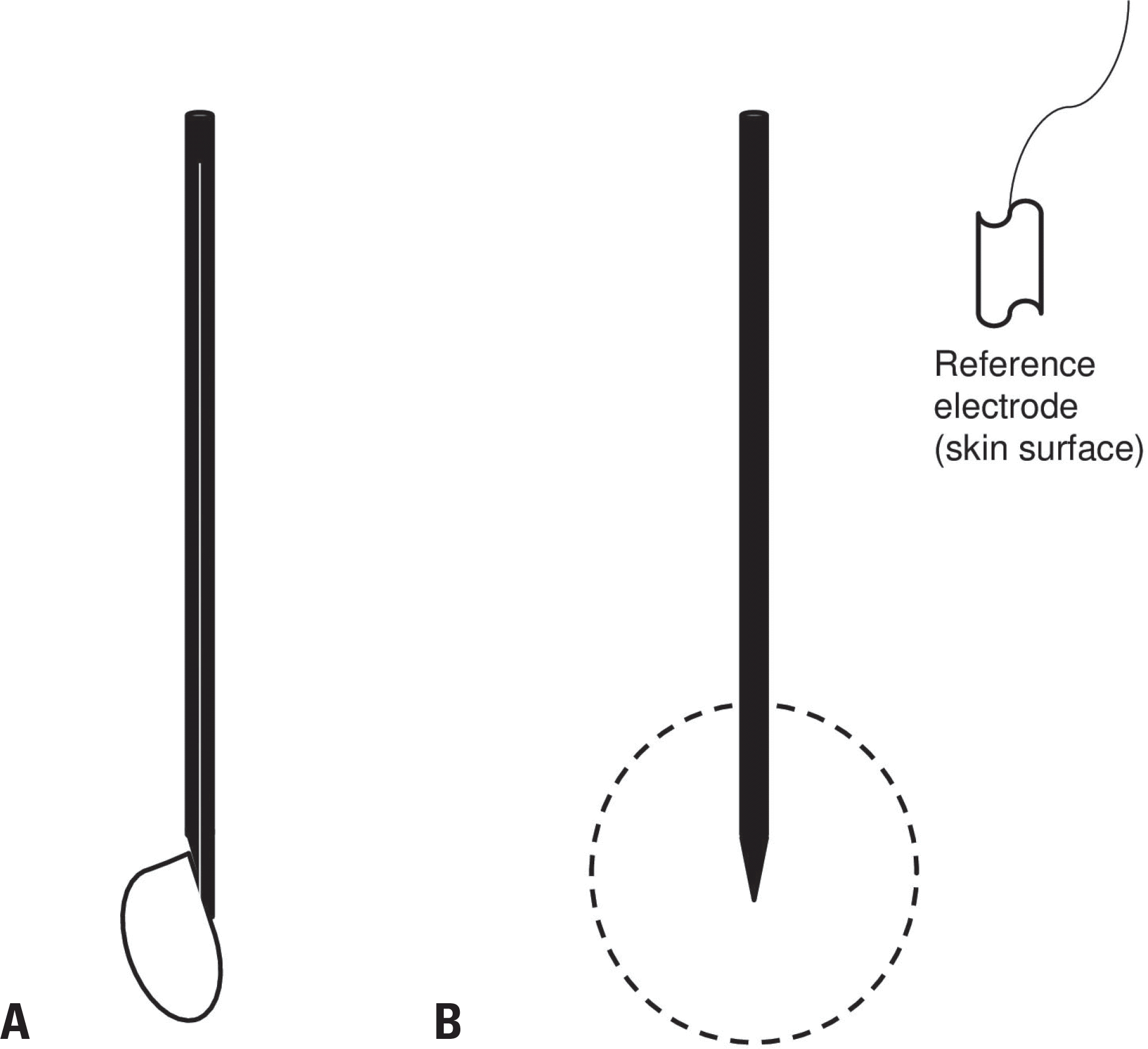
Fig. 1.
Types and recording area of electromyography needles. (A) A concentric needle and its recording field (side view). A wire that acts as the active electrode runs through the center of the needle shaft (white), and the shaft acts as the reference electrode. (B) A monopolar needle and its recording field (side view). The needle tip without a Teflon coating acts as the active electrode, and an additional reference electrode needs to be attached to nearby skin.
 XML Download