

 PDF
PDF ePub
ePub Citation
 Print
Print


INTRODUCTION
Shingles (herpes zoster) may cause a variety of neurological complications in the central and peripheral nervous systems, including postherpetic neuralgia, varicella zoster virus myelitis, segmental weakness, and delayed ischemic cerebral infarction due to zoster virus-associated granulomatous vasculitis.1 Zoster-associated limb paresis (ZALP) is a relatively uncommon complication of herpes zoster characterized by focal motor weakness, which may occur in limbs affected by herpes zoster.2 Zoster-associated mononeuropathy (ZAM) as a cause of herpes zoster-associated paresis is even rarer.3 Here we report a case of a herpes zoster involving the femoral nerve.
CASE
A 79-year-old man was admitted to the dermatology department of our hospital with pain and skin eruptions over the left medial thigh. He was diagnosed as having herpes zoster and was treated with 750 mg of famciclovir daily for 7 days. The patient reported that he could not walk due to pain but did not have weakness in the leg. He was not evaluated for gait difficulty at that time. Two weeks after the treatment, the patient revisited the hospital due to weakness in the left leg and was admitted to our neurology department.
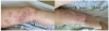
Upon admission he showed scars of herpetic eruption and pigmentation over the left medial thigh and medial calf, but not over the lateral thigh (Fig. 1). Muscle weakness in the left lower limb was mostly noted for hip flexion (MRC grade 2) and knee extension (MRC grade 2). The left tibialis anterior, gastrosoleus group, hip adductors, and abductors were all 5/5. The patient had no left patellar muscle stretch reflex and a normal left Achilles reflex. Sensation was impaired over the left anteromedial thigh and medial calf, but not over the lateral thigh. His right lower limb was entirely normal with respect to strength, sensation, and muscle stretch reflexes.
The findings of routine laboratory studies were normal. Magnetic resonance imaging (MRI) of the lumbar spine showed only small herniations of the intervertebral discs from levels L1/2 to L4/5, mild spinal stenosis at level L4/5, and degenerative minute spondylolisthesis at level L4/5. MRI did not reveal any nerve or spinal cord abnormalities such as prolonged T2-weighted signals, nerve enlargement, or postgadolinium enhancement. An electrodiagnostic evaluation was performed. Compound muscle action potential in femoral nerve conduction study and sensory nerve action potential in medial femoral cutaneous nerve conduction study were unobtainable on the left, while the amplitude responses of those were normal on the right (Fig. 2). The amplitude of the left peroneal motor nerve response was slightly decreased, while the left posterior tibial motor nerve response was normal. Needle electromyography (EMG) revealed positive sharp waves in the left L2-S1 paraspinal, vastus lateralis, and iliacus muscles. The EMG findings suggested left femoral neuropathy with coexistent lumbosacral radiculopathy. We assumed that the lumbosacral radiculopathy was attributable to the preexisting herniations of the intervertebral discs at levels L1 to L5, because a diffuse denervation process was not observed in the corresponding peripheral limb muscles. We thought that herpes zoster accounted for the left femoral neuropathy but not for the radiculopathy.
Combining the history with the findings of the physical examination, imaging studies, and electrodiagnostic evaluation led to a diagnosis of herpes zoster femoral neuropathy with coexisting polyradiculopathy. We treated the patient with intravenous acyclovir at 10 mg/kg/day for 7 days and physical therapy. The patient's muscle strength improved to MRC grade 3 at the time of discharge (2 weeks after starting acyclovir). He could not walk at the time of discharge, but his left leg strength had improved to MRC grade 4 when he visited the outpatient department approximately 2 months after starting acyclovir. However, he still complained of postherpetic neuralgia. The patient refused an electrodiagnostic follow-up examination due to his improving symptoms.
DISCUSSION
Segmental zoster paresis is a rare occurrence. The incidence of segmental limb paresis with cutaneous zoster has been reported at 3% to 5%.3 ZAM as a cause of zoster-associated paresis is even rarer. In one series of 49 patients with ZALP, it was caused by radiculopathy (37%), plexopathy (41%), mononeuropathy (14%), or radiculoplexus neuropathy (8%).3 In another series, ZAM was associated with prolonged symptoms, significant weakness, and a high rate of postherpetic neuralgia.4 Our patient also complained of postherpetic neuralgia at 2 months of follow-up.
ZALP may localize to the root, plexus, or more-peripheral nerve, and is difficult to localize clinically in part because the involved myotomes often do not correspond to the dermatomes affected by the rash.1 For example, mononeuropathies may be difficult to distinguish clinically from more-proximal focal plexopathies or radiculopathies. Electrophysiological studies are useful for correctly diagnosing and evaluating the extent of lesions. Nerve conduction studies usually disclose reduced sensory nerve action potentials and compound muscle action potentials in the affected segments.5 Needle EMG generally reveals abnormal spontaneous activities, such as fibrillations and positive sharp waves in clinically weak muscles. Imaging studies such as magnetic resonance neurography can be invaluable in the identification and localization of affected areas. One recent retrospective study found MRI imaging abnormalities in seven of 10 patients (70%) with zoster-associated plexopathy.1 Jones et al. reviewed the nerve imaging features of 26 patients with zoster paresis: nine of the 14 patients (64%) with postganglionic electrodiagnostic localization (at the plexus or nerve level) demonstrated imaging abnormalities in the affected plexus or nerve, and one patient showed enhancement with gadolinium contrast.3 More commonly, nerve enlargement or increased T2-weighted signals within the affected nerve were observed. None (0%) of the 12 patients with preganglionic localization (at the anterior horn or root level) demonstrated abnormal findings in nerve or spinal cord imaging, but abnormalities have been described in other cases of zoster paresis.36 We did not perform an imaging study to explore the affected femoral nerve in the present patient.
The precise mechanism underlying zoster-associated paresis is poorly understood.4 Virally mediated injury at the level of the anterior horn cell or ventral root has been suggested as the cause of herpes zoster paresis.4 A postmortem observation showed the degeneration of anterior spinal roots with lymphocytic infiltration of the posterior and anterior horns.7 An MRI study revealed contrast enhancement of the anterior roots of affected segments, suggesting the presence of hypervascularity or disruption of the blood-nerve barrier caused by viral-induced inflammation in patients with zoster paresis.8 Anterior spinal roots seem to be common sites of inflammation and degeneration, but anterior horn cells, the brachial or lumbar plexus, and peripheral nerves can also be involved. Individual reports of ZAMs suggest that a distally mediated neuropathic mechanism could underlie herpes zoster paresis in some patients.4
The prognosis of zoster-associated limb paresis is generally good. Patients usually recover most functions, and more than half of them recover completely.2 It remains unclear whether a diagnosis failure affects the prognosis. Mondelli et al.9 reported that antiviral therapy at the appropriate dose and duration was associated with a reduced incidence of segmental zoster paresis. It is unclear whether additional intravenous antiviral or steroid treatment is beneficial once paresis occurs. Further studies of the optimal treatment for promoting functional recovery are necessary. The protective effect provided by the zoster vaccine against zoster-associated paresis would also be of particular interest.

 XML Download