

 PDF
PDF ePub
ePub Citation
 Print
Print


INTRODUCTION
Erythema nodosum (EN) is a delayed-type hypersensitivity reaction that primarily presents as erythematous tender nodules on the shins.12345 Common triggers for EN include infection, drugs, pregnancy, malignancy, and inflammatory conditions, although several cases are idiopathic in nature.145 EN is rare in the pediatric population.23 The most common etiology is infection caused by Streptococcus pyogenes, Mycobacterium, Yersinia, atypical bacteria, or Salmonella.12345
Kawasaki disease (KD) is an acute, febrile systemic vasculitis that primarily occurs in infants and young children.678 Although its etiology is unknown, an aberrant innate immune response in predisposed individuals has been proposed as an important contributor to the development of inflammatory vasculitis and the clinical manifestations of KD.68 Patients with KD primarily present with “polymorphic and nonspecific” skin eruption.7 Although the most common form is a diffuse maculopapular eruption, rashes can show various forms, including urticarial exanthema, scarlatiniform rashes, erythroderma, erythema multiforme-like rash, micropustular, and pustular eruptions.7 However, EN-like lesions in a patient with KD have not been reported.
Here, we report the first case demonstrating an association between KD and EN. EN masked underlying KD, and the patient developed a coronary artery lesion (CAL).
CASE REPORT
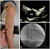
A 3-year-old girl was referred to our hospital with a 3-day history of high fever and leg swelling. She was born to non-consanguineous healthy parents as a full-term infant. At the initial visit, she showed high fever of 40℃, injection of the oropharynx, cervical lymphadenopathy, and red-purple cutaneous nodules, particularly on the lower limbs (Fig. 1A) without gastrointestinal symptoms. She complained of severe pain in the neck and cutaneous lesions. She did not show clinical manifestations of KD, such as conjunctival hyperemia, strawberry tongue, bleeding lips, and erythema at Bacille Calmette-Guérin (BCG) inoculation site. She denied a relevant medication history. Hematological parameters were as follows: leukocyte count, 22.1×10⁹/L with 74.0% segmented neutrophils; hemoglobin concentration, 11.5 g/dL with 34% hematocrit; platelet count, 332×109/L; fibrinogen level, >650 mg/dL; total serum protein, 7.2 g/dL; serum albumin, 3.8 g/dL; total bilirubin, 0.5 mg/dL; aspartate aminotransferase, 22 IU/L; alanine aminotransferase, 13 IU/L; C-reactive protein (CRP), 9.6 mg/dL; and sodium, 137 mmol/L. Rapid diagnostic tests for S. pyogenes and adenovirus were both negative. Antistreptolysin O antibody titers were within reference ranges. She was diagnosed with EN following acute pharyngitis, and ampicillin/sulbactam therapy was initiated intravenously at a dose of 150 mg/kg/day. Stool culture obtained upon admission revealed only a slight increase in Salmonella spp [O9, non H-d (final report was submitted to us on the 10th day of illness)], which appeared to be the causative antigen for EN. Yersinia spp were negative in the stool culture. Although the Salmonella spp were sensitive to ampicillin/sulbactam, she did not improve clinically. On the 5th day of illness, the antibiotic was switched to meropenem at a dose of 100 mg/kg/day. Following defervescence on the 8th day of illness, the related symptoms including cutaneous lesions disappeared. Echocardiography performed to screen for fever of unknown origin revealed medium-sized aneurysms of the left anterior descending artery [4.6 mm (Z: +6.6)] (Fig. 1B) although it did not show any abnormal findings on admission. This finding led to a diagnosis of incomplete KD, and oral aspirin therapy was initiated. She did not show recurrence of fever, her CRP turned negative on the 17th day of illness, and she was discharged. She did not show any gastrointestinal symptoms during hospitalization. Paired serum antibody of Mycoplasma pneumoniae obtained during the hospitalization were both negative. The CAL gradually regressed after discharge. Skin desquamation was not observed during follow-up. Cardiac catheterization performed 2 months after discharge showed complete regression of the CAL (Fig. 1C). Oral aspirin therapy was discontinued after the cardiac catheterization. To date, follow-up echocardiogram has determined no apparent CAL without any cardiovascular events. Written informed consent was obtained from the patient's parents.
DISCUSSION
This is the first case report to describe a patient with KD who presented with EN as the initial manifestation. EN-related symptoms masked those of KD with a consequent delay in the diagnosis of KD and the development of CAL. The CAL regressed spontaneously without any subsequent cardiovascular events in the patient.
The pathogenesis of EN was a major concern in our patient. Initially, the development of EN was attributed to Salmonella spp infection.12345 However, stool culture showed only slight bacterial growth, and the patient showed no gastrointestinal symptoms. Moreover, the patient did not respond to highdose ampicillin/sulbactam to which the Salmonella spp is sensitive. Based on these findings, the development of EN was considered a component of the major manifestations of KD and not a sequela of Salmonella infection. Although EN is histopathologically characterized by septal panniculitis without vasculitis,12 it might occur in patients with KD because skin lesions in KD are typically described as “nonspecific.”789 Moreover, not all symptoms related to KD are explained by vasculitis alone.910 We did not perform a skin biopsy in our patient, which serves as a limitation of this study.
Another concern is that the clinical presentation of EN and KD is indistinguishable. In the present case, prolonged fever, injection of the oropharynx, cervical lymphadenopathy, neck pain, rash, and swelling of the extremities could be observed as features of both conditions.1234578 Conjunctival hyperemia, strawberry tongue, bleeding lips, and/or erythema at the BCG inoculation site could be helpful to distinguish KD from EN; however, these findings might not always be observed in older patients.11
EN in the pediatric population is rare; however, it may occur following various conditions, including KD. KD should be considered in the differential diagnosis of refractory EN in pediatric patients.
 XML Download