

 PDF
PDF ePub
ePub Citation
 Print
Print


INTRODUCTION
Cancer treatment focuses on curing the disease itself, whereas individuals place an emphasis on treatment safety and efficacy. The American Cancer Society estimated that 10,380 new cancer cases and 1,250 deaths from cancer occurred in 2016 among males and females aged 0 to 14 years [1]. Infertility is an important long-term adverse effect in males despite recent advances in treatments for malignancies that may cure young cancer patients [23456]. Increasingly many young adults are long-term survivors of cancer. Patients younger than 15 years of age undergoing cancer treatment are projected to have a 5-year cancer survival rate of 75% [7]. More than 50% of these young male survivors will desire paternity after treatment, including 75% of those who were childless at the time of diagnosis [8].
Although chemotherapy and radiation therapy for malignancies are highly effective, their associated gonadotoxic side effects may severely impair fertility in agent- and dose-dependent manners and may cause temporary or permanent gonadal toxicity in male patients [9]. The resumption of spermatogenesis after various types of therapies is unpredictable, and studies on spermatogenesis in long-term cancer survivors have provided evidence of persistent azoospermia or severe oligozoospermia in up to 24% of cases [10]. Nonetheless, the eventual return of sperm production in many posttreatment cancer patients has prompted the question of whether post-therapy spermatozoa are a suitable option for conception, either naturally or via assisted reproductive technologies.
Spermatogenesis may still continue over several years if the spermatogonial cell population is not completely depleted. If a population of these germ stem cells remains after cancer treatment, the regeneration of spermatozoa may continue for years [11].
Sperm cryopreservation is a well-established technique and is offered before cancer treatment in case of azoospermia in the future. New fertility preservation options, such as the generation of gametes from embryonic stem (ES) or induced pluripotent stem (iPS) cells, have been developed that may change reproductive options for men and boys facing germ-cell loss and sterility. It is very important to inform patients and family facing infertility of this possible side effect of their treatment, as well as all the options available to prevent it, because the recovery of gonadal function after cancer treatment remains unpredictable.
One of the aims of this review is to discuss the pathophysiology of male infertility caused by radiation and/or chemotherapy for cancer treatment. The other aim is to review the use of sperm cryopreservation to maintain fertility and to discuss new fertility preservation strategies.
RADIATION
Radiation therapy remains the main treatment option for many malignant cancers in men of reproductive age. The testis is one of the most radiosensitive organs. Animal data indicate that the fractionation of radiotherapy increases its gonadal toxicity, and evidence suggests that this also occurs in humans. Gonadal damage caused by radiotherapy depends on the gonadal dosage and how radiation is delivered. Damage may be caused during direct irradiation of the testis or, more commonly, from scattered radiation during treatment directed at adjacent tissues.
Testes directly exposed to ionizing radiation exhibit germ cell loss and Leydig cell dysfunction [1213]. The testes do not need to be directly irradiated in order for spermatogenic impairment to occur; if the radiation field is proximal to a testis and the dose is sufficient, sperm production may be diminished even if the testes are shielded [14]. Radiation therapies begin to affect spermatogenesis gradually from 0.1 to 1.2 Gy and induce irreversible gonadal damage at 4 Gy [15]. The function of the testes may be significantly impaired by very low doses of radiation therapy. The effects of low-dose and single-fraction irradiation on spermatogenesis in healthy men have been discussed [16]. Doses as low as 0.1 Gy may cause morphological and quantitative changes to spermatogonia, which are radiosensitive immature cells. At doses of 2 to 3 Gy, spermatocytes are damaged and spermatid numbers decrease. Doses of 4 to 6 Gy result in significant reductions in the numbers of spermatozoa and damage to spermatids.
The dose limits at which azoospermia becomes permanent remain unclear. Doses of more than 1.2 Gy are known to increase the recovery time of spermatogenesis [16]. Reductions in sperm count following damage to the testes by radiation doses of up to 3 Gy have been noted after 60 to 70 days. Radiation doses of 4 Gy reduce sperm concentrations because of damage to spermatids. Radiation doses of less than 0.8 Gy may result in oligospermia, whereas those greater than 0.8 Gy may lead to azoospermia [17]. Another study showed that a radiation dose of 7.5 Gy or higher to the testes for childhood cancer decreased the chance of these patients having their own children [18].
According to a study on boys with acute lymphoblastic leukemia who received radiotherapy for the testis at doses of 12, 15, and 24 Gy, all became azoospermic, while doses less than 24 Gy had no effect on testosterone levels [19]. Elevated gonadotropin levels were observed and subclinical Leydig cell damage was suspected. Leydig cells are considered to be more resistant to radiation-induced damage at a dose of up to 30 Gy [20]. According to a survey, the recovery of spermatogenesis may start at least 9 years after treatment [21]. However, better techniques that enable more accurate dose delivery and protection of the gonads have resulted in earlier complete recovery of spermatogenesis, at 9 to 18 months after radiation at doses up to 1 Gy, 30 months at doses up to 2 to 3 Gy, and 5 years at doses up to 4 Gy.
According to a study presenting findings from longterm follow-up of stage I and IIA seminoma patients after radiation therapy, 64% achieved natural pregnancy and 50% showed complete recovery of spermatogenesis [22].
Although radiation typically acts by killing cells immediately by apoptosis or when they attempt to proliferate or divide, the minimum number of type A spermatogonia after single radiation dose between 0.2 and 4 Gy was not reached rapidly, but as the result of a progressive decline over the course of approximately 21 weeks [232425]. The reasons for this gradual decline currently remain unknown, but may be due in part to some of the non-cycling A stem spermatogonial population only expressing lethal damage when they are recruited into the cycle. Furthermore, the differentiation of spermatogonia into spermatocytes is reduced during this time [26]. This phenomenon has been observed in rats and indicates somatic damage, or at least altered signaling from somatic cells.
The ability to differentiate into spermatocytes and later stages increases approximately 21 weeks after radiation therapy. The number of type A spermatogonia begins to increase at this time, suggesting that self-renewal exceeds cell loss. The timing of the recovery depends on the dose of radiation. It begins 7 months after irradiation with a single dose of 1 Gy and takes 24 months after irradiation with 6 Gy. Complete recovery of the sperm count to the pre-irradiation level requires approximately 2 years after a single dose of 1 Gy.
High doses of radiation therapy may kill all spermatogonial stem cells (SSCs), resulting in permanent azoospermia. Previous studies reported that only approximately 15% of patients recovered their sperm count or fertility after single doses of approximately 10 Gy [2728]. It is important to note that responses to the doses administered above were for single doses of radiation, which have been examined in the greatest detail. Fractionated radiation used for cancer treatments for 3 to 4 weeks causes greater delays in spermatogenic recovery and leads to permanent azoospermia. Sandeman [29] showed that a total gonadal dose of more than 2.5 Gy of fractionated radiation generally resulted in permanent azoospermia.
In a previous study, all patients who received a testicular dose of radiation of 1.2 to 3.0 Gy in 14 to 26 fractions for Hodgkin disease became azoospermic after the treatment [30]. An update of this study revealed that spermatogenesis did not recover in patients receiving doses of 1.4 to 2.6 Gy over a follow-up period ranging between 17 and 43 months. However, fertility returned in 2 patients with testicular radiation doses of 1.2 Gy, suggesting a threshold for permanent testicular damage [31]. Another study reported the effects of radiation on follicle-stimulating hormone (FSH) levels and the sperm count during the treatment of Hodgkin disease [32]. Testicular doses of less than 0.2 Gy had no significant effects on FSH levels or sperm count, whereas doses between 0.2 and 0.7 Gy increased FSH levels and reduced the sperm concentration. Lower doses of radiation to the testes were associated with better recovery rates for spermatogenesis. The recovery length of spermatogenesis after modest radiation therapy that may kill some stem cells is longer than that after a chemotherapy regimen that may not kill stem cells.
The mechanisms by which prostate radiotherapy affects reproductive function have recently been reported. If the dose received by the testes from I125 brachytherapy of the prostate is close to 0.18 Gy, prostate brachytherapy has almost no effect on spermatogenesis. However, due to the prolonged half-life of the isotopes used, attempts at conception were delayed for up to 3 to 12 months after treatment [3334].
CHEMOTHERAPY
In addition to radiation, most chemotherapeutic drugs are considered to be toxic to the gonads, particularly alkylating medications. Many studies have investigated cyclophosphamide, which is used in the treatment of immunological diseases or in combination chemotherapy for hematological and testicular cancers. Most studies have focused on semen analyses and biochemical markers of fertility. Although the end point of the assessment of male factor fertility is the achievement of fatherhood, survivors of cancer may be less likely to find a partner or may not want their own children. This may be because of the psychological effects of the disease or treatment and because of the genetic risk in the offspring of patients subjected to cytotoxic treatments. Female factors that may influence the couple's fertility are also difficult to identify.
Many combination chemotherapies used in the treatment of cancer also produce reductions with a similar time course. Due to toxicity to later stage germ cells, 10- to 100-fold decreases in sperm counts may occur within 1 to 2 months, while azoospermia generally does not occur until after 2 months, when sperm are derived from differentiating spermatogonia [3536]. Although sperm are produced for several months after the start of cytotoxic therapies, pregnancy needs to be avoided during this period because of a higher risk of genetic damage to sperm.
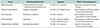
Similar to radiation therapy, Leydig cells may incur damage following chemotherapy, resulting in subsequent hypogonadism [37]. Although side effects have been minimized with advances in the delivery of chemotherapy using synergistic agents at lower toxic doses, a risk of infertility remains. The extent of gonadal damage is largely dependent on the drug type, age of the patient, and amount of the chemotherapeutic agent administered. Table 1 summarizes some chemotherapy drugs and their effects on spermatogenesis.
Alkylating agents, particularly mustards, are among the most potent germ cell mutagens, inducing dominant lethality, heritable (reciprocal) translocations, and specific locus mutations in post-stem cell stages [3839]. Three alkylating anticancer drugs (melphalan, mitomycin C, and procarbazine) have been shown to induce specific locus mutations in SSCs; however, no chemical has yet been shown to induce transmissible chromosomal translocations (dominant lethality and heritable translocations) in stem cells [40]. Chemotherapy with alkylating agents, with or without radiation to sites below the diaphragm, has been associated with a fertility deficit in approximately 60% of men [41]. The duration and permanence of induced azoospermia depends on the dose of the cytotoxic agent and the additive effects of different agents. When cyclophosphamide is given as a single agent, doses of 19 g/m2 are required for prolonged azoospermia [35]. A follow-up of 26 male patients with azoospermia after the cessation of cyclophosphamide showed the return of spermatogenesis in 12 patients within 15 to 49 months (mean, 31 months) [42]. According to another study of 116 males treated with cyclophosphamide alone, 52 showed evidence of testicular dysfunction after the treatment [17]. The incidence of gonadal dysfunction was related to the total dose of cyclophosphamide. More than 80% of postpubertal patients received more than 300 mg/kg of cyclophosphamide. Since busulfan is very effective as a single agent for killing SSCs in rodents and monkeys, it is the alkylating agent with the strongest sterilizing effect [43].
Limited information is currently available on parenthood rates after treatment for Hodgkin disease. According to one study, only 18 out of 101 men who had received chemotherapy, radiotherapy, or both for Hodgkin disease over a 21-year period had fathered a child. Twelve of those men had been treated with radiotherapy only [44]. Another study showed improved fatherhood rates following chemotherapy or radiotherapy for Hodgkin disease. Twenty-five of 51 men (49%) were able to have their own children [45]. The chemotherapy regimens used for the treatment of non-Hodgkin lymphoma (NHL) are generally considered to be less gonadotoxic than those for Hodgkin disease. According to one study, all 71 patients treated with CHOP (cyclophosphamide, doxorubicin, vincristine, and prednisolone)-based chemotherapy were rendered azoospermic during the treatment, while 67% recovered to normospermic levels 5 years after the treatment, with a further 5% being oligospermic [46]. The lower incidence of permanent infertility in men treated for NHL than in Hodgkin disease patients may be related to the absence of procarbazine in the standard regimens used for NHL [47]. Furthermore, the use of lower doses of alkylating agents may also be important. The absence of procarbazine and alkylating drugs is also a likely explanation for the reduced toxicity of ABVD (doxorubicin hydrochloride [Adriamycin], bleomycin, vinblastine, and dacarbazine) [48].
Cisplatin and cyclophosphamide were investigated as the test chemical and positive control, respectively, to assess their cytogenetic effects on spermatogonia in mice 24 hours after a treatment with single exposure. The different doses of the chemicals tested in mice were cisplatin at 2, 3, and 5 mg/kg and cyclophosphamide at 40 mg/kg. This study showed that each dose of cisplatin induced a significant number of chromosomal aberrations, mostly chromatid breaks and fragments [49]. According to an analysis of 170 patients who received cisplatin-based chemotherapy for testicular germ cell cancer for at least 1 year, the post-chemotherapy count was normospermic in 64%, oligospermic in 16%, and azoospermic in 20% of 89 patients whose prechemotherapy counts were normospermic. There was clear evidence for continued recovery after 1 year, as the probability of spermatogenesis increased to 48% by 2 years and 80% by 5 years. The probability of recovery to oligospermic and normospermic count levels was significantly higher in 54 patients treated with carboplatin-based therapy than in those treated with cisplatin-based therapy [50].
According to rat data, vinblastine did not cause any significant changes in the incorporation of [3H] thymidine at any stage in the seminiferous epithelial cycle; however, some time-dependent reductions were observed, particularly at stages I and VIII [51]. Another study on mice showed that spermatogonia were resistant to the actions of vinblastine with no observable loss, even at a dose of 7 mg/kg [52]. However, vinblastine exhibited the ability to induce the arrest of mitotic metaphase in rat spermatogonia [53] and inhibited meiotic division in mouse and rat spermatocytes [5455].
The induction of structural chromosomal aberrations by bleomycin was studied in the bone marrow cells and spermatogonia of mice at doses of 10, 20, 40, and 80 mg/kg. Bleomycin induced genetically important reciprocal translocations in stem-cell spermatogonia, as measured with the spermatocyte test, and the response of bone marrow cells to bleomycin was not markedly different from that of spermatogonia [56].
TREATMENT FOR IMPAIRED SPERMATOGENESIS
Even if a patient's semen analysis shows azoospermia after radiotherapy and/or chemotherapy for malignant cancer, some sperm may still be present in the testis. Spermatozoa were retrieved from the testes by microdissection testicular sperm extraction in 37% of patients who were azoospermic after chemotherapy [57]. According to a previous study, sperm do not survive epididymal transit and do not reach the ejaculate if the human testis contains fewer than 3–4 million sperm [58]. The success rate of retrieving sperm by testicular sperm extraction is related to the presence of residual hypospermatogenesis in the testis. The success rate was higher in patients treated with alkylating agents than in those treated with agents that are toxic to stem cells and/or the somatic environment.
In adolescent males who have already entered puberty, the most established approach to preserving fertility is the cryopreservation of ejaculated sperm. Cryopreserved sperm may be used later in life for intrauterine insemination or in vitro fertilization, with or without an intracytoplasmic sperm injection. In most males, the process of providing sperm for cryopreservation is effective, inexpensive, and non-invasive because most adolescent males are able to provide ejaculated semen. However, options are limited to experimental techniques for prepubertal males. Prepubertal testicular tissue banking under an Institutional Review Board-approved protocol is currently available at several institutions [9].
The option of the preservation of one's own semen before gonadotoxic treatment is not available for prepubertal boys, and in such patients, only the necessary amount of testicular tissue has to be removed for cryopreservation for future offspring. A morphological study estimated that 1 testis of a 10-year-old pre-pubertal boy contains approximately 83×106 germ cells [59].
Regarding male infertility following chemotherapy and/or radiation, stem cell therapy to generate male gametes may represent a promising treatment strategy. Stem cells are defined as having the potential for self-renewal and differentiation. Three major stem cell sources were recently identified for the generation of male differentiated germ cells: ES cells, iPS cells, and SSCs. ES cells are derived from the inner cell mass of developing blastocysts. The first human ES cell line was established in 1998 [60]. Advances have since been made in the derivation of differentiated male germ cells from mouse or human ES cells [616263]. Transcription factors were used to reprogram somatic cells to iPS cells in 2006 [64]. A recent study demonstrated that iPS cells generate haploid spermatids [65]. SSCs have the ability to self-renew and differentiate into male gametes (i.e., mature spermatozoa) in the testis throughout life [66] Recent studies showed that spermatogonia, including SSCs, may be induced to differentiate into differentiated male germ cells, eventually resulting in haploid spermatids.
Although sperm DNA damage occurs following chemotherapy or radiotherapy, an increase in genetic defects or congenital malformations was not detected among children conceived by parents who had previously undergone treatments. However, the use of assisted reproductive technologies and micromanipulation techniques may increase this risk [67].
CONCLUSIONS
Cancer and its cytotoxic treatments may impair male fertility in various manners, thereby excluding these men from the opportunity to father offspring. Improvements in the toxicity of cancer treatment and the selective delivery of therapeutic agents will result in better outcomes, reduced sperm damage, and earlier recovery of spermatogenesis. All men wishing to have their own children after gonadotoxic treatment need to be counselled properly by a fertility specialist regarding the potential risks of their disease and the side effects of therapy, as well as the possibility of fertility recovery.
Male factor infertility is a known side effect of cancer treatment. All patients need to be thoroughly educated about the impact of cancer treatment on their fertility and provided with numerous options to preserve their future fertility potential. It is currently very difficult to predict which patients will recover spermatogenesis and which will remain azoospermic. No parameters help predict which patients will remain permanently sterile.
Therefore, semen analyses to test sperm count and quality before radiotherapy and/or chemotherapy need to be offered to male patients for future family planning. The cryopreservation of sperm before treatment also needs to be suggested. Sperm cryopreservation is the only clinical method currently available. Fertility maintenance is still an issue in younger boys, and extensive efforts are being made to improve techniques for testicular tissue or spermatogonial cryopreservation and transplantation and testis xenografting. However, they are not routinely applied due to clinical and ethical reasons.
The cryopreservation of semen is a safe and effective way of preserving fertility for adolescent and adult males. For adult men with azoospermia, testicular sperm extraction is necessary and is the only option for retrieving sperm. However, fertility preservation options for pre-pubertal males are limited; therefore, patients and their families need to be counseled before treatments by a specialist in fertility preservation. The creation of spermatozoa from ES cells, iPS cells, and SSCs is theoretically an option for eventual reproduction in men who have lost their testicular germ cells. Although still a topic of research, further developments of this technology are expected in the near future.
 XML Download