

 PDF
PDF ePub
ePub Citation
 Print
Print


Relapsing polychondritis (RP) is an uncommon disorder involving various cartilages and other proteoglycan-rich structures including eyes, inner ears and blood vessels [1]. Nervous system involvement is rare, which could be manifested as cranial nerve dysfunction, vasculitic stroke, meningitis and meningoencephalitis [2, 3]. Meningoencephalitis due to RP is potentially fatal and its response to immunosuppressant therapy is good in the majority of cases [2]. Thus, early diagnosis and subsequent appropriate treatments are vital in approaching these patients. Therefore, comprehensive understanding of its clinical characteristics would be of prime importance. In this report, we present a patient revealing distinct cognitive and behavior abnormality due to meningoencephalitis with RP. Serial neuropsychological tests were performed at different clinical conditions in this patient.
CASE REPORT
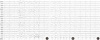
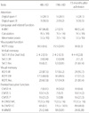
A 54 years old man was admitted to our hospital for headache and dizziness associated with memory loss for three weeks. Characteristic of headache was tightness in bilateral and diffuse but more prominent in occipital region. Dizziness was vertigo in nature and worsened over the period. He also complained to left ear pain. Three months ago, he was treated by opthalmologist due to scleritis. Two months ago, he was admitted to otolaryngology department for his sudden sensorineural hearing loss on his left side and received steroid therapy with dexamethasone, 10 mg a day for one week. On physical examination, the vital signs were normal including body temperature and there was no other abnormality. On neurological evaluation, he was alert and conscious. However, left beating spontaneous nystagmus exacerbated by head shaking test was noted along with catch up saccade on right head turning in head thrust test. Hearing was reduced on his left side. There were no other deficits in the rest of neurologic examinations except Korean version of mini-mental state examination (K-MMSE). He gained 25 points (30 points in full) including not being able to remember two words among three five minutes later. Initial blood tests showed mild macrocytic anemia with elevated ESR (101 mm/hour), high level of C-reactive protein(CRP) (2.69 mg/dL). Other tests including liver function test, renal function test, electrolytes, thyroid function test and urinalysis were found to be within normal limit. Comprehensive neuropsychological test was performed three days later (Table 1). He got a low score on calculation, copying complex figure, verbal learning and retention tests, and frontal executive functions. On brain magnetic resonance imaging, there were no abnormalities. On next day (fifth hospital day), he became aggressive and agitated, complaining of visual hallucinations of seeing his brother who was dead. Electroencephalogram (EEG) was proceeded to find out the reasons and it revealed frequent bilateral frontal spike and slow wave complexes (Fig. 1). Next, we carried out cerebrospinal fluid (CSF) examination and found out lymphocytic pleocytosis, 59 cells/mm3 (98% of lymphocytes) with normal glucose (58.5 mg/dL) and slightly increased protein level (61.9 mg/dL). To find the cause of cognitive impairment Fluorodeoxyglucose (18F) positron emission tomography (FDG-PET) imaging was performed. However, we could not find abnormal glucose metabolism. Based on these findings, viral encephalitis was suspected. Intravenous acyclovir (750 mg, three times a day) was prescribed thereafter (for two weeks in total) with antiepileptic agents (valproate 300 mg, three times a day) although polymerase chain reaction for herpes simplex virus was negative in CSF. In autoimmune screening tests encompassing rheumatoid factor, anti-cyclic citrullinated peptide antibody, antinuclear antibody (ANA), anti-double stranded DNA (anti-ds DNA), anti-neutrophil cytoplasmic antibody (ANCA), and thrombophilia screening tests like lupus anticoagulant, antiphospholipid antibodies, protein C and S were within normal range. On next day (6th HD), he developed fever, cauliflower like swelling of left ear lobe and red eyes. In spite of two days of antiviral and antiepileptic treatments, his aggressive behavior and hallucinations were progressive. He kept on shouting, not having sleep, and he was not oriented to time and place. Under the suspicion of encephalitis due to polychondritis, intravenous steroid therapy was started (7th HD), initially with methylprednisolone 5 mg four times a day for four days replaced by solumedrol pulse therapy for three days and followed by oral prednisolone. With time, his aggressive and agitated behavior recovered slowly and on 19th HD, follow up neuropsychological tests was performed. Compared to the initial test, the cognitive dysfunctions became worse involving more cognitive domains; attention, naming, and praxis in addition to the previous ones (Table 1). Repeat EEG showed intermittent diffuse semirhythmicbifrontal dominant delta slowing, but no definite epileptiform discharges. On 30th HD, he was discharged and followed up regularly at outpatient clinic. With continuous tapering oral prednisolone, azathioprine was added, and now he is on prednisolone 10 mg and azathioprine 150 mg by rheumatologist. He showed continuous improvement in cognitive function and general condition. However, there was an additional episode of chondritis in his hand two months after the discharge. The follow up EEG and neuropsychological test were taken 7.5 months after the admission. At this moment, his abnormal behavior or personality changes like aggression and excessive anxiety disappeared, and his cognitive problems reduced significantly. EEG was noted to be completely normal (Fig. 1). However, definite poor performance was still observed in calculation, copying a complex figure, verbal learning and memory, part of word fluency test despite that all the scores improved substantially. Interestingly, the deficits were found in the same cognitive domains as the earliest tests.
DISCUSSION
Meningoencephalitis due to RP was reported very rarely. Especially in Korea, there have been a few case reports until now [4, 5]. Providing that the correct diagnosis of RP can be delayed by 2.9 years on average [6], the actual incidence of this disorder might be higher. The pattern of cognitive dysfunctions has been described with the results of brief cognition screening tests [2,5,7], but not with comprehensive neuropsychological tests. It might be due to serious conditions related to meningoencephalitis and RP. As a result, we could not perform these tests while our patient was showing the most severe cognitive and behavioral symptoms. Since he complained of cognitive impairment from the earlier time when he was in a better condition, we could evaluate his neuropsychological functions using the same tests throughout. The cognitive impairment was distinct involving different cognitive domains. Concerning the initial test, the deficits in copying, verbal learning and memory, executive dysfunction was more prominent. In the second test performed 12 days after steroid treatment, his deficits became worse both in severity and extent. Interestingly, the visual memory tended to be spared throughout the tests although slight impairment was noted in recognition. On final follow-up neuropsychological test performed long after the two earlier tests, all of the cognitive deficits showed substantial recovery. In particular, improvements in verbal learning and memory and color reading of stroop test were more dramatic. Notably, the remaining neuropsychological deficits were the same as the initial test.
Lymphocytic infiltration in the meninges, brain parenchyma with or without vasculitis are the main mechanism of meningoencephalitis due to RP [8]. We could not find overt abnormalities explaining the cognitive abnormalities of our patient in brain MRI in spite of clear abnormality in EEG and CSF study. It is consistent with the part of prior reports [2]. Thus the lymphocytic infiltrations in parenchyma are thought to be main pathology in our patient. Moreover, disease severity was not well correlated with findings in neuroimages from earlier time. However, since we did not perform followup neuroimages during the worst clinical courses, we could not exclude the possible development of neuroimages abnormalities after initial work-up. In previous case reports, T2 high signals in subcortical white matter, basal ganglia, thalamus, or part of cortex temporal lobe with or without meningeal enhancement were reported as the positive MRI findings [2,4,5,9]. The lesions in brain MRI were reported to disappear completely in some cases [2], but not in most of the cases [5,7] after the appropriate treatments. In most of the reports, the descriptions about the residual cognitive dysfunction after the treatment are so brief. However, as in neuroimaging studies, the abnormalities in cognitive function are thought to be not completely abolished [2,7]. To what extent the cognitive deficits would be recovered is likely to be dependent on time delay to treatment with steroid, and residual disease activity of RP. Thus early interventions with effective immunosuppressants would be the most important.
Our patient's clinical course suffices both diagnostic criteria of RP, from McAdam et al [8], and Damiani and Levine (1979)[10]. He had episodes of auricular chondritis, ocular inflammation, audiovestibular damage, and responsiveness to steroid. The clinical symptoms caused by RP are variable from the simple headache to the mental changes. Early recognition followed by appropriate treatment is of great importance in reducing the mortality and morbidity. Thus when we confront with the patients presenting with sudden development of dementia symptoms, the suspicion of RP as a causative would be necessary.
 XML Download