Journal List > Dement Neurocogn Disord > v.11(2) > 1120801

Yang, Ho, Yi, Seol, and Kim: Misfolded Proteins in Neurodegenerative Dementias: Molecular Mechanisms

Abstract

During recent years, there has been remarkable progress with respect to the identification of molecular mechanisms and underlying pathology of neurodegenerative dementias. The latest evidence indicates that a common cause and pathological mechanism of diverse neurodegenerative dementias can be found in the increased production, misfolding, aggregation, and accumulation of specific proteins such as β-amyloid, tau protein, α-synuclein, prion protein, polyglutamine, transactive response DNA-binding protein (TARDBP or TDP-43), or fused in sarcoma (FUS). The conformational variants of these proteins range from small oligomers to the characteristic pathologic inclusions. However, it is noteworthy that a certain pathology can be a hallmark of a certain dementia, but there is a substantial overlap between different pathologies and different types of dementias. In this review, molecular mechanisms and pathologies of different neurodegenerative dementias will be summarized from the perspective of proteins rather than from the viewpoint of individual dementias. We will also review recent evidence surrounding these protein misfolding disorders, the role of toxic oligomers, cell-to-cell transmission, and the links between the misfolded proteins, along with the general therapeutic strategies for the protein misfolding disorders.

INTRODUCTION

In the last several years, noticeable progress has been made in regard to the identification of underlying molecular and pathologic mechanisms of neurodegenerative disorders in which dementias are the major clinical presentation. Although the exact causes of most dementias have not been fully understood, studies suggest that the increased production, misfolding, aggregation, and accumulation of proteins are a common factor in the development of many neurodegenerative dementias [1-4].
This diverse group includes Alzheimer's disease (AD), dementia with Lewy bodies (DLB), Parkinson's disease with dementia (PDD), multiple system atrophies (MSAs), frontotemporal lobar degeneration (FTLD), progressive supranuclear palsy (PSP), corticobasal degeneration (CBD), Huntington's disease (HD) and related polyglutamine diseases, and prion diseases.
The proteins which are overproduced, misfolded, and pathologically aggregate include β-amyloid (Aβ), tau protein, α-synuclein, prion protein, polyglutamine, transactive response DNA-binding protein (TARDBP or TDP-43), and fused in sarcoma (FUS) protein. Molecular classification based on these proteins comprises β-amyloidopathies, tauopathies, α-synucleinopathies, prionopathies, polyglutaminopathies, TDP-43 proteinopathies, and FUS proteinopathies (Table 1). These are collectively called protein conformational disorders, protein misfolding disorders, proteinopathies, or proteopathies.
Although some protein aggregations are hallmarks of certain dementias, for example, extracellular senile plaques and intracellular neurofibrillary tangles in AD, they are not exclusively found in any singular type of dementia. One type of dementia may have multiple types of protein aggregations and one type of protein aggregation may be found through various dementias.
In this review, we attempted to determine the roles of misfolded proteins in order to understand the pathologic origin of neurodegenerative dementias, and demonstrated the inter-connectedness of misfolded proteins and neurodegenerative dementias.

1. General principles

1) The mechanisms of misfolding and aggregation

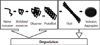
The canonical features of these neurodegenerative disorders is that a specific protein can enfold into an alternative conformation which is stable, precipitates its aggregation, and is deposited in tissues in a fibrillar form [1-3]. These fibrillar deposits share similar morphological and tinctorial characteristics with amyloids. In general, the naive protein consists of α-helices and random coils, whereas the misfolded protein is composed of rich β-structures which are insoluble and implicated in diseases [5, 6]. During misfolding and aggregation, a large conformational change in the proteins takes place, forming a misfolded intermediate with exposed hydrophobic fragments. This intermediate has a tendency to aggregate and stabilize, giving rise to the formation of oligomers, protofibrils, fibrils, and finally aggregates or inclusions (Fig. 1). The exposure of hydrophobic sequences of the misfolded proteins to the surface is considered critical for protein aggregation [2, 3, 6-8]. The aggregation of proteins follows nucleation-dependent polymerization, in which the critical event is oligomer formation that is soluble but serves as a nucleus to direct further elongation of aggregates. This process is characterized by a slow lag phase which is followed by a rapid growth to form polymers [9-11]. An imbalance between production, aggregation, and clearance of proteins brings about accumulation of misfolded proteins. In general, the accumulation of these misfolded proteins causes oxidative and neuroinflammatory damage leading to subsequent neurotransmitter, synaptic, and mitochondrial dysfunctions.

2) Selective neuronal vulnerability

The diverse phenotypes of different protein misfolding disorders may come from the selective vulnerability of neurons or glial cells to the specific misfolded proteins. The relationship between the phenotypes and the brain regions affected is more pronounced in the early stage of the diseases than in the later stages in which there are more extensive and diverse manifestations.
Postmortem analyses of human brains have revealed a characteristic pathological pattern of the deposition of specific proteins. Aβ plaques appear first in the neocortex, followed by the allocortex and the subcortical structures in AD [12-15]. Tau pathologies first appear in the locus coeruleus and transentorhinal area including the entorhinal cortex, and then spread to the amygdala and interconnected neocortical areas in AD [15, 16]. The mutant polyglutamines also have their own specific groups of neurons showing selective vulnerability to each protein in polyglutaminopathies [17]. Huntingtin (HTT) begins to appear in the striatum in HD. α-Synuclein initially accumulates in the lower brainstem centers and ascends to the limbic and cortical association areas in PD [18, 19], while in contrast, α-synuclein first appears in the cortex in DLB [20]. Although why different proteins are prone to accumulate in specific brain areas is still unknown, it has been speculated that local changes in expression or post-translational modifications of the specific proteins may account for the regional selectivity of early involvement [4].
In addition to the regional selectivity, these protein deposits may have different pathological and clinical features, depending on whether the aggregates accumulate predominantly in neurons or in glial cells, or intracellularly or extracellularly (Table 1). Unlike other α-synucleinopathies or protein misfolding disorders, the pathological deposition of α-synuclein in MSA is predominant in oligodendrocytes, forming oligodendroglial inclusions [21]. Aβ and prion proteins accumulate extracellularly, tau and α-synuclein aggregate in the cytoplasm, HTT assembles in the nucleus, and TDP-43 and FUS proteins accumulate primarily in the cytoplasm but are also found in the nucleus [22-24].

2. Basic pathologies of neurodegenerative dementias

1) β-Amyloid (Aβ) and β-amyloidopathies

Under physiological conditions, Aβ may attenuate excitatory transmission at the synapse and suppress neuronal hyperactivity [25]. Aβ peptides are produced by proteolysis of the neuronal transmembrane amyloid precursor protein (APP) through sequential cleavage by β- and γ-secretases, by way of the amyloidogenic pathway. Monomers of Aβ40 are more prevalent than the highly toxic and aggregation-prone Aβ42 species [26-28]. However, the predominant pathway of amyloid metabolism is through the non-amyloidogenic pathway, in which APP is processed by α- and γ-secretases forming a soluble fragment of APP (sαAPP) which may have protective and neurotrophic effects [29]. Multiple genetic and environmental factors may shift this equilibrium toward the increased formation of Aβ42 species which is followed by oligomerization, aggregation, and deposition as insoluble fibers of amyloid plaques. Nonetheless, Aβ also makes up diffuse plaques that contain nonfibrillar deposits of peptides. Neprilysin and insulin-degrading-enzyme (IDE) degrade Aβ monomers and oligomers to maintain the steady-state levels of Aβ [30-32]. Among the various forms of Aβ, soluble oligomers and intermediate fibrils are known to be the most toxic [33].
Although Aβ deposits are one of the pathological hallmarks of AD, amyloid plaques are also found in other dementias including DLB [34, 35], PDD [36], Down syndrome [37-39], Creutzfeldt-Jakob disease (CJD) and Gerstmann-Sträussler-Scheinker disease (GSS) [40-42] (Table 2).

2) Tau and tauopathies

Tau protein, one of the microtubule-associated proteins (MAPs), is abundant in axons and promotes the assembly and stabilization of microtubule and intracellular transport. Hyperphosphorylated tau, which is caused by a combination of excessive kinase and decreased phosphatase activities, becomes insoluble, is detached from microtubules, and self-aggregates into pair helical filaments (PHFs) and straight filaments [43]. Similar to Aβ oligomers, intermediate aggregates are toxic [44]. PHFs sequester toxic intermediate species and are considered to be protective [45, 46].
Neurofibrillary lesions contain aggregated filaments which are formed by abnormal phosphorylation of tau protein. Neurofibrillary lesions include not only neurofibrillary tangles (NFTs), but also neuropil threads and dystrophic neurites. NFTs accumulate around the nuclei and dystrophic neurites and neuropil threads accumulate in axons and dendrites. Dystrophic neurites are usually associated with amyloid plaque cores to form neuritic plaques.
Although there has been a poor correlation between the severity of neuronal loss, dementia, and the distribution of amyloid plaques, several studies supported that NFTs parallel with the severity of AD dementia [15, 47-49].
Neurofibrillary lesions are also characteristically part of the main pathology in several neurodegenerative dementias other than AD, which are termed tauopathies. Some tauopathies also show the combined amyloid plaques, while other tauopathies show only abundant neurofibrillary lesions without amyloid plaques. The former group includes Down syndrome [37-39] and some cases of CJD and Gerstmann-Sträussler-Scheinker disease (GSS) [40-42], while the latter encompasses CBD [50-53], PSP [54-56], MSA [57], neurodegeneration with brain iron accumulation (NBIA, formerly known as Hallervorden-Spatz disease) [58], Pick's disease [59-61], and FTDP-17 [62, 63]. However, MSA, NBIA, some subtypes of AD also have prominent α-synuclein lesions. In AD, tau pathology is largely limited to neurons, whereas some other tauopathies such as MSA, PSP, CBD, and FTDP-17 demonstrate both neuronal and glial inclusions [64-67].

3) α-Synuclein and α-synucleinopathies

α-Synuclein is a 140-residue neuronal protein which is found mostly in the neuronal presynaptic terminal in an unfolded form under normal physiological conditions. This protein was first identified as the precursor protein for non-amyloid constituents of senile plaques in AD [68]. It is presumed to support the regulation of the release of synaptic vesicles and the stabilization of soluble N-ethylmaleimide-sensitive factor attachment protein receptor (SNARE) family proteins including synaptobrevin-2 and vesicle-associated membrane protein 2 (VAMP2) [69, 70]. Genome-wide association studies (GWASs) have shown that SNCA gene which encodes α-synuclein is linked to sporadic PD [71] and the missense point mutations in the SNCA gene and the multiplications of the gene loci have been found in SNCA in families with autosomal dominant PD [72]. Pathological deposits of α-synuclein have been identified within aggregates in the forms of Lewy bodies and Lewy neurites in patients with PD and DLB, and oligodendroglial inclusions in MSA patients, collectively termed α-synucleinopathies [73]. However, AD and neurodegeneration with brain iron accumulation (NBIA, formerly known as Hallervorden-Spatz disease) are known to have abnormal deposition of α-synuclein [74, 75]. The pathogenic mechanisms underlying the aberrant functions of α-synuclein still remain poorly understood, but some possibilities including alteration of neurotransmitter release, lysosomal dysfunction, calcium homeostasis, cytoskeletal effects, and mitochondrial dysfunction have been suggested [76].

4) Prion protein and prionopathies

PrPC (normal cellular prion protein isoform) is the natural prion protein encoded by the PRNP gene. PrPC is a glycoprotein which is expressed in the cells in the central nervous system (CNS) and the immune system [77]. In the brain, the expression of PrPC is notably observed in neuronal synaptic membranes and is also expressed in astrocytes [78]. PrPC has been shown to be involved in signal transduction and to interact with several intracellular signaling proteins [79]. Although the understanding of the biologic functions of PrPC remains elusive, it is known to not be essential for cell survival [80]. However, the conversion of PrPC to the pathogenic PrPSC (PrP scrapie) ends up resulting in neurodegeneration [81, 82]. PrPC acts as the raw material for conversion to PrPSC [83]. Thus, these corrupted molecules easily self-aggregate and elicit neuronal injury. Although the exact mechanism of transmission remains uncertain, it is believed that prions can spread to other cells by synaptic transport [84].
In humans, a variety of prion diseases (prionopathies or transmissible spongiform encephalopathies) are present, including CJD, GSS, fatal familial insomnia, and kuru [83, 85], although the diseases are uncommon. Prion diseases are unique in that the same disease can have genetic, sporadic, and infectious (transmissible) origins and in that prion disease is the only infectious cerebral proteopathy [83, 84]. However, several studies implicated prion-like transmission of protein aggregates or inclusions in the initiation and spread of various neurodegenerative disorders [84, 86-90].

5) Huntingtin and polyglutaminopathies

Huntingtin (HTT) is a cytoplasmic protein which is found to be associated with synaptic vesicles and microtubules. In Huntington's disease (HD), the CAG repeats in the HD gene are suggested to be expanded by dynamic mutation as other trinucleotide repeat expansion disorders [91] and mutant HTT proteins with an expanded polyglutamine tract are produced. Mutant proteins are prone to toxic conformational changes and accumulate into insoluble aggregates as neuronal intranuclear inclusions (NIIs) [92-94]. These processes are common features among polyglutamine diseases which include HD, spinocerebellar ataxia 1, 2, 3, 6, 7, and 17 (SCA1/2/3/6/7/17), dentatorubral-pallidoluysian atrophy (DRPLA), and spinal and bulbar muscular atrophy (SBMA, also known as Kennedy's disease). Among them, SCA17 and DRPLA include dementia as one of the cardinal manifestations, similar to HD [95-97]. The counterparts of HTT are TATA-binding protein gene (TBP) and atrophin 1 in SCA17 and DRPLA, respectively [98-100].

6) Transactive response DNA-binding protein of 43kDa (TDP-43) and TDP-43 proteinopathies

TDP-43 is a ubiquitously expressed 414-residue protein which is mainly localized inside the nucleus under physiological conditions and is able to shuttle between the nucleus and the cytoplasm in a transcription-dependent manner by virtue of the presence of a nuclear localization sequence (NLS) and nuclear export sequence (NES) [101]. It regulates diverse processes of gene expression including transcription and splicing through RNA and DNA binding. It contains two RNA recognition motifs (RRMs) that allow for binding to nucleic acids and a C-terminal glycine-rich domain (GRD) which is important for protein-protein interactions and essential for solubility and cellular localization. Disruption of the RRM changes nuclear distribution by decreasing the level of TDP-43 in the nucleoplasm. Deletion of the C-terminal GRD elicits the formation of nuclear and cytoplasmic aggregates.
TDP-43 pathology is, along with tau pathology, also the main cause for some clinical subtypes of FTLD such as behavioral variant FTD (bvFTD), semantic dementia (SD), and progressive nonfluent aphasia (PNFA) [102, 103] and amyotrophic lateral sclerosis-Parkinsonism-dementia complex of Guam (ALS/PDC) [104].
TDP-43 has been identified as the major protein aggregation in frontotemporal dementia and amyotrophic lateral sclerosis (FDT/ALS), one of the subtypes of frontotemporal lobar degeneration with ubiquitin inclusions (FTLD-U) [105]. Pathologic TPD-43 is ubiquitinated, hyperphosphorylated, and cleaved to produce C-terminal fragments. Remarkably, it is consistently observed that normal nuclear localization of TDP-43 is lacking in inclusion-bearing neurons. In addition to the TDP-43 inclusions, TDP-43 preinclusions, which are observed cell bodies without inclusions, are a common neuropathologic finding. They show diffuse or granular cytoplasmic TDP-43 staining and do not colocalize with ubiquitin [106].
In FTLD, TDP-43 positive aggregates are found in most sporadic cases as well as in familial forms. Familial forms are caused by mutations in the TAR-DNA binding protein (TARDBP) gene [107], progranulin (GRN) genes [105, 108], and valosin containing protein (VCP) genes [109]. Recently, it has been found that an expanded hexanucleotide repeat in chromosome 9 open reading frame 72 (C9ORF72) is the most common mutation in FTD/ALS families with TDP-43 pathology [110, 111]. However, the causative relationship between TDP-43 pathology and the mutation remains unclear at this time.

7) Fused in sarcoma (FUS) and FUS proteinopathies

FUS, also known as translocated in liposarcoma (TLS) or heterogeneous ribonucleoprotein (hnRNP) P2, is a 526-residue protein identified as a proto-oncogene that causes liposarcoma via chromosomal translocation [112]. FUS is an RNA-binding protein like TDP-43 and comprises the RRM domain, GRD, and NLS/NES, which are arranged differently in various proteins from TDP-43. The FUS protein is a nuclear protein and is involved in DNA repair and RNA splicing regulation [113, 114].
Mutations within the FUS gene in the GRD and the C-terminus of FUS protein impair nuclear import and lead to redistribution to the cytoplasm, consequently affecting FUS-dependent RNA metabolism.
FUS pathology is observed in the majority of FTLD cases with ubiquitin-positive, and TDP-43 negative pathology (FTLD-FUS) [115-117]. FTLD-FUS cases are characterized by negative family history, disease onset at young age, presence of bvFTD, and caudate atrophy. FUS-positive inclusions are also found in basophilic inclusion body disease [118] and neuronal intermediate filament inclusion disease [24].
FUS has been identified to misfold and aggregate in distinct subtypes of FTLD-U (FTD/ALS), similar to TDP-43 but with TDP-43 negative pathology [116, 119]. Contrary to TDP-43, no association of post-translational modifications such as ubiquitination, phosphorylation, or truncation has been identified.

3. Links between proteins

Some of the protein misfolding diseases have more than one pathology, as some proteins are commonly found in more than one disease (Table 2). The understanding of the interaction between the misfolded proteins will help elucidate in part the complexity of the protein misfolding disorders.

1) β-amyloid (Aβ) and tau

Several experimental studies support that the accumulation of Aβ precedes and trigger the aggregation of tau [120-123], which is consistent with the amyloid cascade hypothesis. Furthermore, Aβ-induced neuronal dysfunction was prevented by tau reduction [124], and morphological analysis showed that tau-depleted neurons revealed no evidence of degeneration in the presence of Aβ [125]. These results are supported by prior evidence that Aβ promotes the activation of glycogen synthase kinase-3β (GSK3β) through the insulin and Wnt signaling pathways, with subsequent tau phosphorylation [126-129].

2) β-amyloid (Aβ) and α-synuclein

AD patients develop features of PD and vice versa. Moreover, Aβ pathology and α-synuclein pathology can be found in both AD and DLB/PDD. It has been found that Aβ and α-synuclein synergistically interact to cause neurodegeneration in the transgenic mouse model [130]. Aβ promoted the aggregation and intraneuronal accumulation of α-synuclein and the development of motor deficits, supporting that Aβ may contribute to the development of DLB or PDD by promoting α-synuclein aggregation. Although α-synuclein did not affect Aβ pathology, it aggravated the cognitive deficits, suggesting that α-synuclein may augment the Aβ-independent neurotoxicity of Aβ [131, 132].

3) β-amyloid (Aβ) and prion protein

It has been reported that prion protein which Aβ binds is required for the impairment of synaptic plasticity mediated by Aβ oligomers [133]. It has also been found that the region of importance for the interaction between prion proteins and Aβ resides at the extreme amino-terminus of prion protein [134]. The role of prion protein in Aβ-induced toxicity was confirmed by a recent study showing that prion protein is required for disrupting hippocampal synaptic plasticity by Aβ peptides [135].

4) α-Synuclein and tau

GWASs have shown that there are strong associations with PD for SNCA, LRRK2, and MAPT [71], suggesting functional links among these proteins that affect the cytoskeleton. Oligomeric α-synuclein indirectly augments the phosphorylation of tau presumably via GSK-3β or other kinases and destabilizes the microtubules, which in turn may promote the formation of α-synuclein oligomers and cause further disruption of microtubules [136-139]. Additionally, leucine-rich repeat kinase 2 (LRRK2, also known as dardarin) which is encoded by LRRK2, an autosomal dominant inherited PD gene, can increase the phosphorylation of tau through GSK-3β and Ste20 kinase [140-142]. These findings suggest a synergistic interaction between α-synuclein and LRRK2 that involve tau.

5) Transactive response DNA-binding protein (TARDBP or TDP-43) and fused in sarcoma (FUS) protein

Both TDP-43 and FUS are the cytoplasmic RNA-binding proteins which play critical roles in the development of frontotemporal lobar degeneration with ubiquitin-positive inclusions (FTLU-U) and ALS. These proteins are transported to the nucleus via import receptors and also contribute to stress granule formation. Although the exact mechanisms for the accumulation of TDP-43 and FUS and the resultant neurodegeneration are currently unclear [119], it has been proposed that excessive mislocation of the proteins along with ataxin-2 into the cytoplasm causes dysfunction of the RNA quarantine system, inducing a joined cascade of neurodegeneration which is promoted by ataxin-2 [143].

4. Some issues under debate

1) The relationship between soluble oligomers and insoluble inclusions

Determining which particular species of the misfolded proteins are neurotoxic has been under debate. Several experiments are in favor of the toxicity of soluble oligomeric species [33, 44-46, 144]. However, this oligomeric species-induced toxicity does not indicate that the insoluble inclusions are innocent. There has still been evidence that these inclusions are toxic [145, 146], but other data support that the insoluble inclusions may be neuroprotective [45, 46, 147, 148].
Considering that oligomers are found around and within the amyloid plaques and are toxic to adjacent neurons [149-151], it has been suggested that inclusions serve as reservoirs for oligomers that can diffuse away from the inclusions and cause synaptic or neuronal toxicity [152]. The inclusions may initally sequester toxic soluble oligomers, but, eventually the reservoirs are overwhelmed and can no longer be protective. Mass effect of the inclusions on surrounding neurons seems to be plausabe but it has been shown that senile plaques did not exert any mass lesion effects on neighboring cells [153]. Nonetheless, whether it can be the case for other protein misfolding disoders needs to be further investigated.

2) Cell-to-cell disease transmission

Pathological similarities between prion diseases and AD suggest that the prion-like formation and seeding of proteinaceous lesions may be involved in the pathogenesis of disease [154-157].
The deposition of Aβ deposits were found in axonally interconnected areas following the injection of Aβ aggregates into the brain, suggesting it spreads through neuronal pathways [157-159]. The accumulation of abnormal tau starts in the entorhinal cortex (ERC) in the earliest stage of AD, and then spreads to the hippocampus followed by the neocortical regions [15]. However, it has been poorly explored whether tau pathology in the ERC initiates spreading to other structures, or that the pathology in the extrahippocampal areas begins independently. Using a transgenic mouse model expressing the pathological human tau protein primarily in the ERC, it has been demonstrated that tau pathology which began in the ERC spreads out from one neuron to other neurons outside of the ERC across synapses [160]. This study result confirms observations from previous studies suggesting the trans-synaptic cell-to-cell spread hypothesis for AD [161, 162].
α-Synuclein pathology in PD starts in the anterior olfactory nucleus and the lower brainstem centers and ultimately ascends to the cortex [18, 19]. A recent study showed that purified α-synuclein fibrils are internalized into primary mouse hippocampal cells by endocytosis, recruit soluble endogenous α-synuclein, and promote the formation of insoluble Lewy bodies or neurites [163]. In this study, the aggregates of α-synuclein appeared early in the axon terminals and later in the cell body, suggesting propagation along the axon and eventually to other cells. The report reinforces the earlier conclusions suggesting α-synuclein pathology from dying neurons is conveyed to neighboring neurons through cell-to-cell transmission [19, 164, 165], which is similar to the transmission of misfolded prion proteins in CJD [166].
Furthermore, the prion-like cell-to-cell transmission of the lesions has also been demonstrated in polyglutamine and TDP-43 aggregates [167, 168]. However, the mode of cell-to-cell transmission for these proteins remains unclear.

5. General therapeutic strategies for protein misfolding disorders

Despite remarkable progress in the understanding of the pathomechanisms of protein misfolding disorders, there has been no successful disease-modifying therapy for them as this has been the case in AD. Therapeutic strategies targeted for the misfolded and aggregated proteins have been proposed: 1) stabilization of the normal protein conformation; 2) inhibition of the protein misfolding by interfering with post-translational modification; 3) unfolding the misfolded proteins; 4) inhibition of protein oligomerization by compounds binding to monomers; 5) inhibition of protein aggregation with small molecules that bind to aggregates and further interfere with the recruitment of monomers; 6) upregulating molecular chaperones; 7) enhancing the clearance mechanisms by immunization; and 8) gene therapy [4, 169, 170]. The combinations of more than one strategy, particularly in the earliest stages of the diseases are expected to be the most effective treatments for patients with disorders in the near future.

CONCLUSIONS

The misfolding and aggregation of proteins have been regarded as central events in the development of various neurodegenerative dementias. The common pathological mechanisms of these disorders are increased production, misfolding, aggregation, and accumulation of specific proteins, with the conformational variants ranging from small oligomers to the characteristic inclusions. However, there is a substantial overlap between different pathologies and different dementias evidenced by the existence of the interactions between the misfolded proteins, although certain pathologies can be a hallmark of certain dementias. There has been remarkable progress in understanding the role of toxic oligomers and cell-to-cell transmission. The understanding of the pathomechanistic roles of misfolded proteins will be the fundamental basis for the identification of biomarkers in the earliest stage of dementia, and should facilitate the development of effective treatments which can modify the natural course of the dementia.

Figures and Tables

Fig. 1
Pathogenesis of protein misfolding diseases. Naïve proteins are misfolded, self-accumulate, and form oligomers, protofibrils, fibrils, and eventually aggregates or inclusions. An imbalance between production, aggregation, and clearance of proteins brings about accumulation of misfolded proteins. The mature forms are more resistant to degradation than the intermediate species. The common toxic effects of the misfolded proteins may include oxidative stress, neuroinflammatory damage, and disturbed neurotransmitter, synaptic, and mitochondrial functions.
dnd-11-38-g001
Table 1
Misfolded proteins and neuropathologic changes
dnd-11-38-i001

Aβ, β-amyloid; FUS, fused in sarcoma; TDP-43, transactive response (TAR)-DNA-binding protein or TARDBP.

Table 2
Misfolded proteins and dementias
dnd-11-38-i002

√√, major component; √, minor component.

*For ALS/PDC, bvFTD, FTD/ALS, PNFA, and SD, these may have one of the different pathologies depending on each case. It does not mean that different pathologies shown in the table are found simultaneously.

Aβ, β-amyloid; AD, Alzheimer's disease; ALS/PDC, amyotrophic lateral sclerosis-Parkinsonism-dementia complex of Guam; BIBD, bvFTD, behavioral variant FTD; c9FTD/ALS, chromosome 9p-linked frontotemporal dementia and amyotrophic lateral sclerosis; CBD, corticobasal degeneration; CJD, Creutzfeldt-Jakob disease; DLB, dementia with Lewy bodies; DM, Dystrophia myotonica; FFI, fatal familial insomnia; FTD/ALS, frontotemporal dementia and amyotrophic lateral sclerosis; FTDP-17, frontotemporal dementia and parkinsonism linked to chromosome 17; FTLD, Frontotemporal lobar degeneration; FUS, fused in sarcoma; GSS, Gerstmann-Sträussler-Scheinker disease; HD, Huntington's disease; IBMFPD, Inclusion body myopathy, Paget's disease of the bone and frontotemporal dementia; MSA, Multiple system atrophy; NBIA, neurodegeneration with brain iron accumulation; NIFID, neuronal intermediate filament inclusion disease; basophilic inclusion body disease; PDD, Parkinson disease with dementia; PNFA, progressive nonfluent aphasia; PSP, progressive supranuclear palsy; SD, semantic dementia; TDP-43, transactive response (TAR)-DNA-binding protein or TARDBP.

Notes

The work was supported by a grant from Wonkwang University in 2011.

References

1. Carrell RW, Lomas DA. Conformational disease. Lancet. 1997. 350:134–138.
crossref
2. Dobson CM. Protein misfolding, evolution and disease. Trends Biochem Sci. 1999. 24:329–332.
crossref
3. Soto C. Protein misfolding and disease; protein refolding and therapy. FEBS Lett. 2001. 498:204–207.
crossref
4. Soto C. Unfolding the role of protein misfolding in neurodegenerative diseases. Nat Rev Neurosci. 2003. 4:49–60.
crossref
5. Serpell LC, Blake CC, Fraser PE. Molecular structure of a fibrillar Alzheimer's A beta fragment. Biochemistry. 2000. 39:13269–13275.
crossref
6. Serpell LC, Berriman J, Jakes R, Goedert M, Crowther RA. Fiber diffraction of synthetic alpha-synuclein filaments shows amyloid-like cross-beta conformation. Proc Natl Acad Sci U S A. 2000. 97:4897–4902.
crossref
7. Teplow DB. Structural and kinetic features of amyloid beta-protein fibrillogenesis. Amyloid. 1998. 5:121–142.
crossref
8. Tagliavini F, Prelli F, Verga L, Giaccone G, Sarma R, Gorevic P, et al. Synthetic peptides homologous to prion protein residues 106-147 form amyloid-like fibrils in vitro. Proc Natl Acad Sci U S A. 1993. 90:9678–9682.
crossref
9. Jarrett JT, Berger EP, Lansbury PT. The C-terminus of the beta protein is critical in amyloidogenesis. Ann N Y Acad Sci. 1993. 695:144–148.
crossref
10. Scherzinger E, Sittler A, Schweiger K, Heiser V, Lurz R, Hasenbank R, et al. Self-assembly of polyglutamine-containing huntingtin fragments into amyloid-like fibrils: implications for Huntington's disease pathology. Proc Natl Acad Sci U S A. 1999. 96:4604–4609.
crossref
11. Wood SJ, Wypych J, Steavenson S, Louis JC, Citron M, Biere AL. Alpha-synuclein fibrillogenesis is nucleation-dependent. Implications for the pathogenesis of Parkinson's disease. J Biol Chem. 1999. 274:19509–19512.
crossref
12. Thal DR, Rüb U, Orantes M, Braak H. Phases of A beta-deposition in the human brain and its relevance for the development of AD. Neurology. 2002. 58:1791–1800.
crossref
13. Buckner RL, Snyder AZ, Shannon BJ, LaRossa G, Sachs R, Fotenos AF, et al. Molecular, structural, and functional characterization of Alzheimer's disease: evidence for a relationship between default activity, amyloid, and memory. J Neurosci. 2005. 25:7709–7717.
crossref
14. Walker LC, Jucker M. Amyloid by default. Nat Neurosci. 2011. 14:669–670.
crossref
15. Braak H, Braak E. Neuropathological stageing of Alzheimer-related changes. Acta Neuropathol. 1991. 82:239–259.
crossref
16. Braak H, Del Tredici K. The pathological process underlying Alzheimer's disease in individuals under thirty. Acta Neuropathol. 2011. 121:171–181.
crossref
17. Zoghbi HY, Orr HT. Glutamine repeats and neurodegeneration. Annu Rev Neurosci. 2000. 23:217–247.
crossref
18. Braak H, Del Tredici K, Bratzke H, Hamm-Clement J, Sandmann-Keil D, Rüb U. Staging of the intracerebral inclusion body pathology associated with idiopathic Parkinson's disease (preclinical and clinical stages). J Neurol. 2002. 249:Suppl 3. III/1–III/5.
crossref
19. Braak H, Del Tredici K, Rüb U, de Vos RA, Jansen Steur EN, Braak E. Staging of brain pathology related to sporadic Parkinson's disease. Neurobiol Aging. 2003. 24:197–211.
crossref
20. Halliday GM, McCann H. Human-based studies on alpha-synuclein deposition and relationship to Parkinson's disease symptoms. Exp Neurol. 2008. 209:12–21.
crossref
21. Gai WP, Power JH, Blumbergs PC, Blessing WW. Multiple-system atrophy: a new alpha-synuclein disease? Lancet. 1998. 352:547–548.
crossref
22. Forman MS, Trojanowski JQ, Lee VM. Neurodegenerative diseases: a decade of discoveries paves the way for therapeutic breakthroughs. Nat Med. 2004. 10:1055–1063.
crossref
23. Neumann M, Kwong LK, Truax AC, Vanmassenhove B, Kretzschmar HA, Van Deerlin VM, et al. TDP-43-positive white matter pathology in frontotemporal lobar degeneration with ubiquitin-positive inclusions. J Neuropathol Exp Neurol. 2007. 66:177–183.
crossref
24. Neumann M, Roeber S, Kretzschmar HA, Rademakers R, Baker M, Mackenzie IR. Abundant FUS-immunoreactive pathology in neuronal intermediate filament inclusion disease. Acta Neuropathol. 2009. 118:605–616.
crossref
25. Kamenetz F, Tomita T, Hsieh H, Seabrook G, Borchelt D, Iwatsubo T, et al. APP processing and synaptic function. Neuron. 2003. 37:925–937.
crossref
26. Haass C, Selkoe DJ. Soluble protein oligomers in neurodegeneration: lessons from the Alzheimer's amyloid beta-peptide. Nat Rev Mol Cell Biol. 2007. 8:101–112.
crossref
27. Gandy S, Martins R, Buxbaum J. Molecular and cellular basis for anti-amyloid therapy in Alzheimer disease. Alzheimer Dis Assoc Disord. 2003. 17:259–266.
crossref
28. Selkoe D, Schenk D. Alzheimer's disease: molecular understanding predicts amyloid-based therapeutics. Annu Rev Pharmacol Toxicol. 2003. 43:545–584.
crossref
29. Furukawa K, Sopher BL, Rydel RE, Begley JG, Pham DG, Martin GM, et al. Increased activity-regulating and neuroprotective efficacy of alpha-secretase-derived secreted amyloid precursor protein conferred by a C-terminal heparin-binding domain. J Neurochem. 1996. 67:1882–1896.
crossref
30. Kanemitsu H, Tomiyama T, Mori H. Human neprilysin is capable of degrading amyloid beta peptide not only in the monomeric form but also the pathological oligomeric form. Neurosci Lett. 2003. 350:113–116.
crossref
31. Qiu WQ, Walsh DM, Ye Z, Vekrellis K, Zhang J, Podlisny MB, et al. Insulin-degrading enzyme regulates extracellular levels of amyloid beta-protein by degradation. J Biol Chem. 1998. 273:32730–32738.
crossref
32. Leissring MA, Farris W, Chang AY, Walsh DM, Wu X, Sun X, et al. Enhanced proteolysis of beta-amyloid in APP transgenic mice prevents plaque formation, secondary pathology, and premature death. Neuron. 2003. 40:1087–1093.
crossref
33. Walsh DM, Selkoe DJ. A beta oligomers - a decade of discovery. J Neurochem. 2007. 101:1172–1184.
34. Jellinger KA, Attems J. Prevalence and impact of vascular and Alzheimer pathologies in Lewy body disease. Acta Neuropathol. 2008. 115:427–436.
35. Merdes AR, Hansen LA, Jeste DV, Galasko D, Hofstetter CR, Ho GJ, et al. Influence of Alzheimer pathology on clinical diagnostic accuracy in dementia with Lewy bodies. Neurology. 2003. 60:1586–1590.
36. Jellinger KA. Significance of brain lesions in Parkinson disease dementia and Lewy body dementia. Front Neurol Neurosci. 2009. 24:114–125.
crossref
37. Giaccone G, Tagliavini F, Linoli G, Bouras C, Frigerio L, Frangione B, et al. Down patients: extracellular preamyloid deposits precede neuritic degeneration and senile plaques. Neurosci Lett. 1989. 97:232–238.
crossref
38. Flament S, Delacourte A, Mann DM. Phosphorylation of Tau proteins: a major event during the process of neurofibrillary degeneration. A comparative study between Alzheimer's disease and Down's syndrome. Brain Res. 1990. 516:15–19.
crossref
39. Cork LC. Neuropathology of Down syndrome and Alzheimer disease. Am J Med Genet Suppl. 1990. 7:282–286.
crossref
40. Ghetti B, Tagliavini F, Masters CL, Beyreuther K, Giaccone G, Verga L, et al. Gerstmann-Sträussler-Scheinker disease. II. Neurofibrillary tangles and plaques with PrP-amyloid coexist in an affected family. Neurology. 1989. 39:1453–1461.
crossref
41. Tagliavini F, Giaccone G, Prelli F, Verga L, Porro M, Trojanowski JQ, et al. A68 is a component of paired helical filaments of Gerstmann-Sträussler-Scheinker disease, Indiana kindred. Brain Res. 1993. 616:325–329.
crossref
42. Hsiao K, Dlouhy SR, Farlow MR, Cass C, Da Costa M, Conneally PM, et al. Mutant prion proteins in Gerstmann-Sträussler-Scheinker disease with neurofibrillary tangles. Nat Genet. 1992. 1:68–71.
crossref
43. Iqbal K, Alonso AeC, Chen S, Chohan MO, El-Akkad E, Gong CX, et al. Tau pathology in Alzheimer disease and other tauopathies. Biochim Biophys Acta. 2005. 1739:198–210.
crossref
44. Khlistunova I, Biernat J, Wang Y, Pickhardt M, von Bergen M, Gazova Z, et al. Inducible expression of Tau repeat domain in cell models of tauopathy: aggregation is toxic to cells but can be reversed by inhibitor drugs. J Biol Chem. 2006. 281:1205–1214.
crossref
45. Oddo S, Vasilevko V, Caccamo A, Kitazawa M, Cribbs DH, LaFerla FM. Reduction of soluble Abeta and tau, but not soluble Abeta alone, ameliorates cognitive decline in transgenic mice with plaques and tangles. J Biol Chem. 2006. 281:39413–39423.
crossref
46. Lee HG, Perry G, Moreira PI, Garrett MR, Liu Q, Zhu X, et al. Tau phosphorylation in Alzheimer's disease: pathogen or protector? Trends Mol Med. 2005. 11:164–169.
crossref
47. McKee AC, Kosik KS, Kowall NW. Neuritic pathology and dementia in Alzheimer's disease. Ann Neurol. 1991. 30:156–165.
crossref
48. Arriagada PV, Growdon JH, Hedley-Whyte ET, Hyman BT. Neurofibrillary tangles but not senile plaques parallel duration and severity of Alzheimer's disease. Neurology. 1992. 42:631–639.
crossref
49. Neve RL, Robakis NK. Alzheimer's disease: a re-examination of the amyloid hypothesis. Trends Neurosci. 1998. 21:15–19.
crossref
50. Paulus W, Selim M. Corticonigral degeneration with neuronal achromasia and basal neurofibrillary tangles. Acta Neuropathol. 1990. 81:89–94.
crossref
51. Ksiezak-Reding H, Morgan K, Mattiace LA, Davies P, Liu WK, Yen SH, et al. Ultrastructure and biochemical composition of paired helical filaments in corticobasal degeneration. Am J Pathol. 1994. 145:1496–1508.
crossref
52. Mori H, Nishimura M, Namba Y, Oda M. Corticobasal degeneration: a disease with widespread appearance of abnormal tau and neurofibrillary tangles, and its relation to progressive supranuclear palsy. Acta Neuropathol. 1994. 88:113–121.
crossref
53. Wakabayashi K, Oyanagi K, Makifuchi T, Ikuta F, Homma A, Homma Y, et al. Corticobasal degeneration: etiopathological significance of the cytoskeletal alterations. Acta Neuropathol. 1994. 87:545–553.
crossref
54. Bancher C, Lassmann H, Budka H, Grundke-Iqbal I, Iqbal K, Wiche G, et al. Neurofibrillary tangles in Alzheimer's disease and progressive supranuclear palsy: antigenic similarities and differences. Microtubule-associated protein tau antigenicity is prominent in all types of tangles. Acta Neuropathol. 1987. 74:39–46.
crossref
55. Flament S, Delacourte A, Verny M, Hauw JJ, Javoy-Agid F. Abnormal Tau proteins in progressive supranuclear palsy. Similarities and differences with the neurofibrillary degeneration of the Alzheimer type. Acta Neuropathol. 1991. 81:591–596.
crossref
56. Schmidt ML, Huang R, Martin JA, Henley J, Mawal-Dewan M, Hurtig HI, et al. Neurofibrillary tangles in progressive supranuclear palsy contain the same tau epitopes identified in Alzheimer's disease PHFtau. J Neuropathol Exp Neurol. 1996. 55:534–539.
crossref
57. Papp MI, Kahn JE, Lantos PL. Glial cytoplasmic inclusions in the CNS of patients with multiple system atrophy (striatonigral degeneration, olivopontocerebellar atrophy and Shy-Drager syndrome). J Neurol Sci. 1989. 94:79–100.
crossref
58. Eidelberg D, Sotrel A, Joachim C, Selkoe D, Forman A, Pendlebury WW, et al. Adult onset Hallervorden-Spatz disease with neurofibrillary pathology. A discrete clinicopathological entity. Brain. 1987. 110(Pt 4):993–1013.
crossref
59. Perry G, Stewart D, Friedman R, Manetto V, Autilio-Gambetti L, Gambetti P. Filaments of Pick's bodies contain altered cytoskeletal elements. Am J Pathol. 1987. 127:559–568.
crossref
60. Murayama S, Mori H, Ihara Y, Tomonaga M. Immunocytochemical and ultrastructural studies of Pick's disease. Ann Neurol. 1990. 27:394–405.
61. Lieberman AP, Trojanowski JQ, Lee VM, Balin BJ, Ding XS, Greenberg J, et al. Cognitive, neuroimaging, and pathological studies in a patient with Pick's disease. Ann Neurol. 1998. 43:259–265.
62. Wilhelmsen KC, Lynch T, Pavlou E, Higgins M, Nygaard TG. Localization of disinhibition-dementia-parkinsonism-amyotrophy complex to 17q21-22. Am J Hum Genet. 1994. 55:1159–1165.
63. Foster NL, Wilhelmsen K, Sima AA, Jones MZ, D'Amato CJ, Gilman S. Frontotemporal dementia and parkinsonism linked to chromosome 17: a consensus conference. Conference Participants. Ann Neurol. 1997. 41:706–715.
crossref
64. Papp MI, Lantos PL. Accumulation of tubular structures in oligodendroglial and neuronal cells as the basic alteration in multiple system atrophy. J Neurol Sci. 1992. 107:172–182.
crossref
65. Nishimura M, Namba Y, Ikeda K, Oda M. Glial fibrillary tangles with straight tubules in the brains of patients with progress ive supranuclear palsy. Neurosci Lett. 1992. 143:35–38.
crossref
66. Spillantini MG, Bird TD, Ghetti B. Frontotemporal dementia and Parkinsonism linked to chromosome 17: a new group of tauopathies. Brain Pathol. 1998. 8:387–402.
67. Iwatsubo T, Hasegawa M, Ihara Y. Neuronal and glial tau-positive inclusions in diverse neurologic diseases share common phosphorylation characteristics. Acta Neuropathol. 1994. 88:129–136.
68. Uéda K, Fukushima H, Masliah E, Xia Y, Iwai A, Yoshimoto M, et al. Molecular cloning of cDNA encoding an unrecognized component of amyloid in Alzheimer disease. Proc Natl Acad Sci U S A. 1993. 90:11282–11286.
69. Burré J, Sharma M, Tsetsenis T, Buchman V, Etherton MR, Südhof TC. Alpha-synuclein promotes SNARE-complex assembly in vivo and in vitro. Science. 2010. 329:1663–1667.
crossref
70. Vekrellis K, Rideout HJ, Stefanis L. Neurobiology of alpha-synuclein. Mol Neurobiol. 2004. 30:1–21.
crossref
71. Nalls MA, Plagnol V, Hernandez DG, Sharma M, Sheerin UM, Saad M, et al. Imputation of sequence variants for identification of genetic risks for Parkinson's disease: a meta-analysis of genome-wide association studies. Lancet. 2011. 377:641–649.
crossref
72. Hardy J, Lewis P, Revesz T, Lees A, Paisan-Ruiz C. The genetics of Parkinson's syndromes: a critical review. Curr Opin Genet Dev. 2009. 19:254–265.
crossref
73. Kahle PJ. Alpha-Synucleinopathy models and human neuropathology: similarities and differences. Acta Neuropathol. 2008. 115:87–95.
crossref
74. Neumann M, Adler S, Schlüter O, Kremmer E, Benecke R, Kretzschmar HA. Alpha-synuclein accumulation in a case of neurodegeneration with brain iron accumulation type 1 (NBIA-1, formerly Hallervorden-Spatz syndrome) with widespread cortical and brainstem-type Lewy bodies. Acta Neuropathol. 2000. 100:568–574.
crossref
75. Pollanen MS, Dickson DW, Bergeron C. Pathology and biology of the Lewy body. J Neuropathol Exp Neurol. 1993. 52:183–191.
crossref
76. Vekrellis K, Xilouri M, Emmanouilidou E, Rideout HJ, Stefanis L. Pathological roles of α-synuclein in neurological disorders. Lancet Neurol. 2011. 10:1015–1025.
crossref
77. Manson J, West JD, Thomson V, McBride P, Kaufman MH, Hope J. The prion protein gene: a role in mouse embryogenesis? Development. 1992. 115:117–122.
crossref
78. Moser M, Colello RJ, Pott U, Oesch B. Developmental expression of the prion protein gene in glial cells. Neuron. 1995. 14:509–517.
crossref
79. Martins VR, Linden R, Prado MA, Walz R, Sakamoto AC, Izquierdo I, et al. Cellular prion protein: on the road for functions. FEBS Lett. 2002. 512:25–28.
crossref
80. Büeler H, Fischer M, Lang Y, Bluethmann H, Lipp HP, DeArmond SJ, et al. Normal development and behaviour of mice lacking the neuronal cell-surface PrP protein. Nature. 1992. 356:577–582.
crossref
81. Aguzzi A, Heikenwalder M. Prion diseases: Cannibals and garbage piles. Nature. 2003. 423:127–129.
crossref
82. Aguzzi A, Polymenidou M. Mammalian prion biology: one century of evolving concepts. Cell. 2004. 116:313–327.
crossref
83. Prusiner SB. Shattuck lecture--neurodegenerative diseases and prions. N Engl J Med. 2001. 344:1516–1526.
crossref
84. Aguzzi A, Rajendran L. The transcellular spread of cytosolic amyloids, prions, and prionoids. Neuron. 2009. 64:783–790.
crossref
85. Brown K, Mastrianni JA. The prion diseases. J Geriatr Psychiatry Neurol. 2010. 23:277–298.
crossref
86. Walker LC, Levine H, Mattson MP, Jucker M. Inducible proteopathies. Trends Neurosci. 2006. 29:438–443.
crossref
87. Soto C, Estrada L, Castilla J. Amyloids, prions and the inherent infectious nature of misfolded protein aggregates. Trends Biochem Sci. 2006. 31:150–155.
crossref
88. Frost B, Diamond MI. Prion-like mechanisms in neurodegenerative diseases. Nat Rev Neurosci. 2010. 11:155–159.
crossref
89. Brundin P, Melki R, Kopito R. Prion-like transmission of protein aggregates in neurodegenerative diseases. Nat Rev Mol Cell Biol. 2010. 11:301–307.
crossref
90. Goedert M, Clavaguera F, Tolnay M. The propagation of prion-like protein inclusions in neurodegenerative diseases. Trends Neurosci. 2010. 33:317–325.
crossref
91. Richards RI, Sutherland GR. Dynamic mutation: possible mechanisms and significance in human disease. Trends Biochem Sci. 1997. 22:432–436.
crossref
92. Shao J, Diamond MI. Polyglutamine diseases: emerging concepts in pathogenesis and therapy. Hum Mol Genet. 2007. 16 Spec No. 2:R115–R123.
crossref
93. Schaffar G, Breuer P, Boteva R, Behrends C, Tzvetkov N, Strippel N, et al. Cellular toxicity of polyglutamine expansion proteins: mechanism of transcription factor deactivation. Mol Cell. 2004. 15:95–105.
crossref
94. Nagai Y, Inui T, Popiel HA, Fujikake N, Hasegawa K, Urade Y, et al. A toxic monomeric conformer of the polyglutamine protein. Nat Struct Mol Biol. 2007. 14:332–340.
crossref
95. Koide R, Onodera O, Ikeuchi T, Kondo R, Tanaka H, Tokiguchi S, et al. Atrophy of the cerebellum and brainstem in dentatorubral pallidoluysian atrophy. Influence of CAG repeat size on MRI findings. Neurology. 1997. 49:1605–1612.
crossref
96. Bauer P, Laccone F, Rolfs A, Wüllner U, Bösch S, Peters H, et al. Trinucleotide repeat expansion in SCA17/TBP in white patients with Huntington's disease-like phenotype. J Med Genet. 2004. 41:230–232.
crossref
97. Schöls L, Bauer P, Schmidt T, Schulte T, Riess O. Autosomal dominant cerebellar ataxias: clinical features, genetics, and pathogenesis. Lancet Neurol. 2004. 3:291–304.
crossref
98. Koide R, Ikeuchi T, Onodera O, Tanaka H, Igarashi S, Endo K, et al. Unstable expansion of CAG repeat in hereditary dentatorubral-pallidoluysian atrophy (DRPLA). Nat Genet. 1994. 6:9–13.
crossref
99. Nucifora FC, Ellerby LM, Wellington CL, Wood JD, Herring WJ, Sawa A, et al. Nuclear localization of a non-caspase truncation product of atrophin-1, with an expanded polyglutamine repeat, increases cellular toxicity. J Biol Chem. 2003. 278:13047–13055.
crossref
100. Nakamura K, Jeong SY, Uchihara T, Anno M, Nagashima K, Nagashima T, et al. SCA17, a novel autosomal dominant cerebellar ataxia caused by an expanded polyglutamine in TATA-binding protein. Hum Mol Genet. 2001. 10:1441–1448.
crossref
101. Ayala YM, Zago P, D'Ambrogio A, Xu YF, Petrucelli L, Buratti E, et al. Structural determinants of the cellular localization and shuttling of TDP-43. J Cell Sci. 2008. 121:3778–3785.
crossref
102. Seelaar H, Rohrer JD, Pijnenburg YA, Fox NC, van Swieten JC. Clinical, genetic and pathological heterogeneity of frontotemporal dementia: a review. J Neurol Neurosurg Psychiatry. 2011. 82:476–486.
103. Nakano I. Frontotemporal lobar degeneration (FTLD) concept and classification update. Rinsho Shinkeigaku. 2011. 51:844–847.
104. Geser F, Winton MJ, Kwong LK, Xu Y, Xie SX, Igaz LM, et al. Pathological TDP-43 in parkinsonism-dementia complex and amyotrophic lateral sclerosis of Guam. Acta Neuropathol. 2008. 115:133–145.
105. Neumann M, Sampathu DM, Kwong LK, Truax AC, Micsenyi MC, Chou TT, et al. Ubiquitinated TDP-43 in frontotemporal lobar degeneration and amyotrophic lateral sclerosis. Science. 2006. 314:130–133.
crossref
106. Brandmeir NJ, Geser F, Kwong LK, Zimmerman E, Qian J, Lee VM, et al. Severe subcortical TDP-43 pathology in sporadic frontotemporal lobar degeneration with motor neuron disease. Acta Neuropathol. 2008. 115:123–131.
crossref
107. Benajiba L, Le Ber I, Camuzat A, Lacoste M, Thomas-Anterion C, Couratier P, et al. TARDBP mutations in motoneuron disease with frontotemporal lobar degeneration. Ann Neurol. 2009. 65:470–473.
crossref
108. Cairns NJ, Neumann M, Bigio EH, Holm IE, Troost D, Hatanpaa KJ, et al. TDP-43 in familial and sporadic frontotemporal lobar degeneration with ubiquitin inclusions. Am J Pathol. 2007. 171:227–240.
crossref
109. Neumann M, Mackenzie IR, Cairns NJ, Boyer PJ, Markesbery WR, Smith CD, et al. TDP-43 in the ubiquitin pathology of frontotemporal dementia with VCP gene mutations. J Neuropathol Exp Neurol. 2007. 66:152–157.
crossref
110. DeJesus-Hernandez M, Mackenzie IR, Boeve BF, Boxer AL, Baker M, Rutherford NJ, et al. Expanded GGGGCC hexanucleotide repeat in noncoding region of C9ORF72 causes chromosome 9p-linked FTD and ALS. Neuron. 2011. 72:245–256.
crossref
111. Renton AE, Majounie E, Waite A, Simón-Sánchez J, Rollinson S, Gibbs JR, et al. A hexanucleotide repeat expansion in C9ORF72 is the cause of chromosome 9p21-linked ALS-FTD. Neuron. 2011. 72:257–268.
crossref
112. Crozat A, Aman P, Mandahl N, Ron D. Fusion of CHOP to a novel RNA-binding protein in human myxoid liposarcoma. Nature. 1993. 363:640–644.
crossref
113. Vance C, Rogelj B, Hortobágyi T, De Vos KJ, Nishimura AL, Sreedharan J, et al. Mutations in FUS, an RNA processing protein, cause familial amyotrophic lateral sclerosis type 6. Science. 2009. 323:1208–1211.
crossref
114. Kwiatkowski TJ, Bosco DA, Leclerc AL, Tamrazian E, Vanderburg CR, Russ C, et al. Mutations in the FUS/TLS gene on chromosome 16 cause familial amyotrophic lateral sclerosis. Science. 2009. 323:1205–1208.
crossref
115. Urwin H, Josephs KA, Rohrer JD, Mackenzie IR, Neumann M, Authier A, et al. FUS pathology defines the majority of tau- and TDP-43-negative frontotemporal lobar degeneration. Acta Neuropathol. 2010. 120:33–41.
crossref
116. Neumann M, Rademakers R, Roeber S, Baker M, Kretzschmar HA, Mackenzie IR. A new subtype of frontotemporal lobar degeneration with FUS pathology. Brain. 2009. 132:2922–2931.
crossref
117. Seelaar H, Klijnsma KY, de Koning I, van der Lugt A, Chiu WZ, Azmani A, et al. Frequency of ubiquitin and FUS-positive, TDP-43-negative frontotemporal lobar degeneration. J Neurol. 2010. 257:747–753.
crossref
118. Munoz DG, Neumann M, Kusaka H, Yokota O, Ishihara K, Terada S, et al. FUS pathology in basophilic inclusion body disease. Acta Neuropathol. 2009. 118:617–627.
crossref
119. Mackenzie IR, Rademakers R, Neumann M. TDP-43 and FUS in amyotrophic lateral sclerosis and frontotemporal dementia. Lancet Neurol. 2010. 9:995–1007.
crossref
120. Oddo S, Caccamo A, Kitazawa M, Tseng BP, LaFerla FM. Amyloid deposition precedes tangle formation in a triple transgenic model of Alzheimer's disease. Neurobiol Aging. 2003. 24:1063–1070.
crossref
121. Oddo S, Caccamo A, Shepherd JD, Murphy MP, Golde TE, Kayed R, et al. Triple-transgenic model of Alzheimer's disease with plaques and tangles: intracellular Abeta and synaptic dysfunction. Neuron. 2003. 39:409–421.
crossref
122. Götz J, Chen F, van Dorpe J, Nitsch RM. Formation of neurofibrillary tangles in P301l tau transgenic mice induced by Abeta 42 fibrils. Science. 2001. 293:1491–1495.
crossref
123. Lewis J, Dickson DW, Lin WL, Chisholm L, Corral A, Jones G, et al. Enhanced neurofibrillary degeneration in transgenic mice expressing mutant tau and APP. Science. 2001. 293:1487–1491.
crossref
124. Roberson ED, Scearce-Levie K, Palop JJ, Yan F, Cheng IH, Wu T, et al. Reducing endogenous tau ameliorates amyloid beta-induced deficits in an Alzheimer's disease mouse model. Science. 2007. 316:750–754.
crossref
125. Rapoport M, Dawson HN, Binder LI, Vitek MP, Ferreira A. Tau is essential to beta -amyloid-induced neurotoxicity. Proc Natl Acad Sci U S A. 2002. 99:6364–6369.
crossref
126. Townsend M, Mehta T, Selkoe DJ. Soluble Abeta inhibits specific signal transduction cascades common to the insulin receptor pathway. J Biol Chem. 2007. 282:33305–33312.
crossref
127. Magdesian MH, Carvalho MM, Mendes FA, Saraiva LM, Juliano MA, Juliano L, et al. Amyloid-beta binds to the extracellular cysteine-rich domain of Frizzled and inhibits Wnt/beta-catenin signaling. J Biol Chem. 2008. 283:9359–9368.
crossref
128. Ishiguro K, Shiratsuchi A, Sato S, Omori A, Arioka M, Kobayashi S, et al. Glycogen synthase kinase 3 beta is identical to tau protein kinase I generating several epitopes of paired helical filaments. FEBS Lett. 1993. 325:167–172.
crossref
129. Alvarez G, Muñoz-Montaño JR, Satrústegui J, Avila J, Bogónez E, Díaz-Nido J. Lithium protects cultured neurons against beta-amyloid-induced neurodegeneration. FEBS Lett. 1999. 453:260–264.
crossref
130. Masliah E, Rockenstein E, Veinbergs I, Sagara Y, Mallory M, Hashimoto M, et al. beta-amyloid peptides enhance alpha-synuclein accumulation and neuronal deficits in a transgenic mouse model linking Alzheimer's disease and Parkinson's disease. Proc Natl Acad Sci U S A. 2001. 98:12245–12250.
crossref
131. Mucke L, Masliah E, Yu GQ, Mallory M, Rockenstein EM, Tatsuno G, et al. High-level neuronal expression of abeta 1-42 in wild-type human amyloid protein precursor transgenic mice: synaptotoxicity without plaque formation. J Neurosci. 2000. 20:4050–4058.
crossref
132. Halliday G, Brooks W, Arthur H, Creasey H, Broe GA. Further evidence for an association between a mutation in the APP gene and Lewy body formation. Neurosci Lett. 1997. 227:49–52.
crossref
133. Laurén J, Gimbel DA, Nygaard HB, Gilbert JW, Strittmatter SM. Cellular prion protein mediates impairment of synaptic plasticity by amyloid-beta oligomers. Nature. 2009. 457:1128–1132.
crossref
134. Chen S, Yadav SP, Surewicz WK. Interaction between human prion protein and amyloid-beta (Abeta) oligomers: role OF N-terminal residues. J Biol Chem. 2010. 285:26377–26383.
crossref
135. Freir DB, Nicoll AJ, Klyubin I, Panico S, Mc Donald JM, Risse E, et al. Interaction between prion protein and toxic amyloid β assemblies can be therapeutically targeted at multiple sites. Nat Commun. 2011. 2:336.
crossref
136. Chen L, Jin J, Davis J, Zhou Y, Wang Y, Liu J, et al. Oligomeric alpha-synuclein inhibits tubulin polymerization. Biochem Biophys Res Commun. 2007. 356:548–553.
crossref
137. Jensen PH, Hager H, Nielsen MS, Hojrup P, Gliemann J, Jakes R. Alpha-synuclein binds to Tau and stimulates the protein kinase A-catalyzed tau phosphorylation of serine residues 262 and 356. J Biol Chem. 1999. 274:25481–25489.
crossref
138. Qureshi HY, Paudel HK. Parkinsonian neurotoxin 1-methyl-4-phenyl-1,2,3,6-tetrahydropyridine (MPTP) and alpha-synuclein mutations promote Tau protein phosphorylation at Ser262 and destabilize microtubule cytoskeleton in vitro. J Biol Chem. 2011. 286:5055–5068.
crossref
139. Haggerty T, Credle J, Rodriguez O, Wills J, Oaks AW, Masliah E, et al. Hyperphosphorylated Tau in an α-synuclein-overexpressing transgenic model of Parkinson's disease. Eur J Neurosci. 2011. 33:1598–1610.
crossref
140. Zach S, Felk S, Gillardon F. Signal transduction protein array analysis links LRRK2 to Ste20 kinases and PKC zeta that modulate neuronal plasticity. PLoS One. 2010. 5:e13191.
crossref
141. Lin CH, Tsai PI, Wu RM, Chien CT. LRRK2 G2019S mutation induces dendrite degeneration through mislocalization and phosphorylation of tau by recruiting autoactivated GSK3β. J Neurosci. 2010. 30:13138–13149.
crossref
142. Melrose HL, Dächsel JC, Behrouz B, Lincoln SJ, Yue M, Hinkle KM, et al. Impaired dopaminergic neurotransmission and microtubule-associated protein tau alterations in human LRRK2 transgenic mice. Neurobiol Dis. 2010. 40:503–517.
crossref
143. Ito D, Suzuki N. Conjoint pathologic cascades mediated by ALS/FTLD-U linked RNA-binding proteins TDP-43 and FUS. Neurology. 2011. 77:1636–1643.
crossref
144. Conway KA, Lee SJ, Rochet JC, Ding TT, Williamson RE, Lansbury PT. Acceleration of oligomerization, not fibrillization, is a shared property of both alpha-synuclein mutations linked to early-onset Parkinson's disease: implications for pathogenesis and therapy. Proc Natl Acad Sci U S A. 2000. 97:571–576.
crossref
145. Trojanowski JQ, Lee VM. Aggregation of neurofilament and alpha-synuclein proteins in Lewy bodies: implications for the pathogenesis of Parkinson disease and Lewy body dementia. Arch Neurol. 1998. 55:151–152.
crossref
146. Michalik A, Van Broeckhoven C. Pathogenesis of polyglutamine disorders: aggregation revisited. Hum Mol Genet. 2003. 12 Spec No 2:R173–R186.
crossref
147. Saudou F, Finkbeiner S, Devys D, Greenberg ME. Huntingtin acts in the nucleus to induce apoptosis but death does not correlate with the formation of intranuclear inclusions. Cell. 1998. 95:55–66.
crossref
148. Sisodia SS. Nuclear inclusions in glutamine repeat disorders: are they pernicious, coincidental, or beneficial? Cell. 1998. 95:1–4.
crossref
149. Koffie RM, Meyer-Luehmann M, Hashimoto T, Adams KW, Mielke ML, Garcia-Alloza M, et al. Oligomeric amyloid beta associates with postsynaptic densities and correlates with excitatory synapse loss near senile plaques. Proc Natl Acad Sci U S A. 2009. 106:4012–4017.
crossref
150. Knowles RB, Wyart C, Buldyrev SV, Cruz L, Urbanc B, Hasselmo ME, et al. Plaque-induced neurite abnormalities: implications for disruption of neural networks in Alzheimer's disease. Proc Natl Acad Sci U S A. 1999. 96:5274–5279.
crossref
151. Shankar GM, Li S, Mehta TH, Garcia-Munoz A, Shepardson NE, Smith I, et al. Amyloid-beta protein dimers isolated directly from Alzheimer's brains impair synaptic plasticity and memory. Nat Med. 2008. 14:837–842.
crossref
152. Selkoe DJ. Resolving controversies on the path to Alzheimer's therapeutics. Nat Med. 2011. 17:1060–1065.
crossref
153. Casanova MF, Hill WD, Pourdihimi B. Senile plaques exert no mass lesion effect on surrounding neurons. J Neurosci Methods. 2001. 110:125–133.
crossref
154. Brown P, Salazar AM, Gibbs CJ, Gajdusek DC. Alzheimer's disease and transmissible virus dementia (Creutzfeldt-Jakob disease). Ann N Y Acad Sci. 1982. 396:131–143.
crossref
155. Prusiner SB. Some speculations about prions, amyloid, and Alzheimer's disease. N Engl J Med. 1984. 310:661–663.
crossref
156. Gajdusek DC. Spontaneous generation of infectious nucleating amyloids in the transmissible and nontransmissible cerebral amyloidoses. Mol Neurobiol. 1994. 8:1–13.
crossref
157. Eisele YS, Bolmont T, Heikenwalder M, Langer F, Jacobson LH, Yan ZX, et al. Induction of cerebral beta-amyloidosis: intracerebral versus systemic Abeta inoculation. Proc Natl Acad Sci U S A. 2009. 106:12926–12931.
crossref
158. Kane MD, Lipinski WJ, Callahan MJ, Bian F, Durham RA, Schwarz RD, et al. Evidence for seeding of beta -amyloid by intracerebral infusion of Alzheimer brain extracts in beta -amyloid precursor protein-transgenic mice. J Neurosci. 2000. 20:3606–3611.
crossref
159. Walker LC, Callahan MJ, Bian F, Durham RA, Roher AE, Lipinski WJ. Exogenous induction of cerebral beta-amyloidosis in betaAPP-transgenic mice. Peptides. 2002. 23:1241–1247.
crossref
160. Liu L, Drouet V, Wu JW, Witter MP, Small SA, Clelland C, et al. Trans-synaptic spread of tau pathology in vivo. PLoS One. 2012. 7:e31302.
crossref
161. Braak H, Del Tredici K. Alzheimer's pathogenesis: is there neuron-to-neuron propagation? Acta Neuropathol. 2011. 121:589–595.
crossref
162. Blaizot X, Meguro K, Millien I, Baron JC, Chavoix C, Blaizot AX. Correlations between visual recognition memory and neocortical and hippocampal glucose metabolism after bilateral rhinal cortex lesions in the baboon: implications for Alzheimer's disease. J Neurosci. 2002. 22:9166–9170.
crossref
163. Volpicelli-Daley LA, Luk KC, Patel TP, Tanik SA, Riddle DM, Stieber A, et al. Exogenous α-synuclein fibrils induce Lewy body pathology leading to synaptic dysfunction and neuron death. Neuron. 2011. 72:57–71.
crossref
164. Desplats P, Lee HJ, Bae EJ, Patrick C, Rockenstein E, Crews L, et al. Inclusion formation and neuronal cell death through neuron-to-neuron transmission of alpha-synuclein. Proc Natl Acad Sci U S A. 2009. 106:13010–13015.
crossref
165. Kordower JH, Chu Y, Hauser RA, Freeman TB, Olanow CW. Lewy body-like pathology in long-term embryonic nigral transplants in Parkinson's disease. Nat Med. 2008. 14:504–506.
crossref
166. Olanow CW, Prusiner SB. Is Parkinson's disease a prion disorder? Proc Natl Acad Sci U S A. 2009. 106:12571–12572.
crossref
167. Furukawa Y, Kaneko K, Watanabe S, Yamanaka K, Nukina N. A seeding reaction recapitulates intracellular formation of Sarkosyl-insoluble transactivation response element (TAR) DNA-binding protein-43 inclusions. J Biol Chem. 2011. 286:18664–18672.
crossref
168. Ren PH, Lauckner JE, Kachirskaia I, Heuser JE, Melki R, Kopito RR. Cytoplasmic penetration and persistent infection of mammalian cells by polyglutamine aggregates. Nat Cell Biol. 2009. 11:219–225.
crossref
169. Cohen FE, Kelly JW. Therapeutic approaches to protein-misfolding diseases. Nature. 2003. 426:905–909.
crossref
170. Rochet JC. Novel therapeutic strategies for the treatment of protein-misfolding diseases. Expert Rev Mol Med. 2007. 9:1–34.
crossref
TOOLS
Similar articles