

 PDF
PDF ePub
ePub Citation
 Print
Print


Neurological involvement in Behçet's disease (BD) can be a predictive factor of poor prognosis regarding long-term morbidity and mortality [1]. Central nervous system (CNS) involvement in BD is generally divided into two categories, parenchymal and nonparenchymal. Based on clinical features, parenchymal neuro-Behçet's disease (NBD) is classified into brainstem, diffuse ("brainstem plus"), cerebral hemispheric, spinal cord, and asymptomatic types [2].
Although neuropsychiatric manifestations have been reported in patients with NBD, those presenting with disinhibition has rarely been described [3, 4]. Furthermore, correlation of the neuroanatomy with behavioral changes has barely been documented in literature. In a male patient who presented with progressive disinhibition, we investigated the pathomechanism of disinhibition in NBD through clinicoradiologic correlation using magnetic resonance imaging (MRI).
CASE REPORT
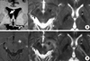
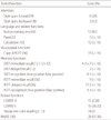
A 48-year-old man with diabetes was admitted to the department of neurology due to progressive behavioral changes. Several years ago, he experienced reddish skin in one of his legs after receiving an operation for varicose veins. Two years ago, he suffered from uveitis in his right eye and was administered with steroids. His family history was unremarkable. He was noted to have recurrent oral ulcers without genital ulcers. He was a calm and timid man who managed a pharmacy, and was very meticulous about his work. About four months ago, he became short-tempered and impatient. He had a tendency to wander around and often chuckled to himself without any reason. His family members and relatives found his behaviors childish. When he worked out in the gym, he would stare at other women, and during daytime, he made visits to a sex toy shop, which, according to his wife, was extremely unusual for his premorbid personality. He acquired reckless driving habits which his wife found unnerving when she rode in the car beside him. On one occasion, the patient drove recklessly around a curve at high speed which led to a traffic accident. At his father's birthday party, immediately after dinner was over, he insisted that everyone join him to go out and eat black-bean-sauce noodles. Although, in the past, he had shown interest in the stock market, by this time, he would sit in front of a computer for several hours trading stocks online. Neurological examination showed no focal signs except relative afferent papillary defect in the right eye. The score of Korean Mini-Mental State Examination was 29/30 and the frontal assessment battery score was 18/18, suggestive of normal function. Formal neuropsychological evaluation showed a deficit of attention and frontal/executive function with retrieval type of memory impairment (Table 1). Brain magnetic resonance imaging (MRI) showed a lesion in the right caudate nucleus with a smaller lesion in the left internal capsule (Fig. 1A). One month later, he developed diplopia. On examination he showed ptosis and mydriasis in the left eye and hypertropia in the right eye. In order to evaluate the brain lesions, he underwent follow-up MRI. The second MRI showed new lesions in the left midbrain, left posterior internal capsule, and left thalamus which enhanced after administration of gadolinium (Fig. 1B). Laboratory tests including routine test, thyroid function test, and HLA B51test revealed nonspecific findings except increased ESR (65 mm/hr, normal range: 0-20 mm/hr) and increased CRP (3.51 mg/dL, normal range: 0.02-0.80 mg/dL). The pathergy test was negative. Cerebrospinal fluid (CSF) analysis showed two of WBCs, 33 mg/dL of protein, 0.41 of IgG index without oligoclonal band.
DISCUSSION
This describes a patient with NBD who presented with prominent behavioral changes, specifically, disinhibition and impulsive behaviors, both of which are associated with the caudate nucleus. Based on the International Behçet's disease study group criteria [5], he was diagnosed with BD because he had recurrent oral ulceration, uveitis, and suspicious skin lesions. In addition, Brain MRI showed lesions characteristic of BD, where an upper brainstem lesion was seen extending into the thalamus and basal ganglia. Given the clinical features and imaging findings, he could be diagnosed with NBD even in the absence of a positive result from the pathergy test and lesions in the skin and genitalia.
Disinhibition and impulsive behaviors in this patient may have been associated with dysfunction of the orbitofrontal circuit [6]. The orbitofrontal circuit projects from the orbitofrontal cortex to the ventromedial caudate nucleus, and projects to the medical dorsomedial globus pallidus and rostromedial substantia nigra. This circuit further extends to the ventral anterior and dorsomedial thalamic nuclei which, in turn, leads back to the orbitofrontal cortex [7]. In addition, the ventral subthalamic nucleus is likely to be a component of the circuit and may be associated with the ventral caudate nucleus [8]. In the present case, the lesion in the right ventral caudate nucleus may have been responsible for disinhibition. Initially, he only showed disinhibition and impulsive behaviors, and MRI preformed at the time demonstrated a lesion restricted to the caudate nucleus. From a former study including 12 NBD patients, six showed disinhibition. Based on the descriptions by the authors, neuroimaging of these patients revealed lesions in the right midbrain, bilateral thalami and midbrain, right basal ganglia, right thalamus, left pedunculothalamic and corona radiate regions, and right mesencephalothalamic region [4]. Considering the quality of MRI or CT in the late 1980s and early 1990s, the lesions described in this report such as those in the thalamus, midbrain, or basal ganglia may have actually been components of the orbitofrontal circuit and, specifically, the mesencephalothalamic lesion may have involved the ventral subthalamic nucleus. Although Kawakita et al. reported psychiatric symptoms in 48% of BD patients, disinhibition was never described [9]. In another study, behavioral changes were found in 54% of the patients, two-thirds of whom showed disinhibition [10]. However, these studies did not include imaging studies for these patients.
In accordance with the previous results [8, 10], neuropsychological evaluation showed dysfunction of attention and frontal lobe with retrieval type of memory deficit. This finding seems to be associated with the lesion in the dorsal caudate nucleus. The dorsolateral prefrontal circuit projects to the dorsolateral head of caudate nucleus, which projects to dorsomedial globus pallidus and rostral substantia nigra. Dorsomedial globus pallidus sends pallidothalamic projections to the ventroanterior thalamus [7]. A previous report showed that cognitive dysfunction may be present in patients with NBD without parenchymal lesion [4]. However, in our patient, even though MRI suggested the involvement of the ventral caudate nucleus, frontal/executive dysfunction may possibly be attributed to the involvement of the dorsal caudate nucleus.
The patient did not show any signs of extrapyramidal dysfunction, although parkinsonism [11] or chorea [10, 12] has been reported in NBD with basal ganglia involvement. The follow up MRI revealed new lesions in the left midbrain, left thalamus, and posterior internal capsule. However, the patient only demonstrated 3rd nerve palsy on the left side without parkinsonism or dystonia. Extrapyramidal manifestations in BD were rare in spite of common involvement of the basal ganglia [2].
We found that progressive disinhibition could be one of presenting symptoms in BD and might be associated with the caudate nucleus. This finding suggests that the involvement of the basal ganglia in BD prior to the involvement of the brainstem could result in unique clinical features such as behavioral changes without extrapyramidal signs.
 XML Download