

 PDF
PDF ePub
ePub Citation
 Print
Print


Abstract
Background
The interlocking pentagon drawing test, a part of the Mini-Mental State Examination (MMSE), is a widely used clinical practice to measure visuoconstructional ability of dementia patients. We investigated the anatomical structures of brain associated with pentagon drawing in subjects with mild to moderate Alzheimer's disease (AD) by using voxel-based morphometry (VBM).
Methods
Medical records of forty-four AD patients were reviewed and a 1.5 T SPGR 3D image data were used for VBM analysis. A voxel-based multiple regression analysis was used to investigate correlation between gray matter loss and pentagon drawing performance of AD patients. The correlations between pentagon drawing score and MMSE score were evaluated by Spearman correlation analysis.
Results
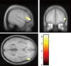
There was a positive correlation between the interlocking pentagon copying scores and the MMSE scores (r=0.448, p=0.002). The lower the scores of interlocking pentagon copying were, the more severe the atrophy of right inferior frontal gyrus became ([x, y, z]=[52, 39, 3], Broadmann area 45, and z score=3.86).
Figures and Tables


References
1. Busatto GF, Diniz BS, Zanetti MV. Voxel-based morphometry in Alzheimers disease. Expert Rev Neurother. 2008. 8:1691–1702.

2. Whitwell JL. Voxel-based morphometry: an automated technique for assessing structural changes in the brain. J Neurosci. 2009. 29:9661–9664.
3. Friston KJ. Commentary and opinion: II. Statistical parametric mapping: ontology and current issues. J Cereb Blood Flow Metab. 1995. 15:361–370.
4. Wright IC, McGuire PK, Poline JB, Travere JM, Murray RM, Frith CD, et al. A voxel-based method for the statistical analysis of gray and white matter density applied to schizophrenia. Neuroimage. 1995. 2:244–252.
5. Ashburner J, Friston KJ. Voxel-based morphometry-the methods. Neuroimage. 2000. 11:805–821.
6. Johnson DK, Storandt M, Morris JC, Galvin JE. Longitudinal study of the transition from healthy aging to alzheimer disease. Arch Neurol. 2009. 66:1254–1259.
7. Godefroy O, Bogousslavsky J. The behavioral and cognitive neurology of stroke. 2007. New York: Cambridge University Press;254–268.
8. Ala TA, Hughes LF, Kyrouac GA, Ghobrial MW, Elble RJ. Pentagon copying is more impaired in dementia with Lewy bodies than in Alzheimer's disease. J Neurol Neurosurg Psychiatry. 2001. 70:483–488.
9. Cormack F, Aarsland D, Ballard C, Tovée MJ. Pentagon drawing and neuropsychological performance in Dementia with Lewy Bodies, Alzheimer's disease, Parkinson's disease and Parkinson's disease with dementia. Int J Geriatr Psychiatry. 2004. 19:371–377.
10. Bourke J, Castleden CM, Stephen R, Dennis M. A comparison of clock and pentagon drawing in Alzheimer's disease. Int J Geriatr Psychiatry. 1995. 10:703–705.
11. Thomann P, Toro P, Santos V, Essig M, Schröder J. Clock drawing performance and brain morphology in mild cognitive impairment and Alzheimer's disease. Brain Cogn. 2008. 67:88–93.
12. Pereira JB, Junque C, Marti MJ, Ramirez-Ruiz B, Bargallo N, Tolosa E. Neuroanatomical substrate of visuospatial and visuoperceptual impairment in Parkinson's disease. Mov Disord. 2009. 24:1193–1199.
13. Kim Y, Lee K, Choi B, Sohn E, Lee A. Relation between the clock drawing test (CDT) and structural changes of brain in dementia. Arch Gerontol Geriatr. 2009. 48:218–221.
14. Shiino A, Watanabe T, Maeda K, Kotani E, Akiguchi I, Matsuda M. Four subgroups of Alzheimer's disease based on patterns of atrophy using VBM and a unique pattern for early onset disease. Neuroimage. 2006. 33:17–26.
15. Takao H, Abe O, Ohtomo K. Computational analysis of cerebral cortex. Neuroradiology. 2010. 52:691–698.
16. Antshel KM, Peebles J, AbdulSabur N, Higgins AM, Roizen N, Shprintzen R, et al. Associations between performance on the Rey-Osterrieth Complex Figure and regional brain volumes in children with and without velocardiofacial syndrome. Dev Neuropsychol. 2008. 33:601–622.
17. Harrington GS, Farias D, Davis CH, Buonocore MH. Comparison of the neural basis for imagined writing and drawing. Hum Brain Mapp. 2007. 28:450–459.
18. Ino T, Asada T, Ito J, Kimura T, Fukuyama H. Parieto-frontal networks for clock drawing revealed with fMRI. Neurosci Res. 2003. 45:71–77.
19. Hampshire A, Chamberlain SR, Monti MM, Duncan J, Owen AM. The role of the right inferior frontal gyrus: inhibition and attentional control. Neuroimage. 2010. 50:1313.
 XML Download