

 PDF
PDF ePub
ePub Citation
 Print
Print


Abstract
Purpose
To report a patient stung by a bee, who was diagnosed with sterile endopthalmitis and another patient diagnosed with optic neuritis, with decreasing visual acuity, after refined bee venom injection around the orbital tissue.
Case summary
A 82-year-old female visited our hospital for decreased visual acuity in the right eye and ocular pain due to a bee sting. The bee sting penetrated the sclera into the vitreous. In the anterior segment, severe cornea edema and anterior chamber cells were seen. Using ultrasonography, inflammation was seen around the intravitreal area. After 3 months, intravitreal inflammation regressed but the patient's visual acuity was light perception negative, and corneal opacity, neovascularization, and phthisis bulbi were detected. A 55-year-old male visited our hospital for ocular pain in the right eye and decreasing visual acuity after refined bee venom injection around the orbital tissue. The best-corrected visual acuity in the right eye was 15/100, there was moderate injection on the conjunctiva. A relative afferent pupillary defect, abnormal color vision test results, and a defect in the visual field test were observed. There was no pain during external ocular movement, and other general blood tests, and a brain MRI were normal. Based on these symptoms, methylprednisolone megatherapy was started for treatment of optic neuritis. After treatment, visual acuity of the right eye was 9/10 and all other clinical optic neuritis symptoms regressed.
Figures and Tables
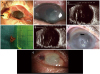 | Figure 1Slit-lamp biomicroscopic photograph and vertical ultrasonography scan images of case 1 in the right eye. (A) Severe mucopurulent keratoconjunctivitis with chemosis and bee's body was seen at the 9 o'clock position of the conjunctiva (black arrow) and the sting was penetrated into intraocular space. (B) Cornea edema with Descemet's membrane folds and large epi-defect was detected. (C) Ultrasonography B-scan image showed vitreous opacity which was considered as bee venom and inflammation and retina was flat. (D) Bee's sting length was calculated to be 4 mm and bee's body length was calculated to be 6 mm. (E) Ultrasonography B-scan image showed stationary vitreous opacity at 10 days after the bee sting injury. (F) Cornea edema, epi-defect, Descemet's membrane detachment (black arrow) and neovascularization on the cornea was detected at 1 month after the bee sting injury. (G) Phthisis bulbi was detected at 3 months after the bee sting injury.
|
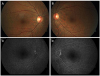 | Figure 2Fundus photography and fluorescein angiography of case 2. (A, B) Fundus photograph showed no specific lesion including papilledema in both eyes. (C, D) Fluorescein angiography showed no specific lesion including leakage from optic disc in both eyes.
|
References
1. Vetter RS, Visscher PK, Camazine S. Mass envenomations by honey bees and wasps. West J Med. 1999; 170:223–227.


2. Kim HJ, Shin JH, Moon SW. Prognosis of ocular injury caused by wasp sting: case reports. J Korean Ophthalmol Soc. 2016; 57:1981–1986.

3. Banks BE, Shipolini RA. Chemistry and pharmacology of honey-bee venom. In : Piek T, editor. Venoms of the hymenoptera: Biochemical, pharmacological and behavioural aspects. 1986. p. 329–416.
4. Kavoussi B, Ross BE. The neuroimmune basis of anti-inflammatory acupuncture. Integr Cancer Ther. 2007; 6:251–257.
5. Bäcker M, Grossman P, Schneider J, et al. Acupuncture in migraine: investigation of autonomic effects. Clin J Pain. 2008; 24:106–115.
6. Uchida S, Hotta H. Acupuncture affects regional blood flow in various organs. Evid Based Complement Alternat Med. 2008; 5:145–151.
7. Chen CJ, Richardson CD. Bee sting-induced ocular change. Ann Ophthalmol. 1986; 18:285–286.
8. Lin PH, Wang NK, Hwang YS, et al. Bee Sting of the cornea and conjunctiva: management and outcomes. Cornea. 2011; 30:392–394.
9. King TP, Spangfort MD. Structure and biology of stinging insect venom allergens. Int Arch Allergy Immunol. 2000; 123:99–106.
10. Song HS, Wray SH. Bee sting optic neuritis. A case report with visual evoked potentials. J Clin Neuroophthalmol. 1991; 11:45–49.
11. Koburova KL, Michailova SG, Shkenderov SV. Further investigation on the antiinflammatory properties of adolapin--bee venom polypeptide. Acta Physiol Pharmacol Bulg. 1985; 11:50–55.
12. Smolin G, Wong I. Bee sting of the cornea: case report. Ann Ophthalmol. 1982; 14:342–343.
13. Sharma P, Sharma R. Toxic optic neuropathy. Indian J Ophthalmol. 2011; 59:137–141.
14. Kerrison JB. Optic neuropathies caused by toxins and adverse drug reactions. Ophthalmol Clin North Am. 2004; 17:481–488.
 XML Download