

 PDF
PDF ePub
ePub Citation
 Print
Print


INTRODUCTION
Urinary tract infection (UTI) is a frequent condition observed in clinical settings and can be managed with proper antibiotic treatment. UTIs involving antibiotic-resistant bacteria that are not susceptible to empirical antibiotics are currently increasing; however, prevention or treatment in these cases is not yet established [12]. Alternative treatments including Lactobacillus have been proposed as means to decrease the misuse of antibiotics and prevent recurrent UTIs [3].
Intestinal bacteria are a common cause of UTI, and among them Escherichia coli are the most frequent culprits [45]. After the death of the bacteria, the E. coli lipopolysaccharide (LPS) reacts with inflammatory cells within the body (i.e., macrophages, monocytes, and neutrophils) to elicit the inflammatory response mediated by tumor necrosis factor-alpha (TNF-α), interleukin-1 (IL-1), prostaglandin, leukotriene, and platelet activating factors [6]. Jerde et al. [7] recently proposed the bladder inflammation index (BII) to assess the histopathology of edema and white blood cell infiltration in a mouse model of cystitis induced by bladder LPS injection. TNF-α acts during the acute phase of the inflammatory reaction [8], and its level is known to increase in the mouse model of LPS-induced cystitis [9]. LPS-mediated immune responses are known to be associated with IL-18 [10]. IL-18 levels are also elevated in urine and bladder tissues in the rat model of cyclophosphamide-induced cystitis [11].
We hypothesized that oral administration of Lactobacillus could prevent cystitis. Thus, this study aimed to determine whether feeding Lactobacillus fermentation extract (LFE) affects the inflammatory reaction in the bladder wall in a mouse model of LPS-induced cystitis based on BII measurement, immunological staining, and analysis of TNF-α and IL-18 levels compared to the controls.
MATERIALS AND METHODS
Twenty-four 8-week old female black mice (C57BL/6; Orient Bio, Seongnam, Korea) were acclimatized for one week. The mice were kept in groups of two in cages with sawdust bedding. Water and feed was supplied freely, and temperature (21–24℃) and humidity (50–60%) conditions were controlled. The mice were subjected to 12-h light/12-h dark cycles. This study was approved by the Ewha Womans University School of Medicine Animal Experimentation Ethics Committee (ESM 14-0272).
Animals were divided into two groups as follows: 1. Control group (n=12): Mice were orally administered normal saline 5 times/week for 2 weeks. On day 14, bladders were injected with LPS and harvested after 24 hours.
2. Study group (n=12): Mice were orally administered SL 16 (B&S Corporation, Tokyo, Japan) 5 times/week for 2 weeks. On day 14, bladders were injected with LPS and harvested after 24 hours.
SL 16 comprised 66.5% distilled water, 33% LFE (soy milk 32%, Lactobacillus curvatus, Lactobacillus casei, Lactobacillus acidophilus, Lactobacillus plantarum, Lactobacillus fermentum, Lactobacillus salivarius, Lactobacillus brevis, and Lactobacillus rhamnosus), 1% citrate, and 0.5% lactate.
Mice were anesthetized with Zoletil (0.6 g/kg; Virbac S.A., Carros, France) and Ropun (0.4 g/kg; Bayer AG, Leverkusen, Germany) prior to bladder injection. A sterilized polyethylene vascular catheter (24 G, 19 mm, Angiocath Plus; Becton Dickinson Medical(s) Pvt. Ltd., Tuas, Singapore) was inserted through the urethra into the bladder and the bladder was emptied by applying pressure to the abdomen. Next, 150 µl of E. coli LPS strain 55:B5 mixture (100 µg/mL; Sigma-Aldrich, St. Louis, MO, USA) was injected through the catheter in both groups. For enhanced effects, this injection was repeated after 30 minutes and the catheter was blocked with a 1 ml syringe to avoid urine leakage for 30 minutes before removal.
The animals were sacrificed by CO2 inhalation and the lower abdomen was disinfected with 70% ethanol, after which the abdominal cavity was exposed through a vertical midline incision of 5 cm. The bladder tissue was harvested and divided vertically. Half of the specimen was preserved at −70℃, and the other half was preserved in 10% formaldehyde.
The tissue fixed in formaldehyde was used to prepare 5-µm vertical tissue sections after paraffin embedding. Hematoxylin and eosin (H&E) staining was performed per standard procedure. TNF-α immunostaining was done using TNF-α antibody (Anti-TNF-alpha antibody ab6671; Abcam, Cambridge, UK) as recommended by the manufacturer, with eosin staining as the control. Histopathology and TNF-α immunostaining of the bladder wall was observed through optical microscopy.
The severity of bladder inflammation was categorized according to the BII as proposed by Jerde et al. [7], by considering leukocyte infiltration into the lamina propria and interstitial tissue edema. Leukocyte infiltration was assessed in 1-mm2 areas of the tissue sections. The score was considered 0 when there was no infiltration, 1 when there were less than 20 leukocytes, 2 when there were 20 to 45 leukocytes, and 3 when there were more than 45 leukocytes. Regarding edema in the interstitial tissue, scoring was as follows: a score of 0 was given for no edema; 1 for mild edema, thinner than the mucosa; 2 for moderate edema, thinner than twice the thickness of the mucosa; and 3 for severe edema, thicker than twice the thickness of the mucosa. The scores for leukocyte infiltration and interstitial edema were added and the average scores from three sites (bladder neck, lateral wall, and bladder dome) in each of the bladder specimens were compared.
Frozen bladder specimens were prepared as described previously [12]. Buffer solution containing 2.66% Tris (hydroxymethyl) aminomethane HCl, 0.985% Tris (hydroxymethyl) aminomethane, 0.5 mM pepstatin A, 0.3 M aprotinin (all obtained from Tocris, Bristol, UK), phenylmethylsulfonyl fluoride, and 1 M leupeptin (both from EMD M illipore, T emecula, CA, U SA) was applied; the specimen was pulverized on ice and incubated at 4℃ for 30 minutes. This preparation was subjected to centrifugal separation at 10,000×g for 10 minutes at 4℃. The TNF-α and IL-18 levels in the supernatant were measured using the MILLIPLEX MAP Non-Human Primate Cytokine Magnetic Bead Panel-Immunology Multiplex Assay Kit (Merck Millipore, Billerica, MA, USA) and Luminex (Luminex, Austin, TX, USA), according to the manufacturers' recommendations.
SPSS software ver. 21.0 (IBM Co., Armonk, NY, USA) was used for all statistical analyses, and all experimental values are presented as mean±standard deviation. BII and TNF-α levels were compared between groups using a non-parametric Mann-Whitney test, and differences were considered statistically significant at p-values <0.05. Sample number and statistical analyses were assessed under the advice of a medical statistician at Ewha Clinical Trial Center.
RESULTS
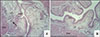
Cystitis induced by transurethral injection of LPS was observed histopathologically. H&E staining revealed moderate to severe submucosal edema and leukocyte infiltration in the LPS-injected control group. Mucosal desquamation and extravascular bleeding were observed in some specimens. In the study group, most samples showed mild submucosal edema and leukocyte infiltration. Two animals each in the control and study groups did not show an inflammatory response (Fig. 1).
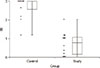
In the control group, the edema score was 2.7±0.6, the leukocyte score was 2.7±0.5, and the BII was 2.7±0.5. In the study group, the edema score was 1.1±0.7, the leukocyte score was 1.1±0.8, and the BII was 1.1±0.7. The average score of the study group was lower than that of the control group (Mann-Whitney test, p<0.001) (Fig. 2).
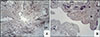
TNF-α immunohistochemical staining revealed dense staining of thick, edematous lamina propria in the control group. Strong chromatogenous enhancement was also observed in the leukocyte concentrated area and the extravascular hemorrhage area. Irregular and weak staining of the antibody was found in the study group (Fig. 3).
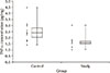
The TNF-α concentration in the control group was 2.82±1.35 pg/mg, and that in the study group was 1.55±0.56 pg/mg. This difference between the control and study groups was statistically significant (p=0.007) (Fig. 4). IL-18 concentration in the control group was 30.89±27.22 pg/mg. However, in the study group, the IL-18 concentration was below the lower limit of detection and could not be measured.
DISCUSSION
This study aimed to determine whether LFE administration could have a preventive effect in cystitis, using a mouse model of cystitis induced by LPS injection into the bladder. The study group was administered LFE and showed decreased BII compared to the control group. Immunostaining and H&E staining revealed increased TNF-α in areas with an inflammatory tissue response.
Transurethral injection of LPS into the bladder was used to induce cystitis. No adverse effects were noted during the experiment. Intraperitoneal injection has also been used to induce cystitis. But Lewis et al. [13] found that, even when the technique was conducted by competent staff, the material was injected into the gastrointestinal tract, subcutaneously, retroperitoneally, or into the urinary bladder in 19.6% of cases. There is also a possibility of infection, pain, local irritation, chemical peritonitis, or perforation of abdominal organs and blood vessels. Thus, we directly inserted a smooth, soft polyethylene tube through the urethra and noted drainage of urine through the catheter. There was slight bleeding during catheter insertion in some cases. We anticipate that transurethral injection is less invasive and induces fewer adverse effects than transperitoneal injection.
The normal flora in the female vagina generally comprises Lactobacillus species. These are known to suppress the growth of other bacteria through a probiotic effect [14]. This has led some investigators to examine the effect of direct application of Lactobacillus into the bladder or vagina to prevent or treat UTI [1516]. Kontiokari et al. [17] compared oral administration of cranberry juice and a Lactobacillus GG drink in women and concluded that Lactobacillus had no effect on recurrent cystitis and assumed that infrequent consumption was the reason. Lee et al. [15] claimed that intravaginal or intravesical administration can provide a more precise volume of medication than oral administration. However, a lack of studies comparing different routes of administration renders it difficult to ascertain the most effective route. Direct injection may be hindered by resistance from the patient and has the potential to cause iatrogenic side effects. Oral administration is typically the method of choice for long-term application and results in decreased inflammatory responses. However, this study and comparable studies [151819] revealed residual inflammation in histology. While it is difficult to know the reason for this, it is evident that administration of Lactobacillus decreases inflammation. Dose or length of treatment may affect this residual inflammation. Thus, long-term observation of treatment might show complete prevention of inflammation.
Oral administration of Lactobacillus may have antiinflammatory effects other than the probiotic effect. Although the process has not yet been elucidated, some studies [202122] suggest possible effects of Lactobacillus on cytokines. Several pre-clinical and clinical studies have shown that Lactobacillus and other probiotic bacteria decreased TNF-α levels [23]. Therefore, we expect that decreasing TNF-α may play a key role in the antiinflammatory effect of Lactobacillus. In the present study, Lactobacillus administration prior to induction of cystitis decreased the inflammatory signs, as well as the TNF-α activity. This suggests the possible use of Lactobacillus in preventing cystitis.
Lactobacillus is reported to be helpful due to its probiotic effects and through indirect effects mediated by the metabolites (biogenics) it produces [24]. Lactobacillus creates various peptides and exopolysaccharides through the process of fermentation. These act as potential modulators of many regulatory processes in the human body, even in the absence of Lactobacillus [25]. For example, Leblanc et al. [26] reported that peptides from Lactobacillus fermentation mediate humoral immune responses in E. coli infection. Thomas et al. [27] reported that histamine derived from Lactobacillus suppresses TNF via modulation of protein kinase A and extracellular signal-regulated kinase. SL-16 is a Lactobacillus fermentation mixture made from soybean and contains metabolites. We therefore considered the possibility that these yet unidentified metabolites may play protective roles in LPS-induced inflammation in the mouse bladder by inhibiting TNF-α-mediated inflammation.
IL-18 is a cytokine that actively controls the innate and adaptive immune response. In the control group, one specimen had IL-18 levels below the lower detection limit, whereas in the study group, all but two samples had IL-18 levels below the measurable range. This may be due to experimental error, but it is difficult to be certain.
This study has some limitations. First, as histopathological examination was performed through visual inspection, results may vary depending on the examiner. To minimize this possibility, each bladder specimen was observed in three separate regions. BII, a verified method of quantification, was also used. After recording the scores assigned by two independent examiners, the average score was calculated and used in the analysis. TNF-α concentrations from cytokine analysis were consistent with the histopathological findings, suggesting that the results of histopathological examination were obtained objectively and were reliable.
Second, LPS injection into the mouse bladder does not induce uniform inflammation on the bladder wall, and in 17.5% of cases, cystitis is not induced [28]. Two specimens in each group did not show an inflammatory response and had almost normal or healthy tissue. It was difficult to judge whether this was due to ineffective LPS injection or the protective effect of LFE. As there were some subjects with concomitant healthy and inflamed tissues, subjects with little evidence of inflammation were not excluded. Excluding such cases would constitute investigator bias, so the analysis for this study includes all subjects, regardless of significant inflammatory findings, which may have impacted the results.
Third, this study only used LPS from one strain of E. coli to induce inflammation in the bladder. As more than 150 strains of E. coli exist, the generalization of the results to all forms of bacterial cystitis is limited. A more solid basis may be established with further studies investigating oral administration of Lactobacillus in cystitis induced by more strains of E. coli bacteria.
Among the few animal studies of Lactobacillus and UTI, Asahara et al. [16] injected L. Casei Shirota into mouse bladders followed by bladder injection of E. coli after 24 hours. They observed anti-bacterial effects in the group that underwent repeated transurethral Lactobacillus injection after injection of E. coli. Lee et al. [15] studied bacterial cultures and histopathological findings in rats after bladder injection of L. rhamnosus after E. coli injection. They found that the Lactobacillus blocked E. coli growth. Compared to these studies, the present study has some strengths; we examined oral administration of Lactobacillus and noted a preventive effect. Moreover, our study included histopathology and cytokine levels for a more thorough investigation.
Our study was designed to compare only the study and control groups, which simplified the experimental process and empowered statistical analysis. To strengthen the hypothesis regarding the preventive effect of oral Lactobacillus administration, we are planning additional experiments using mice orally administered normal saline or SL-16. A study on the effect of bladder instillation of LPS on TNF-α will also be conducted. A comparative study of transurethral injection of Lactobacillus or oral administration of other drugs known to effectively prevent recurrent cystitis (for example, cranberry juice or E. coli extract [18]) may be conducted, as well. Additional immunohistological staining and cytokine analysis of inflammation markers (e.g., IL-6 and interferon-γ) associated with cystitis will be included in further investigations.
CONCLUSIONS
In conclusion, oral administration of LFE reduced the inflammatory response in the bladder tissue and decreased TNF-α expression. This suggests that Lactobacillus has antibiotic properties that may be protective against cystitis. Through further investigations into the mechanism of action and utility, this finding may be developed as a treatment and preventive measure for recurrent cystitis.
 XML Download