

 PDF
PDF ePub
ePub Citation
 Print
Print


Abstract
Purpose
To discuss the clinical course and diagnosis of corneal dysplasia in a xeroderma pigmentosum patient based on a genetic evaluation.
Case summary
A 42-year-old female visited our clinic for decreased left visual acuity and corneal opacity. She had undergone several surgeries previously due to the presence of basosquamous carcinoma in the left lower eyelid, neurofibroma, and malignant melanoma of the facial skin. The patient showed repeated corneal surface problems, with a suspicious dendritic lesion; however, antiviral therapy was ineffective, and herpes simplex virus polymerase chain reaction results were negative. Despite regular follow-ups, the patient showed neovascularization around the corneal limbus and an irregular corneal surface. We performed corneal debridement with autologous serum eye drops for treatment. The patient's visual acuity and corneal surface improved after the procedure. The impression cytology result was corneal dysplasia. In whole exome sequencing, two pathogenic variants and one likely pathogenic variant of the POLH gene were detected.
Conclusions
This is the first genetically identified xeroderma pigmentosum case with ophthalmological lesions of the eyelid and cornea in Korea. Debridement of the irregular corneal surface and autologous serum eye drop administration in xeroderma pigmentosum could be helpful for improving visual acuity.
Figures and Tables
 | Figure 1Facial photos of the patient. (A) Multiple freckle-like hyperpigmented macules and cancer-suspicious skin lesions were found. (B) Left lower eyelid shows ulcerated lesion which suggested basal cell carcinoma.
|
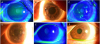 | Figure 2Anterior segment photos of the patient. (A) The patient's right cornea surface was relatively smooth and clear under fluorescein staining compared with the opposite side. (B) Under scleral scatter, there was no opacity. (C) On the patient's left cornea, irregular corneal surface under fluorescein staining was found before corneal surface debridement. (D) Under scleral scatter, central and peripheral corneal subepithelial opacity was found on the left cornea. (E) After corneal surface debridement on the patient's left eye, we could see a dramatically smoothened corneal surface. (F) Although small subepithelial opacities were still present, corneal opacity was improved.
|
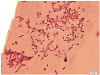 | Figure 3Impression cytology of the patient (periodic acid-Schiff [PAS] stain, ×200). The image shows limbal dysplasia exhibiting nuclear alteration with spiral-shaped or twin nuclei. Corneal epithelial cells are squamoid in shape with metachromic change in the cytoplasm and demonstrate a low nuclear-cytoplasmic ratio. There was no definite goblet cell presence.
|
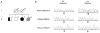 | Figure 4Pedigree of the xeroderma pigmentosum (XP)-affected family and POLH gene mutation analysis for the proband (II-3) and proband's brother (II-1). (A) The open symbols indicate no signs or symptoms of XP. The filled symbol represents an affected individual. The half-closed symbol indicates an individual who likely has XP but who was not tested for the POLH mutation. The arrow indicates the proband (II-3). (B) Sequencing analysis identified that the patient has compound heterozygous variants c.660+2T>A and c.725C>G (p.Ser242) (arrow).
|
Notes
References
1. Brooks BP, Thompson AH, Bishop RJ, et al. Ocular manifestations of xeroderma pigmentosum: long-term follow-up highlights the role of DNA repair in protection from sun damage. Ophthalmology. 2013; 120:1324–1336.


2. Chaurasia S, Mulay K, Ramappa M, et al. Corneal changes in xeroderma pigmentosum: a clinicopathologic report. Am J Ophthalmol. 2014; 157:495–500.e2.

3. Lehmann AR, McGibbon D, Stefanini M. Xeroderma pigmentosum. Orphanet J Rare Dis. 2011; 6:70.
4. Kim MH, Shin JL, Cho BC. A case of limbal squamous cell carcinoma appearing in xeroderma pigmentosum. J Korean Ophthalmol Soc. 1978; 19:473–477.
5. Lee YS, Myong YW, Baek NH. A case of ocular manifestation in xeroderma pigmentosum. J Korean Ophthalmol Soc. 1984; 25:555–559.
6. Yoon BJ, Jun HS. A case of xeroderma pigmentosum with ocular manifestation. J Korean Ophthalmol Soc. 1985; 26:859–864.
7. Cleaver JE. Cancer in xeroderma pigmentosum and related disorders of DNA repair. Nat Rev Cancer. 2005; 5:564–573.
8. Fassihi H, Sethi M, Fawcett H, et al. Deep phenotyping of 89 xeroderma pigmentosum patients reveals unexpected heterogeneity dependent on the precise molecular defect. Proc Natl Acad Sci U S A. 2016; 113:E1236–E1245.
9. Inui H, Oh KS, Nadem C, et al. Xeroderma pigmentosum-variant patients from America, Europe, and Asia. J Invest Dermatol. 2008; 128:2055–2068.
10. Ortega-Recalde O, Vergara JI, Fonseca DJ, et al. Whole-exome sequencing enables rapid determination of xeroderma pigmentosum molecular etiology. PLoS One. 2013; 8:e64692.
11. Geerling G, Maclennan S, Hartwig D. Autologous serum eye drops for ocular surface disorders. Br J Ophthalmol. 2004; 88:1467–1474.
12. Azari AA, Rapuano CJ. Autologous serum eye drops for the treatment of ocular surface disease. Eye Contact Lens. 2015; 41:133–140.
13. Roberson MC. Corneal epithelial dysplasia. Ann Ophthalmol. 1984; 16:1147–1150.
14. Opletalova K, Bourillon A, Yang W, et al. Correlation of phenotype/genotype in a cohort of 23 xeroderma pigmentosum-variant patients reveals 12 new disease-causing POLH mutations. Hum Mutat. 2014; 35:117–128.
15. DiGiovanna JJ, Kraemer KH. Shining a light on xeroderma pigmentosum. J Invest Dermatol. 2012; 132(3 Pt 2):785–796.
 XML Download