

 PDF
PDF ePub
ePub Citation
 Print
Print


Abstract
Purpose
To report a case of chemical injury of the cornea caused by high-dose ethanol during orbital wall fracture repair.
Case summary
A 56-year-old male presented with pain after blowout fracture repair surgery. During the surgery, 2% hexethanol solution (2% chlorhexidine and 72% ethanol mixture), which was used for disinfection of the face, flowed into the left eye. Conjunctival injection in the left limbus, a large corneal epithelial defect, and severe stromal edema were subsequently observed. The patient was treated with topical antibiotics, steroids, and autologous serum eye drops. After 1 week, the corneal epithelial defect was improved, but at the second month of therapy, recurrent corneal erosion with deterioration of the endothelial cell function occurred. Anterior stromal puncture and laser keratectomy were performed. The corneal epithelial defect and erosion improved, but the endothelial cell density was severely decreased.
Conclusions
The 2% hexethanol solution is usually used for preoperative skin disinfection, but it contains a high concentration of ethanol. The surgeon should be aware that high concentrations of ethanol may result in severe corneal damage, including corneal endothelial dysfunction and limbal cell deficiency.
Figures and Tables
 | Figure 1Slit-lamp photographs of the cornea in the left eye with chemical injury at the initial presentation. (A) Anterior segment photograph shows perilimbal injection, severe stromal edema, Descemet's membrane folding, and opacity in his left eye. (B) Anterior segment photograph shows about 11 × 6 mm sized corneal epithelial defect combined with perilimbal conjunctival epithelial defect in his left eye.
|
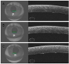 | Figure 2Anterior segment optical coherence tomography images of the cornea in the left eye with chemical injury at the initial presentation. (A–C) showing high density of endothelium layer, swelling of multiple endothelial cells.
|
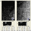 | Figure 3Qualitative and quantitative analysis of central corneal endothelial cell in the left eye with chemical injury. (A) Corneal endothelium after 3 days following chemical injury of cornea is shown. The endothelial cell density was 1,316 cells/mm2. (B) The endothelial cell density of the damaged left cornea was 530 cells/mm2. The coefficient of variation in cell size was 40 and the percentage of hexagonal cells was 31% in corneal endothelial cells of chemical injury of cornea after 4 weeks following phototherapeutic keratectomy in the left eye. CD = cell density; SD = standard deviation; CV = coefficient of variation; AVE = average; NUM = number; PACHY = pachymeter.
|
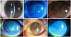 | Figure 4Slit-lamp photographs of the cornea in the left eye with chemical injury after treatment. (A, B) Corneal epithelial defect and corneal edema were decreased a week after the initial treatment. The patient complained visual disturbance and pain in his left eye 2 months after the first recovery. Corneal epithelial defect and stromal edema were recurred. (C, D) Corneal stromal edema and Descemet's membrane folding at the inferior aspect of the cornea have recurred. (E, F) Corneal epithelial defect with punctate epithelial erosions were decreased after anterior stromal puncture and phototherapeutic keratectomy.
|
Notes
References
1. Roth S, Thisted RA, Erickson JP, et al. Eye injuries after nonocular surgery. a study of 60,965 anesthetics from 1988 to 1992. Anesthesiology. 1996; 85:1020–1027.

2. Yu HD, Chou AH, Yang MW, Chang CJ. An analysis of perioperative eye injuries after nonocular surgery. Acta Anaesthesiol Taiwan. 2010; 48:122–129.

3. Liu HY, Yeh PT, Kuo KT, et al. Toxic keratopathy following the use of alcohol-containing antiseptics in nonocular surgery. JAMA Ophthalmol. 2016; 134:449–452.
4. Mac Rae SM, Brown B, Edelhauser HF. The corneal toxicity of presurgical skin antiseptics. Am J Ophthalmol. 1984; 97:221–232.
5. Oh JY, Yu JM, KO JH. Analysis of ethanol effects on corneal epithelium. Invest Ophthalmol Vis Sci. 2013; 54:3852–3856.
6. Chang SW, Chou SF, Wang YH. Ethanol treatment induces significant cell death in porcine corneal fibroblasts. Cornea. 2006; 25:1072–1079.
7. Eslani M, Baradaran-Rafii A, Movahedan A, Djalilian AR. The ocular surface chemical burns. J Ophthalmol. 2014; 2014:196827.
8. Baradaran-Rafii A, Eslani M, Haq Z, et al. Current and upcoming therapies for ocular surface chemical injuries. Ocul Surf. 2017; 15:48–64.
9. Perkins TW, Kumar A, Kiland JA. Corneal decompensation following bleb revision with absolute alcohol: clinical pathological correlation. Arch Ophthalmol. 2006; 124:738–741.
10. Fish R, Davidson RS. Management of ocular thermal and chemical injuries, including amniotic membrane therapy. Curr Opin Ophthalmol. 2010; 21:317–321.
11. Avni Zauberman N, Artornsombudh P, Elbaz U, et al. Anterior stromal puncture for the treatment of recurrent corneal erosion syndrome: patient clinical features and outcomes. Am J Ophthalmol. 2014; 157:273–279.e1.
12. Ko BY, Lee GW. Clinical results of phototherapeutic keratectomy for refractory recurrent corneal erosion. J Korean Ophthalmol Soc. 2011; 52:392–400.
13. Joyce NC. Proliferative capacity of the corneal endothelium. Prog Retin Eye Res. 2003; 22:359–389.
14. Tamori Y, Deng WM. Compensatory cellular hypertrophy: the other strategy for tissue homeostasis. Trends Cell Biol. 2014; 24:230–237.
 XML Download