Abstract
Lauraceae is a family medicinal plant whose tubers possesses antimicrobial, and cytotoxic, such as antiparasitic and anti-inflammatory special effects and has been used for the medicine in the cure of hepatitis and rheumatism. The antimicrobial activities of bioactive compounds including one neolignan; kunstlerone (1) and two alkaloids include isocaryachine (2) and noratherosperminine (3) as well as crude hexane, methanol and dichloromethane extracts were evaluated. Additionally, the effect of compounds 1, 2 and 3 were evaluated on A549, PC-3, A375, HT-29 and WRL-68 cell lines. In conclusion, kunstlerone 1 showed moderate cytotoxicity against various cancer cell lines such as A549, PC-3, A375, HT-29 and WRL-68, respectively with EC50 values of 28.02, 26.78, 33.78, 33.65 and 16.46 µg/mL. The crude methanol extract showed antigrowth activity against S. pyogenes II and B. cereus, with MICs of 256 µg/mL. The compounds kunstlerone (1), isocaryachine (2) and noratherosperminine (3) showed complete inhibition against P. shigelloides, with MIC ≤60 µg/mL compare to ampicillin, as a positive control, which showed antigrowth activity against P. shigelloides at MIC 10 µg/mL.
There are more than 2500 species belonging to the Lauraceae family all over the world, spread within the subtropics and tropics of eastern Asia and South and North America.1 Many plants of Lauraceae have been affianced in folk medicine for their attractive bioactivities. For illustration, Beilschmiedia species is a source which can be treat arthritis, rheumatism, etc.234567
In the relations of Lauraceae, most of the alkaloids are in the right place to the isoquinoline class.7 Lauraceae family is generally taking place in Southeast Asia and tropical America with 40 genera and over 2000 species.8 In Malaysia, its contribution is about 213 species, from 16 genera.9 Aporphine alkaloids are collectively in presence but there are also a small number of indole alkaloids and quinoline alkaloids.10 This genus is over and above known to formulate a large number of biologically active compounds demodulating attractive skeletons.101112 The lignans and neolignans consist of a class of natural plant products which are consequential from cinnamic acid derivatives and which are related biochemically to phenylalanine metabolism,1314 in addition of alkaloids, flavonoids, and endiandric acid derivatives, lignans, neolignans and benzamides extracted and recognized by the Beilschmiedia species.15 Furthermore, this species has been the subject of a number of encyclopaedic articles.101116 Moreover, some of compounds such as the endiandric acid derivatives and Epoxyfuranoid lignans have shown strong antibacterial results and antitubercular activities.121718 In this study, our objective was to determine the prospective bioactivities of the crude hexane, methanol and dichloromethane extract including two alkaloids and one neolignan for cytotoxic activity on cancer cell lines and antimicrobial study.
The optical rotations were recorded on a Jasco (Japan) P-1020 Polari meter equipped with a Sodium lamp; MeOH as solvent. LC-MS were obtained on an Agilent Technologies 6530 Accurate-Mass Q-TOF LC-MS. The ultraviolet spectra were obtained in MeOH on a Shimadzu UV-310 ultraviolet-visible spectrometer. The Fourier Transform Infrared (FTIR) spectra were obtained with CHCl3 (NaCl window technique) on a Perkin Elmer 2000 instrument. The 1H-NMR and 13C-NMR spectra were recorded in deuterated chloroform on a JEOL 400MHz (unless stated otherwise) instrument; chemical shifts are reported in ppm on 8 scale, and the coupling constants are given in Hz. Silica gel 60, 70 – 230 mesh ASTM (Merck 7734) was used for column chromatography. Mayer's reagent was used for alkaloid screening. TLC Aluminum sheets and PTLC (20 × 20 cm Silica gel 60 F254) were used in the TLC analysis. The TLC and PTLC spots were visualized under UV light (254 and 366 nm) followed by spraying with Dragendorff's reagent for an alkaloid detection. All solvents, except those used for bulk extraction are AR grade.
Beilschmiedia kunstleri Gamble (Lauraceae), collected from Hutan Simpan sungai Tekam, Jerantut, Pahang, Malaysia was identified by Mr. Teo Leong Eng. A voucher specimen (KL 5627) is deposited at the Herbarium of the Department of Chemistry, University of Malaya, Kuala Lumpur, Malaysia and at the Herbarium of the Forest Research Institute, Kepong, Malaysia. Beilschmiedia kunstleri Gamble is a tall tree up to 12 m tall and 14 cm diameter, bark reddish brown, striated; inner bark pale brown; young shoots reddish brown; leaves simple, alternate or spinally arranged, coriaceous, from elliptic lance late to ablanceolate, apex blunt to rounded, base obtuse, 25 – 60 cm × 10 – 18 cm, bright green above, paler below, drying brown to deep brown, midrib raised above, secondary nerves 20 pairs, arching near the margin; P.T.O.
Air-dried bark (1.50 kg) of Beilschmiedia kunstleri Gamble were ground and extracted exhaustively with hexane (5.00 L) for 72 hours. The residual plant material was dried and left for 5 h after moistening with 10% NH4OH. It was then macerated with CH2Cl2 (10.00 L) for 5 days. After filtration, the supernatant was concentrated to 500 mL at room temperature (30 ℃) followed by acidic extraction with 5% HCl until a negative Mayer's test result was obtained. The aqueous solution was made alkaline to pH 11 with NH4OH and re-extracted with CH2Cl2. This was followed by washing with distilled H2O, dried over anhydrous sodium sulphate, and evaporation to give an alkaloid fraction (2.90 g). We repeat the extraction of alkaloids by using MeOH solvent and after acid base extraction obtained another (35.00 g) of crude alkaloid. The crude alkaloid from CH2Cl2 and MeOH was submitted to exhaustive column chromatography over silica gel using CH2Cl2 gradually enriched with methanol to yield fractions. These Fractions afforded kunstlerone (1) as neolignan, isocaryachine (2) and noratherosperminine (3) as alkaloids.
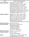
Antimicrobial activity of the extracts and isolates were investigated using the method of agar dilution.19 Provisionally, the tested compounds dissolved in either CH2Cl2 or MeOH were individually diverse with Muller Hinton (MH) broth to obtain a final volume of 2 mL. Two-fold dilution was prepared and the solution was then transferred to the agar solution of MH to yield the final concentrations ranging from 256 µg/mL, Twenty seven strains of microorganisms, refined in MH broth at 30 ℃ for 24 hrs, were diluted with 0.9% normal saline solution to regulate the cell density of 108 CFU/mL.
The organisms were inoculated against each plate using a multipoint inoculators and further incubated at 30 ℃ for 18 – 48 hrs. Compounds which possessed high efficacy to inhibit bacterial cell growth were analyzed (Table 3). As aspect, these microorganism strains has assume that which strain has ability for interchanging and which sourcing the strains from different culture folders has not any effect against result the media quality results and qualification test methods.
All the cells that applied in this study were obtained from American Type Cell Collection (ATCC) and maintained in a 37 ℃ incubator with 5% CO2 saturation. A375 human melanoma, HT-29 human colon adenocarcinoma cells and WRL-68 normal hepatic cells were maintained in Dulbecco's modified Eagle's medium (DMEM). Whereas A549 non-small cell lung cancer cells and PC-3 prostate adenocarcinoma cells were maintained in RPMI medium. Both medium were supplemented with 10% fetus calf serum (FCS), 100 units/mL penicillin, and 0.1 mg/mL streptomycin.
Different cell types from above were used to find out the inhibitory effect of compounds kunstlerone (1), isocaryachine (2), noratherosperminine (3) on cell growth using the MTT assay. Briefly, cells were seeded at a density of 1 × 10⁵ cells/mL in a 96-well plate and incubated for 24 hours at 37 ℃, 5% CO2. Next day, cells were treated with the compounds respectively and incubated for another 24 hours. After 24 hours, MTT solution at 2 mg/mL was added for 1 hour. Absorbance at 570 nm was measured and recorded using Plate Chameleon V microplate reader (Hidex, Turku, Finland). Results were expressed as a percentage of control giving percentage cell viability after 24 hours exposure to test agent. The potency of cell growth inhibition for each test agent was expressed as an EC50 value, defined as the concentration that caused a 50% loss of cell growth. Viability was defined as the ratio (expressed as a percentage) of absorbance of treated cells to untreated cells.20
In additional search for bioactive compounds and interesting chemical entities from Malaysian flora, we have performed a phytochemical study on the bark of a Malaysian Lauraceae, Beilschmiedia kunstleri Gamble, which has led to the isolation of one neolignan, kunstlerone (1), and two alkaloids consist of isocaryachine (2) and noratherosperminine (3) (Fig. 1) were isolated from the bark of this species. Recently, we have reported some constituents from the leaves and bark of Beilschmiedia species.2122 These compounds were obtained from the CH2Cl2 and CH3OH extract of the bark of B. Kunstleri.
The crude dichloromethane, hexane and methanol extracts, and the isolates (1 – 3) from bark of B. kunstleri Gamble were experienced and tested for antimicrobial activity against 15 strains of microorganisms by means of the agar dilution method.23 The domino effect (Table 1) give us an idea about that all the tested extracts entirely inhibit the expansion of the Gram-positive bacterium C. diphtheriae NCTC 10356 with MIC 256 µg/mL. This strain was the most sensitive, as it turned out the only one inhibited by the crude hexane and dichloromethane extracts. Other microorganisms such as A. xylosoxidan ATCC 2706, S. aureus ATCC 25923, M. lutens ATCC 10240, B. cereus and A. hydrophila were wholly inhibited by the crude dichloromethane extract, with MIC 256 µg/ mL. In addition, the crude methanol extract displays antigrowth activity against S. pyogenes II and B. cereus, with MICs of 256 µg/mL.
In the current research, the antimicrobial activity of pure compounds (1 – 3) isolated from the dichloromethane and methanol extract was similarly evaluated. It was found (Table 1) that compounds 1, 2 and 3 displayed complete inhibition against P. shigelloides, with MIC ≤60 µg/mL. In evaluation ampicillin, a positive be in charge, showed antigrowth doings against P. shigelloides at MIC 10 µg/mL.
To evaluate the cytotoxic activity, each different compound namely kunstlerone (1), isocaryachine (2) and noratherosperminine (3) were tested with a series of different doses on A549, PC-3, A375, HT-29 and WRL-68, respectively (Fig. 2). After 24 hours, cell viability was determined by the MTT assay. Test agents induced cell cytotoxicity in a concentration dependent manner. These dose titration curves allowed determining EC50 for the test agents towards different cell lines (Table 2).
From Fig. 2, isocaryachine (2) and noratherosperminine (3) showed no cytotoxic effect on various cell lines. On the other hand, kunstlerone (1) also showed cytotoxic effect on several of the cancer cell lines with different EC50 valuesin a concentration dependent manner. These dose titration curves allowed determining EC50 for the various compounds towards different cell lines. Kunstlerone (1) demonstrated dose-depended cytotoxic effects with EC50 values of 33.78 ± 3.4, 28.02 ± 1.7, 33.65 ± 0.9, 26.78 ± 2.3 and 16.46 ± 0.7 µg/mL; in A375, A549, HT-29, PC-3 and WRL-68, respectively. These results indicate that cell lines differ in their sensitivity to the same test agent, which may be determined by multiple cell type-specific signalling cascades and transcription factor activities.
Cytotoxic screening models make available important preliminary data to select plant extracts or natural compounds with potential anticancer properties. In this study, the cytotoxic effect of kunstlerone (1) was investigated by the addition of the MTT tetrazolium salt to various treated cancer cell lines. Taken together, the cytotoxic effects exerted by kunstlerone (1) as a promising compound and suggest its potential as anti proliferation agent. To our knowledge, the cytotoxic potentials of kunstlerone (1) have not been examined and the underlying molecular mechanisms remain to be discovered.
In conclusion, the constituents of Beilschmiedia kunstleri grown in the Pahang, Malaysia was found to possess antimicrobial and cytotoxic activities. The crude methanol extract displays antigrowth activity and kunstlerone (1), isocaryachine (2) and noratherosperminine (3) displayed complete inhibition against P. shigelloides. In evaluation ampicillin, a positive be in charge of this folder, showed antigrowth against P. shigelloides. Kunstlerone (1) showed also moderate cytotoxicity against various cancer cell lines such as A549, PC-3, A375, HT-29 and WRL-68. Therefore, the antimicrobial and cytotoxic activity study revealed that this plant has potential bioactivity.
Figures and Tables
Fig. 2
Dose-response curves (using GraphPad Prism) tested with kunstlerone (1), isocaryachine (2), noratherosperminine (3) and Doxorubicin (positive control) in the MTT assays towards A) A375, B) A549, C), HT-29, D) PC-3 and E) WRL-68.

Acknowledgements
The authors acknowledge the financial support provided by University of Malaya Research Grant (UMRG 045/11BIO), Centre of Natural Products and Drugs Development (CENAR), Postgraduate Research Grant of University of Malaya (PS366/2010B) and High Impact Research (HIR) Grant of University of Malaya (F000009-21001). We also acknowledge the support provided by Faulty of science, Qom University of Technology, and Din Mat Nor and Rafly Syamsir for the plant samples and also Hairin Taha for editorial assistance.
References
1. Simi A, Sokovi MD, Risti M, Gruji-Jovanovi S, Vukojevi J, Marin PD. Phytother Res. 2004; 18:713–717.

2. Pudjiastuti P, Mukhtar MR, Hadi AH, Saidi N, Morita H, Litaudon M, Awang K. Molecules. 2010; 15:2339–2346.


3. Lenta BN, Tantangmo F, Devkota KP, Wansi JD, Chouna JR, Soh RC, Neumann B, Stammler HG, Tsamo E, Sewald N. J Nat Prod. 2009; 72:2130–2134.

4. Earl EA, Altaf M, Murikoli RV, Swift S, O'Toole R. BMC Complement Altern Med. 2010; 10:25.
7. Saxton JE, Bentley KW. The Alkaloids: The isoquinoline alkaloids. The chemical Society;2007.
8. Lajis NHJ, Ahmad R. Stud Nat Prod Chem. 2006; 33:1057–1090.
9. Guinaudeau H, Leboeuf M, Cave A. J Nat Prod. 1979; 42:325–360.
10. Harborne JB, Mendez J. Phytochemistry. 1969; 8:763–764.
12. Yang PS, Cheng MJ, Chen JJ, Chen IS. Helv Chim Acta. 2008; 91:2130–2138.
13. Sovová H, Opletal L, Bártlová M, Sajfrtová M, Krenková M. The Journal of supercritical fluids. 2007; 42:88–95.
14. Jackson DE, Dewick PM. Phytochemistry. 1984; 23:1029–1035.
15. Funasaki M, Lordello ALL, Viana AM, Santa-Catarina C, Floh EIS, Yoshida M, Kato MJ. J Braz Chem Soc. 2009; 20:853–859.
16. Chouna JR, Nkeng-Efouet PA, Lenta BN, Devkota KP, Neumann B, Stammler HG, Kimbu SF, Sewald N. Phytochemistry. 2009; 70:684–688.

17. Engler TA, Wei D, Letavic MA. Tetrahedron Lett. 1993; 34:1429–1432.
18. Engler TA, Wei DD, Letavic MA, Combrink KD, Reddy JP. J Org Chem. 1994; 59:6588–6599.
20. Braga PA, Dos Santos DA, De Silva MF, Vieira PC, Fernandes JB, Houghton PJ, Fang R. Nat Prod Res. 2007; 21:47–55.

21. Mollataghi A, Hadi AHA, Awang K, Mohamad J, Litaudon M, Mukhtar MR. Molecules. 2011; 16:6582–6590.
22. Mollataghi A, Hadi AHA, Cheah SC. Molecules. 2012; 17:4197–4208.


 PDF
PDF ePub
ePub Citation
 Print
Print


 XML Download