

 PDF
PDF ePub
ePub Citation
 Print
Print


INTRODUCTION
Early-onset Alzheimer's disease (EOAD) is defined as the AD that developed before the age of 65-year-old.1 The arbitrary cut-off age of 65-year-old was based on the usual retirement age. However, the clinical characteristics of EOAD described are distinct from that of late-onset AD (LOAD). The early intense neuropsychiatric symptom and non-amnesic presentation are more frequent in EOAD.123
Recently, we experienced a case of atypical EOAD dementia in a patient who presented visuospatial deficit and language problem. Through stepwise evaluation of dementia, from the standard neuropsychological test, brain magnetic resonance imaging (MRI), (18)F-florbetapir positron emission tomography (PET), and the measurements of 3 established cerebrospinal fluid (CSF) AD biomarkers, we diagnosed the patient as AD with atypical clinical presentation. In this report, we described the clinical, laboratory, and radiologic characteristics of the patient to raise awareness of the recent issue related to the diagnosis of atypical EOAD.
CASE REPORT
A 54-year-old, right handed man visited our memory clinic complaining of inability to perform his daily job adequately. He had 9 years of formal education and was a product delivery truck driver for 20 years. He was good in a health without any diseases or trauma history except chronic alcohol intake for 30 years. From 3 months prior, the patient experienced gradual forgetfulness in loading products and problems in navigating to required destinations. He was found to frequently make mistakes in calculation for account book records. Therefore, he was recommended to resign from the company. Since then, he remained home bound or at his wife's store, because he easily got lost on unfamiliar roads. His family noticed that he became silent and the content of his speech was poor with use of limited words. Further, he was found awkward in using the cellular phone, avoided spontaneous use, and was often depressed.
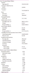
On evaluation, he looked healthy, but was less concerned about his cognitive deficits when asked about his difficulties, in response to his family's report on his recent problems. He was the fifth child in his family. His mother had sudden death at 60-year-old due to unidentified cause, and his father suffered from unknown type of dementia since the age of 70 years. None of his siblings had any kinds of neurological disorders. On neurologic examination, his muscle tone, speed of fine movement, and gait were normal. Other abnormal neurologic signs were not evident. For further evaluation, we performed the comprehensive neuropsychological, and language test. He got 16 points in Korean Mini-Mental State Examination, lost scores in temporal orientation (-2), spatial orientation (-1), registration (-2), serial -7's test (-3), remote memory (-1), and stage command (-3), repetition (-1), and writing a complete sentence (-1). Seoul Neuropsychological Battery for the comprehensive cognitive evaluation.4 His cognitive performance was the far below the normal range in all cognitive domains that were validated by digit span, Korean version of Boston Naming Test (K-BNT), calculation, ideomotor praxis, Rey complex figure copy, Seoul Verbal Learning Test, recall of Rey complex figure, controlled oral association word recall test, and Stroop test (Table 1). On Korean version of Western Aphasia Battery for validation of language deficits, his speech was fluent, but circumstance not focused, repetition of syllable, paraphagic error, and mild stuttering were noted. His comprehension was slightly impaired, especially on sequential commands. However, the responses on yes/no question and auditory word recognition were good. The repetition was most seriously impaired with ability to repeat only up to 2 short phrases. The naming was decreased, as identified in K-BNT. The collective results were suggestive of conduction aphasia.5
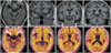
The blood chemistry, thyroid function test, and other laboratory test did not show other combined systemic disorder. Brain MRI revealed mild atrophy in the right side of medial temporal lobe, but marked atrophy in the left side (Fig. 1). The widening of sylvian fissure was evident in left side due to the diminished volume of adjacent temporal and inferior frontal and parietal lobe. And the sulcal widening of bilateral parietal lobe was also suspected. An (18)F-florbetapir-PET (Fig. 1) revealed a high uptake in the whole hemisphere. The stronger (18)F-florbetapiruptake was noted in bilateral parietal lobe.
Subsequently, the CSF AD biomarkers were checked after lumbar puncture under the informed consent from the patient and his family. The enzyme-linked immunosorbent assay using INNOTEST kit (Fujirebio, Ghent, Belgium) was used for analysis. The concentration of Aβ42, total Tau, and phospho-Tau181 (pTau181) in CSF was 323.3 pg/mL, 696.1 pg/mL, and 93.8 pg/mL, respectively. These fulfill the diagnostic criteria of AD.6 His apolipoprotein genotype was identified as ε3/ε3, and genetic study for causative mutation are planned. After diagnosis of AD, he has been on cholinesterase inhibitor and speech therapy for the past 2 months.
DISCUSSION
The dominant symptom of EOAD in our patient was distinct, demonstrating visuospatial, language and calculation deficits. His neuropsychological performance was extensively diminished involving all tested cognitive domains, and much worse than expected considering that his cognitive deficits became evident only 3 months prior. Although controversial, it is generally accepted that EOAD is more rapidly progressive and demonstrates atypical presentation compared with LOAD, as in our patient.1237 Thus, the AD diagnosis is often challenging in EOAD. The value of biomarker in AD diagnosis is proven and therefore incorporated into the revised diagnostic criteria of AD.89 Especially, the concentrations of Aβ42, tTau, and pTau181 in CSF are the currently superior AD biomarkers since they reflect the AD-related biochemical change of brain.101112 The international cooperative group has reached a consensus on the clinical usefulness of CSF biomarkers.13 Especially, 3 situations are suggested as an indication of the CSF biomarker analysis; early-onset dementia, mild cognitive impairments in case that the patient wants to know the result, and atypical clinical presentation.13 This indications were applicable to our patient who demonstrated early-onset dementia with atypical presentation. Among the dominant dementia symptoms, the conduction aphasia was the most unexpected feature of AD manifestation in our patient.14 The marked widening of left sylvian fissure due to the atrophy of the bordering structures is a relevant MRI lesion of the conduction aphasia.15 We diagnosed the case as AD with the help of AD biomarker study, amyloid imaging and CSF biomarker. The amyloid PET imaging and decreased CSF Aβ level indicated increased Aβ burden. In addition, the profiles of tau proteins were of diagnostic value. The markedly increased tTau and pTau181 level are indicative of ongoing severe axonal damage and increased tangle pathology in the patient's brain.10 These results were in agreement with his neuropsychiatric characteristics. The cognitive impairments were extensive and rapidly progressing. As reported in network study in EOAD,1617 wide destruction of functional network of the brain was suspected in our patient.
In summary, we recently diagnosed EOAD in a patient with the help of AD biomarker study, both Aβ imaging and CSF biomarkers. The atypical clinical characteristics of the patient involved various non-amnesic deficits from the earlier time of dementia. This case report emphasizes the importance of biomarker study in AD diagnosis, especially in early onset dementia with atypical clinical presentation.
 XML Download