

 PDF
PDF ePub
ePub Citation
 Print
Print


An 83-year-old male was admitted to the emergency department with two days of cognitive impairment. On initial presentation, his neurologic examination showed him to be alert but disoriented with a mini-mental status examination score of zero. An initial CT and diffusion-weighted image of the brain showed no abnormalities. A chest X-ray showed a 3×3 cm mass on left upper lobe. Biopsy confirmed mass to be a squamous cell carcinoma. Examination of the cerebrospinal fluid (CSF) was within normal limits.
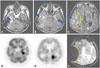
Fluid attenuated inversion recovery images (FLAIR) showed suspicious high signal intensity lesion on left medial temporal lobe and hyperintense CSF on right temporal area (Fig. 1A, B, and C). Axial T1- and T2-weighted images showed no abnormality on corresponding same region. Whole-body positron emission tomography (PET) showed glucose uptake on left medial temporal lobe indicating limbic encephalitis. At first glance, hyperintense CSF lesion on right temporal lobe were thought to be more remarkable than left medial temporal lobe lesions, causing confusion to diagnosis. With PET showing glucose uptake on left medial temporal lobe, we could suspect limibic encephalitis.
The term "hyperintense CSF" is used to describe failed suppression, or hyperintensity, of CSF on FLAIR of brain.1 Pathologic conditions associated with hyperintense CSF includes subarachnoid hemorrhage, meningitis, and acute infarction following gadolinium administration.1 Some non-pathologic conditions showing hyperintense CSF are images obtained during oxygen inhalation, CSF flow-related artifact.1 Gradient echo imaging showed magnetic susceptibility artifact matching hyperintense CSF seen in FLAIR image (Fig. 1). We reached an agreement that the lesion on FLAIR image is an artifact in the process of nulling CSF signal during acquiring FLAIR image. Knowledge of pathologic conditions of hyperintense CSF needed for correct diagnosis.
 XML Download