

 PDF
PDF ePub
ePub Citation
 Print
Print


INTRODUCTION
Alzheimer's disease (AD) can damage several central nervous structures associated with autonomic dysfunction, including hypothalamus, locus ceruleus, limbic system, insular cortex, and the lower brainstem.1234 AD patients also exhibit various circadian autonomic patterns, including circadian body temperature dysfunction,56 sleep-wake alternation,7 and disturbed day-night blood pressure (BP) dipping.8 However, autonomic involvement in AD is still poorly understood. Chronobiological changes with distinct parasympathetic impairment and increased sympathetic function have been reported in AD.91011 However, other studies have found no change in the short-term and long-term circadian BP or heart rate variability in AD.1213
Clinical stages preceding AD have been identified. Epidemiological studies have proposed that cohorts at risk for developing AD is comprised of older individuals with mild cognitive impairment (MCI) characterized by cognitive decline intermediate between normal aging and dementia.1415 In addition, recent studies have further proposed that subjects with memory impairment and normal cognitive performance [subjective memory impairment (SMI)] are an at-risk of developing AD.161718 The model tested in this study is based on the hypothesis that autonomic dysfunction in Alzheimer's pathology will become worse in the cascade of Alzheimer's spectrum. The parasympathetic and sympathetic functions of heart can be characterized by evaluating time-domain heart rate variability and BP variability. In this study, we determined whether heart rate and BP variability were associated with AD. We compared heart rate and BP variability among AD patients, normal controls, patients with SMI, and those with MCI.
METHODS
Subjects
This study was approved by the ethics committee of Seoul St. Mary's Hospital. Each patient gave written informed consent before participating. Patients with newly diagnosed AD from the Department of Neurology of Seoul St. Mary's Hospital were enrolled in this study. They were recruited using clinical diagnostic criteria for MCI and AD as well as comprehensive neuropsychological tests addressing five cognitive domains including attention, memory, language, visuospatial function, and frontal/executive function.19202122 SMI was diagnosed when there was complaint of memory decline although there was no objective neuropsychological explanation in neuropsychological tests.2324 A total of 25 healthy elderly subjects without any neurological or psychiatric history were included as controls. Patient data such as age, sex, education status, disease duration, history of hypertension, diabetes mellitus, or dyslipidemia, and smoking status were collected. Laboratory tests that may reflect cognitive function were performed, including homocysteine, vitamin B12, and folate. Patients' thyroid, hepatic, and renal functions were also assessed. Patients with any laboratory abnormalities were excluded from the study. Those with atrial fibrillation, atrial flutter, frequent ectopies, and those who were on permanent pacemakers were also excluded. General cognitive status and dementia severity were evaluated using the Korean version of Mini-Mental Status Examination. BP and heart rate monitoring were performed simultaneously after discontinuing any antihypertensive medications for 7 days. During the period, no serious clinical problem was observed.
Heart rate variability analysis
In all subjects, 24-h Holter monitoring was conducted using an Aria recorder. The recordings were subsequently analyzed using Impresario Solo system (Delmar Reynolds, Hertford, UK). While being monitored, participants lived their normal lives and recorded their activities in diaries. They also provided information on hours of rest at night. Time domain analyses of heart rate variation (HRV) were also conducted for all participants.
The time domain HRV analysis was derived from a direct measurement of normal-to-normal R-R over a 24-hour period, including day time (from 8 am to 9 pm) and night time (from 11 pm to 6 am). The following time domain HRV parameters were calculated: mean of all normal RR intervals (mRR), standard deviation of N-N intervals (SDNN), mean of 5 min standard deviation of NN interval durations (SDNN index), root mean square successive NN deltas (RMS-SD), percent of difference between adjacent normal RR intervals greater than 50 ms (pNN50) and standard deviation of 5 min means of NN intervals (SDANN).25 The first three parameters were used to reflect sympathetic and parasympathetic functions. The next two parameters were used because they were associated with parasympathetic function. The last parameter was used because it was related to sympathetic function alone.25
Blood pressure variability analysis
Automated 24-hour BP recording instruments (Mobil-O-Graph NG, I.E.M., Stolberg, Germany) were used to measure BP every 15 min during the daytime and every 30 min at night. The following parameters were evaluated: average systolic and diastolic BP and heart rate for the day time, night time, and 24-h periods. Any nocturnal falls in BP and heart rate were calculated as percent changes with daytime and nighttime mean values. Subjects with less than 10% nocturnal fall in mean BP were considered "non-dippers" and those with pathological increase in nocturnal BP were categorized as "reverse dippers".2627 Nocturnal hypertension was defined according to the 2007 European Hypertension Society/European Cardiology Society guidelines (i.e., average nighttime BP ≥120/70 mm Hg).2829 BP variability was calculated as within-subject standard deviation of mean systolic blood pressure during the day time and night time.303132
Data analysis
One-way analysis of variance or analysis of covariance (with Bonferroni post-hoc testing) were used to compare means among groups. Pearson's χ 2tests were used to compare the frequencies of categorical variables. Statistical software SPSS 22.0 (SPSS Inc., Chicago, IL, USA) was used for all analyses. Statistical significance was considered when p value was less than 0.05.
RESULTS
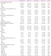
Of the 103 subjects (28 males and 75 females; mean age ±SD: 69.9±8.9 years; mean education duration: 11.4±5.4 years), 25 were normal controls, 17 had SMI, 24 had MCI, and 37 had AD. These groups had similar baselines of sex distribution, symptom duration, education, hypertension, diabetes mellitus, and cigarette smoking (Table 1). The mean age of AD patients was higher than that of the controls and subjects with SMI. However, AD patients had lower Mini-Mental State Examination scores than SMI patients, MCI patients, and controls (Table 1).
AD patients also had higher pulse pressures than the controls. Nocturnal BP dipping in patients seemed to be reversely associated with decreasing cognitive status in the order of AD, MCI, SMI, and normal controls (Table 2). The frequency of non-dippers was not significantly different among groups. However, nocturnal BP reverse dippers were more prevalent in the AD group than those in other groups. There was no significant difference in daytime and nighttime BP variations among groups. However, the AD group had greater variations in RMS-SD, SD of successive NN delta, and pNN50 domains compared to those in the other groups, especially at night. These variations reflected parasympathetic dysfunction during nighttime and at asleep (Table 3).
DISCUSSION
In line with the Braak schema for pathogenetic sequences in AD, two main brain structures (insular cortex and brainstem) can be involved progressively in autonomic controls.33 A deficit in central cholinergic function could likely lead to autonomic dysfunction.34 These structures might be affected by neurodegeneration in a "preclinical stage". The autonomic dysfunction might be present before the onset of clinical symptoms of dementia. This raises the question of whether cardiovascular autonomic impairment is related in stepwise pattern to cognitive spectrum of AD.35 In this study, we found that diurnal sympathetic and parasympathetic cardiac variability were significantly disturbed in AD patients compared to healthy controls, but not in MCI or SMI patients.
Numerous studies have reported that patients with AD have experienced fluctuations in their circadian functions, include arousal and sleep architectures, core body temperature, and hormones such as cortisol and melatonin.567 This suggests that AD will not only affect cognitive function, but also endocrine-vegetative function that mediates the circadian rhythmicity.36 However, studies on circadian BP and heart rate variability in AD have produced contradictory results. Some reports have suggested that there is a loss in circadian periodicity in most AD patients, while others have found that BP and heart rate chronological rhythmicity are not abolished in AD patients.910111213
In this study, AD patients experienced blunted day-night dips in their mean BP compared to that of SMI patients or controls. Although the prevalence of non-dipping was not different between the control group and patient groups, there were tendencies of higher prevalence of nocturnal hypertension and reverse dippers in the AD group than that in the control group. In addition, 24-h Holter monitoring revealed disruption or enhancement in the parasympathetic function of AD patients. If the autonomic manifestations of clinical "AD" are AD-specific, these results suggest that such autonomic manifestations are also stage-specific. They might be limited to AD's clinical stages.
In the general population, abnormal BP and heart rate variability have been associated with cardiovascular morbidity and mortality. They are also independent risk factors of target organ damage, including renal impairment and cognitive dysfunction.30373839 Cardiovascular autonomic failure related to generalized cholinergic dysfunction is a potential peripheral marker of AD.1 In AD patients, relative depression of parasympathetic activity has been reported to be associated with cognitive dysfunction.91011 However, increased or unchanged sympathetic functions have also been reported in AD.111213 Our results added more information to the debate involving the association between chronobiological BP, heart rate changes, and cognitive dysfunction in mild AD. Increased parasympathetic dysfunctions at night and flattened day-night BP dips reflecting suppressed sympathetic function may be related to sleep-arousal system disorder. Such result might be an indirect sign of disturbed integrity of the sleep-wake cycle in mild AD.
In this study, pulse pressure was significant greater in AD patients compared to that of controls. Higher pulse pressure in the elderly is a recognized marker of increased arterial stiffness, atherosclerosis, white matter change, cognitive dysfunction, and ultimately dementia.4041 Increased pulse pressure in the elderly resulting from increased systolic pressure and decreased diastolic pressure may be related to hypoperfusion-related ischemia and alteration in white matter integrity.
This study had several strengths. One major strength of this study was its inclusion of patients with relatively early AD who had never taken cholinesterase inhibitors. Cholinesterase inhibitors can reduce heart rate variability. They can also affect sleep and arousal.42 Because the participants in this study were never exposed to these medications, our results were less likely to be affected by such a confounder. Therefore, we can suggest that the association occurred independently of such treatment. Another strength of this study was its enrollment of patients with a comprehensive spectrum of memory disorders, including SMI and MCI. Although clinical diagnosis has always been criticized, we minimized selection bias by only including patients who had fulfilled the clinical diagnostic criteria for each disease.1920212324
Although this study had several strengths, it also had several limitations. First, the relationships among SMI, MCI, and AD were not well understood. Even clinical AD phenotype's relationship to AD pathology was unclear given that the vast majority of neuropathology with AD did not have evidence of dementia. In addition, up to 20% of clinically diagnosed AD cases may lack amyloid-deposition.4344 Second, this study enrolled relatively mild and first-visit patients. Therefore, the autonomic function test results might have been skewed. Third, the proportion of hypertension in the AD group was higher than that in the control group. Most hypertensive patients were taking anti-hypertensive medications. Therefore, these data must be carefully interpreted as anti-hypertensive medications can contribute to BP and heart rate variability. In addition, we did not assess any other mechanisms that might have played a pathophysiological role in BP and heart rate variability. Finally, the interpretation of heart rate variability as a direct quantitative marker of cardiac autonomic dysfunction is controversial. No sensitive measurement methods were determined.
In summary, our results revealed that diurnal sympathetic and parasympathetic cardiac variability were significantly disturbed in cholinesterase-naïve AD patients. Further studies are needed to clarify the difference between mild AD patients and advanced AD patients and to assess the longitudinal changes during the progress of the disease.


 XML Download