

 PDF
PDF ePub
ePub Citation
 Print
Print


INTRODUCTION
Idiopathic Parkinson's disease (PD) is characterized by various motor symptoms, including bradykinesia, rigidity, postural instability, and resting tremors. As a common neurodegenerative disorder, PD affects 1% to 2% of the general po-pulation over 65 years of age.1 In addition to major motor symptoms, a variety of non-motor symptoms appear throughout the course of PD.2 These non-motor symptoms comprise a variety of cognitive, neuropsychiatric, sleep, autonomic, and sensory disorders. Among different non-motor symptoms, cognitive disorder such as dementia is a frequent manifestation of advanced PD.3 Dementia is the most devastating non-motor feature that causes severe decline in quality of life. It increases caregiver burden and mortality as well as institutionalization. The prevalence of dementia in PD ranges from 24% to 50%. PD dementia accounts for about 3-4% of all dementia in the population.3 Therefore, identifying biomarkers that can predict the development of PD dementia is important as they could help clinician select high-risk patients for appropriate counseling and plans for future treatment.
Although the pathophysiologic mechanisms involved in the development of dementia in PD remain unknown, post-mortem and in vivo studies as well as epidemiological and pre-clinical studies have suggested that neuro-inflammation may play an important role in the pathogenesis of PD, especially in the non-motor symptoms such as cognitive impairment and depression.456 In addition, it has been reported that anti-inflammatory agents such as non-steroid anti-inflammatory drugs and cyclooxygenase 2 inhibitors may delay the onset or slow the progression of Alzheimer's disease (AD), supporting the role of neuro-inflammation in the development of dementia in PD patients.167
High sensitivity C-reactive protein (hs-CRP) is one of the most widely studied biomarkers of systemic inflammation. It has been reported that hs-CRP is associated with cognitive impairment in AD patients.78 Furthermore, many previous reports have suggested that high concentrations of hs-CRP are associated with increased risk of cardiovascular disease, stroke, and cognitive impairment including dementia.678 However, despite that fact that hs-CRP is the most studied biomarker of systemic inflammation, few studies have evaluated the association between hs-CRP levels and the progression of cognitive decline in PD. Therefore, the clinical implication of hs-CRP in the development of dementia in PD patients is poorly defined. Consequently, the objective of this study was to clarify the clinical value of elevated hs-CRP levels in the development or progression of dementia in PD.
METHODS
A total of 215 consecutive PD patients [112 PD without dementia (PD-D) and 103 PD wih dementia (PD+D)] admitted to the Movement Disorder and Parkinson's Disease Unit of the Department of Neurology at our hospital between October 2012 and May 2015 were enrolled in this study. Ninety-four control subjects who visited our hospital were also enrolled in this study. The control subjects did not have any history or symptoms of PD, memory impairment, or other cognitive impairment, according to the results of dementia screening questionnaire. In addition, the controls did not have history of other neurological diseases such as head trauma, epilepsy, cerebrovascular disease, or brain surgery. This study was approved by the local ethics committee. All participants provided written informed consent.
All participants in this study had detailed medical history, physical and neurological examinations, neuropsychological assessments, laboratory tests, and magnetic resonance imaging of the brain. The medical and neurological histories of patients were obtained from patients and their family members or other caregivers. All PD patients were diagnosed using the United Kingdom PD Society Brain Bank Clinical Diagnosis Criteria for PD, 123I-n-fluoropropyl-2bcarbomethoxy-3b-(4-iodophenyl) nortropane (FP-CIT) positron emission tomography (PET) scans, and the Diagnostic and Statistical Manual of Mental Disorders 4th edition revision (DSM-IVR) criteria for dementia.910 All PD patients exhibited reduction in their striatal dopamine transporter uptake levels as determined by FP-CIT PET. All PD patients did not have any history or symptoms of memory impairment or other cognitive dysfunctions according to the dementia screening questionnaire or any cerebrovascular lesions according to neuroimaging results. Patients with clinical dementia rate (CDR) of ≥0.5 and mini-mental status examination (MMSE) score of ≤24 points were included in the PD+D group of this study. In all PD patients with dementia, the onset of PD preceded the development of dementia by at least 12 months. The dose and duration of levodopa intake were determined for each patient by chart review and verified by self-reporting of each patient in all cases. The severity of motor impairment in PD patients was evaluated according to the staging system of Hoehn and Yahr.11
We excluded patients who displayed markedly fluctuating cognition with pronounced variations in attention and alertness or recurrent vivid hallucinations suggesting the presence of dementia with Lewy bodies. We also excluded patients who took medications (e.g., anticholinergic agents) that could influence cognition and memory, patients who presented any signs of atypical Parkinsonism, and those who fulfilled the DSM-IVR criteria for delirium or amnestic disorders and depressive disorders.1012 In addition, we excluded patients with secondary causes of Parkinsonism (e.g., Wilson's disease, neuroleptic drug users, and psychiatric diseases) that could possibly impede the safety during the study. None of the subjects recruited in this study had a history of recent infection, surgery, trauma within the previous month, cardiovascular disease, cerebrovascular disease, malignancy, or use of NSAIDs as these can affect serum hs-CRP levels. We also assessed the presence of hypertension, diabetes mellitus (DM), hypercholesterolemia, and cigarette smoking based on patients' medical histories and laboratory findings as these could influence hs-CRP levels. Hypertension was defined as a systolic blood pressure of 140 mm Hg, a diastolic blood pressure of 90 mm Hg, and/or current use of anti-hypertensive agents. DM was defined as a history of fasting glucose level of 110 mg/dL or current use of hypoglycemic agents. Hypercholesterolemia was defined as total cholesterol concentration of 220 mg/dL or current use of lipid-lowering agents. Cigarette smoking was considered to be present if the patient reported smoking at least once within the previous five years.
Serum hs-CRP was measured routinely for all patients and control subjects. Venous blood samples from all subjects were collected in tubes containing ethylenediaminetetraacetic acid. These blood samples were separated immediately by centrifugation at 3000 rpm for 10 minutes. Separated sera were kept at -70℃ until laboratory evaluations. Laboratory data were evaluated by an examiner who was blinded to the clinical details and patient information.
Statistical analyses were performed using SPSS software package version 18.0 (SPSS Inc., Chicago, IL, USA). Results were expressed as mean±standard deviation. Kruskal-Wallis test was used to compare continuous variables between the PD group and the control group. Mann-Whitney U test was used to compare continuous variables between PD+D and PD-D groups. A Pearson's chi-square test was used to compare categorical variables. Statistical significance was considered when p value was less than 0.05.
RESULTS
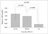
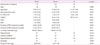
The demographic and clinical characteristics of the study populations are summarized in Table 1. Age, gender, and education level were not significantly (p>0.05) different between the PD group and control subjects. Atherosclerosis risk factors such as hypertension, DM, hypercholesterolemia, and cigarette smoking were not significantly (p>0.05) different either between the PD-D group and the PD+D group. Serum hs-CRP levels in the PD-D group and the PD+D group were 1.76±3.62 mg/dL and 1.44±2.78 mg/dL, respectively, which were significantly (p=0.02) higher than that (0.41±1.06 mg/dL) in the control group (Table 1). However, the levels of hs-CRP were not significantly (p>0.05) different between the PD-D group and the PD+D group (Fig. 1).
The MMSE score was 16.23±3.87 in the PD+D group, which was significantly (p=0.001) lower than that in the PD-D group (27.48±1.48) or in the control group (28.18±1.62). In addition, CDR and sum of box of CDR were significantly (p=0.001) different between the PD-D group and the PD+D group (CDR: 0.37±0.25 vs. 0.91±0.43; sum of box of CDR: 1.11±0.62 vs. 4.92±2.09). The duration of PD treatment in the PD+D group was 25.51±23.76 months, which was significantly (p=0.025) longer than that in the PD-D group (18.61±20.89 months). Daily levodopa dose in the PD+D group was 594.09±354.58 mg, which was significantly (p=0.005) higher than that in the PD-D group (459.83±266.54 mg). In addition, motor impairment in the PD+D group was more severe than that in the PD-D group (stage of 2.38±1.08 vs. 2.09±0.93, p=0.033). However, the duration of PD symptoms was not significantly (p>0.05) different between the PD-D group and the PD+D group.
DISCUSSION
Some protection against PD is offered by long-term antiinflammatory medications, although the pathogenesis of neurodegenerative diseases including PD and dementia remains unknown.2 Therefore, the possibility of an association between neuro-inflammation and PD pathogenesis has received strong support from recent epidemiological findings that chronic use of nonsteroidal anti-inflammatory drugs can reduce the risk of PD by about 45%.6131415
We evaluated the relationship between serum hs-CRP levels and the risk of developing dementia in PD patients. Our results revealed that serum hs-CRP levels were significantly higher in both PD-D and PD+D patients compared to control subjects. However, our study did not reveal a significant elevation in hs-CRP levels in PD+D patients compared to PD-D patients. Our results suggest that neuro-inflammation might contribute to the pathogenesis of PD occurrence. However, it does not significantly contribute to the development of dementia in PD patients.
Several lines of evidence from previous studies have suggest that neuro-inflammation is mediated mostly by activated microglial cells and peripheral immune cells, triggering deleterious events such as oxidative stress and cytokine-receptormediated apoptosis that can eventually lead to death of dopaminergic neurons and hence disease progression.46 In addition, it has been reported that the expression of pro-inflammatory cytokines such as tumor necrosis factor α, interleukin (IL)-1β, IL-2, and IL-6 in biological fluids (serum or cerebrospinal fluid) is increased in patients with PD.456 The hs-CRP plays a crucial role in human immune system. It has been widely considered to be a sensitive biomarker for systemic inflammation. CRP is mainly synthesized in the liver. However, post-mortem studies on patients with AD and intra-cerebral hemorrhage suggest that CRP can be produced from within the brain by neurons and glial cells.1617 In addition, an animal study has found that microglial cultures can produce CRP, suggesting that microglial cells may be the source of CRP in the central nervous system (CNS).18 These studies suggest that CRP levels in peripheral blood might mirror inflammation in CNS.161718 Several longitudinal and cross-sectional studies have reported that high hs-CRP levels in plasma or serum are associated with increased risk of cognitive deterioration and AD.78192021 A prior report has also indicated that CRP does not cross the blood-brain barrier (BBB) in trace amounts although it can increase paracellular permeability of the BBB when the blood CRP concentration exceeds the threshold required to impair the BBB function.22 The threshold dose of CRP can be easily reached during systemic inflammation or obesity. In other words, the interactions between CRP and BBB involve a two-phase process. Increased paracellular permeability occurring at a high dose of CRP will enable the entry of CRP into the CNS. The entry of CRP then induces reactive gliosis and impairs CNS function.22
Recently, Lindqvist et al.5 have evaluated the association between CRP levels in CSF and dementia in PD patients. In their study, the mean CRP level in CSF was significantly higher in PD patients with dementia compared to PD patients without dementia.5 In contrast to their results, our results revealed that the measured levels of hs-CRP were not significantly different between the PD-D group and the PD+D group, although the level of hs-CRP in both PD-D and PD+D groups was significantly higher than that in the control subjects. The discrepancy between their results and our results might be due to differences in study populations, such as age and gender distribution because they are important factors for the development of dementia in PD. We also thought that neuro-inflammation maybe have no role in the development of dementia in PD because loss of dopamine cell has been estimated to be 60 to 70 percent at the onset of symptoms in PD patients.23 In addition, a small number of PD dementia patients being evaluated in the previous study may account for such discrepancy.5 The study of Lindqvist et al.5 had a possibility of unbalanced statistical comparison analysis because the number of PD without dementia (n=76) was higher than the number of PD with dementia (n=16). Therefore, the finding of the previous study demonstrating that CRP levels in the CSF were significantly different between PD dementia patients and PD patients without dementia should be verified in additional studies.
An interesting finding of the present study was that the time span of treatment in the PD+D group was significantly longer than that in the PD-D group, although the duration of Parkinsonian symptoms was not significantly different between the two groups. In addition, the Hoehn & Yahr stage representing the severity of motor impairment in PD in the PD+D group was significantly higher than that in the PD-D group. Furthermore, PD+D patients were taking levodopa at a significantly higher dose compared to PD-D patients. These results suggest that PD+D patients had more advanced motor symptoms than PD-D patients. PD patients with dementia might have experienced a more rapid progression of Parkinsonian symptoms, considering that the symptom duration was not significantly different between the two PD groups. Hence, additional investigations on factors that contribute to rapid progression may be necessary to delineate the mechanism( s) underlying the development of dementia in PD patients.
There are several limitations of this study, including its relatively small sample size and a cross-sectional design. Therefore, future large-scale longitudinal studies are needed to verify our findings. In addition, our study was limited due to the lack of neuropathological information to allow confirmation of the presence of Lewy bodies because subjects enrolled in the present study were still alive at the time of the study. Furthermore, it was difficult to distinguish PD dementia patients from dementia patients with Lewy bodies based on clinical criteria, particularly in the early phases of the disease.
In conclusion, our study demonstrated that the levels of hs-CRP were significantly higher in PD patients compared to control subjects, which was consistent with the hypothesis that inflammatory responses might be involved in the pathogenesis of PD. However, hs-CRP levels were not significantly different between the PD+D group and the PD-D group, implying an absence of a meaningful relationship between peripheral inflammatory proteins and the development or progression of dementia in PD patients. This finding suggests that neuro-inflammation may not significantly influence the pathogenesis of dementia in PD patients. However, we were unable to clearly confirm the role of neuro-inflammation in development of dementia in PD patient because this study had a cross-sectional design with small subject size. In addition, serum hs-CRP cannot represent the entire neuro-inflammation. Therefore, large-scale longitudinal studies are required to clarify whether hs-CRP indeed has clinical significance in predicting dementia in PD patients.
 XML Download