

 PDF
PDF ePub
ePub Citation
 Print
Print


INTRODUCTION
The Korean orthographic system consists of both phonograms (Hangul) and ideograms (Hanja). Hangul is a phonetic alphabet comprised of consonants and vowels that are grouped together to form syllables that generally exhibit regular correspondences between graphemes and phonemes. On the other hand, Hanja is derived from complex Chinese characters with distinct meanings. In this respect, Hanja and Hangul are similar to Kanji (ideograms) and Kana (phonograms), respectively, of the Japanese language. Many Japanese language studies have described dissociations between the neural substrates involved in the reading of Kanji and Kana. For example, difficulty in Kana reading has been associated with lesions of the left angular gyrus, adjacent lateral occipital gyri, deep perisylvian temporoparietal area, and posterior superior temporal gyrus, whereas lesions involving the fusiform gyrus and left posterior inferior temporal cortex have been identified in individuals with difficulty in Kanji reading.1234
The results of some Korean studies on the dissociation between Hangul and Hanja reading have concurred with previous Japanese findings.567 However, the exact brain regions involved in the processing of Hangul and Hanja reading have not been firmly established.8910
The authors conducted serial functional magnetic resonance imaging (fMRI) to identify the areas of the brain associated with Hanja reading by investigating a patient exhibiting Hangul/Hanja reading dissociation after an acute ischemic stroke.
CASE REPORT
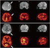
A 63-year-old right handed man was admitted because of memory impairment. His past medical history included hypertension and angina of ten years duration, for which he had been placed on regular antihypertensive medication. The patient denied a history of diabetes mellitus, episodes of stroke, or neuropsychological symptoms. He had been educated for 12 years and had learned Hanja at school. His wife said he had no problems reading or writing Hanja before admission. During neurological examinations, he was fully conscious and had a Korean Mini-Mental State Examination score of 22/30. Deficits were observed mainly in the domains of attention, calculation, and immediate memory recall. Interestingly, we found that he was unable to read or write Hanja, but could read Hangul. Hangul writing was partially impaired. In his wife's statement, he did not have any problems reading Hanja and Hangul before this stroke event. Conventional brain MRI revealed an infarct involving the left frontal and parietal lobe including the angular gyrus. Fluorodeoxyglucose positron emission tomography scans showed decrease uptake in these infarcted regions (Fig. 1). There were no additional hypometabolic regions. fMRI and simple language tests were performed two weeks, six weeks, and six months after stroke onset to identify activated brain regions associated with Hanja and Hangul reading in each scan. An implicit reading task was performed during each fMRI scan. Three sets of test were prepared for the fMRI experiment. Each set included ten two-syllable Hanja and Hangul words and each test included 20 words: 10 Hanja and 10 Hangul words, and a total of 30 Hanja and Hangul words were tested in one visit (Supplementary Table 1 in the onlineonly Data Supplement). After the fMRI, an independent reading and writing task was performed to obtain response times and hit-rates. Written consent was obtained from the study subject, and the study protocol was approved by the Institutional Review Board (GIRBD 0024-2012) of Gil Medical Center. Detailed fMRI scan protocol and analysis method are described in the Supplementary data (Supplementary data in the online-only Data Supplement).
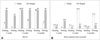
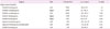
Hit-rates for Hangul reading were perfect (30/30) at every session for language tasks. The mean response time for Hangul reading improved from 2.8 seconds per letter [standard deviation (SD) 2.1 sec] to 1.03 seconds per letter (SD 0.2 sec) over the 6-month experimental period. In contrast, Hanja reading, Hanja writing and Hangul writing was markedly impaired initially. However, the number of correct hits increased from 8 to 28, and the mean response time for Hanja reading shortened from 3.3 to 1.5 seconds after six months. Hangul agraphia also improved to almost normal; however, Hanja agraphia persisted. The overall results are provided in Fig. 2.
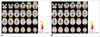
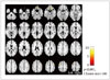
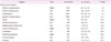
fMRI scans during Hanja reading showed activation of the occipital regions bilaterally including the lingual gyrus, extending to the parietal cortex bilaterally from the prefrontal area, and both insula at the initial time point and six months after the onset of symptoms (Fig. 3, Table 1 and 2). Although the fusiform gyrus was partially activated bilaterally (Fig. 3A, Supplementary Table 2 in the online-only Data Supplement), the patient could not read Hanja. At six months after symptom onset, his ability to read Hanja greatly improved; however fMRI revealed no significant changes in the fusiform gyrus region (Fig. 3B). Instead, both middle frontal gyri showed significantly greater activation after six months (Fig. 4, Table 3).
DISCUSSION
Several models have been proposed to explain the mechanisms related to reading and brain localization. The dual route of phonogram-ideogram processing is one such model, which has been predominantly studied for Japanese.1
Two different orthographic systems exist in the Japanese language [i.e., Kanji (ideogram) and Kana (phonogram)]. Although the correlation between Kanji and Kana is not straightforward, it is generally believed that the anatomical substrates mediating ideograms and phonograms differ. Thus Japanese alexia patients sometimes show dissociative disturbances when reading Kanji or Kana. Lesion studies have shown that angular gyrus involvement more frequently leads to difficulty reading Kana, whereas patients with lesions in the posterior inferior temporal lobe, including the fusiform gyrus, are likely to develop difficulty reading Kanji. These studies also suggest that the anatomical pathways mediating ideograms and phonograms differ, that is, the ventral pathway (posterior portions of the middle and inferior temporal gyri) is involved in reading ideograms241112 and the dorsal pathway (inferior parietal lobe) is used in reading phonograms.1313 Similar findings have been reported on the dissociative reading disturbances seen in lesion case studies regarding Korean orthography. Two patients with Hanja alexia had a lesion in the left posterior inferior temporal lobe,67 while one case of Hangul alexia was associated with a lesion in the inferior parietal lobule, insula and cingulate gyrus.7 Another fMRI study in Korean speaking volunteers showed that the right fusiform gyrus and adjacent temporo-occipital region seem to be more specifically involved in processing Hanja script.8
Our patient also showed a dissociative disturbance between reading Hanja and Hangul. MRI scans showed two distinct ischemic lesions in the left middle frontal gyrus and left inferior parietal lobe with preservation of the posterior inferior temporal cortex, which is supposed to be the anatomical substrate for ideogram reading. Nevertheless, he initially presented with severe Hanja alexia and normal Hangul reading. The areas with increased signal on fMRI during Hanja reading were mainly occipital and frontal regions bilaterally rather than the inferior occipito-temporal region, such as the fusiform gyrus. In addition, as the patient's Hanja alexia improved, activation increased in both middle frontal gyri.
These findings, 1) initial profound Hanja alexia with a destructive lesion involving the left frontal lobe and 2) improved Hanja alexia with increased fMRI activation in both middle frontal gyri, suggest the possibility that the frontal lobe, as well as the fusiform gyrus, could contribute to mediating Korean ideogram reading.
Differences exist between the usage, exposure rate, and age of acquisition of the Korean and Japanese ideographic systems. In contrast to Japanese orthography which heavily incorporates the usage of Kanji, the Korean system depends less on Chinese derived characters, and most Korean words and sentences can be communicated without the use of any ideograms (Hanja). Also the same ideograms in Japanese can be pronounced in different ways, which is not the case in Korean.57 For these reasons, the two language systems may show different patterns of brain activation when ideograms and phonograms are read.
It is difficult to designate the exact location associated with Hanja alexia in this study, due to the fact that two ischemic lesions were concomitantly present. An fMRI study conducted on normal Korean subjects revealed strong activation in the left lateralized middle frontal cortex during Chinese character reading.910 Chinese investigators observed a wide area of activation associated with Chinese letter reading including the left frontal and temporal cortices, the right visual system including the fusiform gyrus, the right parietal lobe and cerebellum.141516 Different regions where associated with reading Chinese characters by Korean (Hanja) and Chinese native speakers; however, both groups showed activation in the left frontal area.
Taken together, these studies indicate that different cerebral regions might be associated with reading Chinese characters by Korean (Hanja), Japanese (Kanji) and Chinese native speakers. In the case of Korean orthography, different results have been reported regarding the areas involved in processing ideograms, which are similar to Japanese678 and Chinese910 populations. Our case study supports that the frontal lobe might play a role in reading Korean ideograms.
Increasing activation in the bilateral middle frontal gyri in parallel with Hanja reading improvements may also represent the activation of language restorative processes from perilesional areas or compensatory processes from contralateral neural circuits post stroke.17 However, because this study was conducted on one subject, we cannot comment on the statistical significance of our findings.
Our study has some limitations. First, the presence of two lesions in the left cerebral hemisphere makes it difficult to conclude which is causative of his dissociative alexia. The analysis of additional subjects with single lesions associated with ideographic alexia would be needed to answer these questions. Second, there were no fMRI data on a control group to determine the normal substrates dissociating the two systems. Third, detailed error analysis of mistaken words was not fully performed. However, our subject showed either fully normal reading or no responses attempted and error analysis was inadequate. Finally, functional and neurophysiological changes after ischemic stroke, for example diaschisis, need to be considered when interpreting the fMRI results.
In conclusion, we found that not only fusiform gyrus, but also the frontal lobe may be involved in reading Hanja (Korean ideograms) after analyzing serial fMRI scans of a patient presenting with dissociative Hanja alexia after an acute ischemic stroke.

 XML Download