

 PDF
PDF ePub
ePub Citation
 Print
Print


INTRODUCTION
Although Parkinson's disease (PD) is primarily characterized by motor symptoms, its association with dementia has been increasingly reported in a number of epidemiological and pathophysiological studies.1 PD patients have a six-fold increased risk of developing dementia when compared with individuals without PD.2 The problems related to PD dementia (PDD) include higher care costs, increased disability and mortality, and reduced quality of life.345 Thus, cognitive decline and subsequent development of dementia have been widely considered as a major aspect of PD.1
For the treatment of PDD, cholinesterase inhibitors have been tested since patients with PDD showed substantial cholinergic deficits.6 Although rivastigmine improved cognitive function and behavioral symptoms, not all PDD patients responded to it and high rates of adverse events such as nausea, vomiting, and tremor were reported.7 On the other hand, impairment in glutamatergic activity was found in dementia with Lewy bodies.8 Based on this rationale, previous studies in PDD patients have investigated the efficacy of the N-methyl D-aspartate (NMDA) receptor antagonist memantine that alleviates the toxic effects of increased levels of the excitatory neurotransmitter glutamate, and have provided evidence of significant treatment advantages including improved attention and episodic recognition memory.91011 However, the follow-up period in these studies was 24 weeks and changes in brain structure or function were not assessed with in vivo neuroimaging. Our group recently published a single photon emission computed tomography (SPECT) study which showed that treatment of PDD patients with memantine for 12 months prevented deterioration of not only cognitive and neuropsychiatric symptoms but also brain perfusion.12
On the basis of this background, the current prospective study aimed to investigate the changes in brain perfusion, severity of dementia, cognitive performance, and neuropsychiatric symptoms in PDD patients after 18 months of memantine administration, using 99mTc hexamethylpropylene amine oxime (HMPAO) SPECT and neuropsychological assessment.
METHODS
Participants
Patients with PDD who had no history of head trauma, epilepsy, stroke, and other neurological or psychiatric disorders were recruited at Incheon St. Mary's Hospital (Incheon, Korea). The onset of PD preceded the development of dementia by at least 12 months in all patients. Participants were excluded if they had remarkable fluctuations in cognitive functions; recurrent vivid hallucinations; any signs of atypical parkinsonism; delirium; amnestic disorders; or cerebrovascular lesions on magnetic resonance imaging. Patients who were taking psychotropic medications were also excluded. Healthy control participants without major medical conditions were matched for age and gender with the patient group.
The initial dose of memantine was 5 mg in the morning and it was gradually increased to 20 mg (10 mg in the morning and 10 mg in the evening) from week 4. This maintenance dose remained unchanged throughout the study period. Safety assessments included physical and neurological examinations, vital signs, electrocardiography, laboratory tests (blood chemical values, hematologic tests, and urinalysis), and adverse events.
Written informed consent was obtained from all participants and the study protocol was approved by the Research Ethics Committee.
Clinical assessment
Clinical assessments were conducted at baseline and follow-up. Assessment of the medical history and physical and neurological examinations were performed by board-certified neurologists. PDD was diagnosed according to the United Kingdom Parkinson's Disease Society Brain Bank Clinical Diagnostic Criteria for PD, and Diagnostic and Statistical Manual of Mental Disorders-4th edition criteria for dementia.13 Fluorinated N-3-fluoropropyl-2β-carbomethoxy-3β-(4-iodophenyl) nortropane positron emission tomography (18F-FP-CIT PET) was also used for the diagnosis of PD.
The severity of PD symptom was evaluated with the Hoehn-Yahr Scale.14 The Clinical Dementia Rating (CDR)15 and Global Deterioration Scale (GDS)16 were used to determine the overall severity of dementia. Global cognitive function and neuropsychiatric disturbances were assessed with the Mini-Mental State Examination (MMSE)17 and Neuropsychiatric Inventory (NPI),18 respectively.
Image acquisition and analysis
Brain SPECT scans were performed at baseline and follow-up. The participants were injected intravenously with 1110 MBq of HMPAO in a dark and quiet room, while lying supine with eyes open. After 40 minutes, images were acquired with a dual-head gamma camera (NM640, GE Healthcare, Milwaukee, WI, USA) equipped with a low-energy, fan-beam collimator. All images were attenuation corrected and reconstructed in a 128×128 matrix with a voxel size of 3.9×3.9×3.9 mm (field of view=240 mm) using filtered back projection.
Statistical Parametric Mapping 8 (SPM8; Wellcome Department of Cognitive Neurology, Institute of Neurology, London, UK) was used for image processing and statistical modeling. All images were spatially normalized to the SPM SPECT template (Montreal Neurological Institute, McGill University, Montreal, Canada) and resliced to a voxel size of 2×2×2 mm3. The binary brain mask in SPM8 was applied to remove extracerebral signal. The images were then smoothed with a 16 mm full-width half-maximum isotropic Gaussian kernel.
Two-sample t-test was used to investigate the differences in regional cerebral blood flow (rCBF) between the patient group and healthy controls at baseline. Perfusion scaling was performed using the reference cluster normalization that provides a substantial increase of statistical power in neurodegenerative diseases.19 In brief, a standard global mean normalized analysis was conducted to detect rCBF increases in the patient group using a threshold of t>2.0.20 The mean values of raw rCBF were extracted from each image using MarsBar toolbox version 0.44 (http://marsbar.sourceforge.net/) and used as a scaling factor for the detection of rCBF decreases in the patient group. The statistical threshold was set at family-wise error (FWE) corrected p<0.05 at both voxel and cluster levels. Voxel-wise paired t-test was performed to analyze the changes in rCBF between baseline and follow-up in the PDD group using the same procedure and statistical threshold described above.
Statistical analysis
Differences in age and gender distribution between PDD patients and healthy controls at baseline were assessed with independent t-test and chi-square test, respectively. Changes in MMSE, CDR, sum of box of CDR, GDS, and NPI scores between baseline and follow-up in the patient group were evaluated with paired t-test or Wilcoxon signed-rank test. A two-tailed p value of less than 0.05 was considered statistically significant. All analyses were conducted with Stata version 13.1 (StataCorp., College Station, TX, USA).
RESULTS
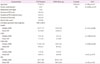
A total of 15 patients with PDD and 11 healthy participants were recruited into this study. Demographic and clinical characteristics of the participants are presented in Table 1. At baseline, the two groups did not differ in age (t=1.50, p=0.15) and gender ratio (χ2=0.001, p=0.97). The mean interval between baseline and follow-up was 1.5±0.9 years. In the patient group, changes in MMSE (z=-0.51, p=0.61), CDR (z=-1.00, p=0.32), sum of box of CDR (z=-0.69, p=0.49), GDS (t=0.27, p=0.79), and NPI (t=1.25, p=0.23) were not significant at follow-up.
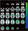
Results from the SPM analysis at baseline are demonstrated in Fig. 1. Two clusters with decreased rCBF were found in the PDD group. The first cluster encompassed the cerebellum and most of the cortical and subcortical areas including the temporal occipital fusiform cortex, postcentral gyrus, and insula (peak voxel: t=8.55; voxel level FWE-corrected p<0.001; x, y, z coordinates=-28, -60, -12; cluster size=27786 voxels). Hypoperfusion in the left lingual gyrus was also found in the patient group (peak voxel: t=7.83; voxel-level FWE-corrected p<0.001; x, y, z coordinates=-10, -98, 0; cluster size=809 voxels). However, areas with increased rCBF were not detected. In addition, in the comparison between baseline and follow-up in the patient group, significant changes in rCBF were not found.
DISCUSSION
The current brain perfusion SPECT study investigated the changes of rCBF in PDD patients after administration of a NMDA receptor antagonist for approximately 18 months. We compared the differences in rCBF between the patient group and healthy controls at baseline, and then analyzed the changes in rCBF at follow-up in the PDD group. Changes in cognitive function, severity of dementia, neuropsychiatric symptoms were also examined.
At baseline, the patient group showed rCBF decreases in most of the cortical and subcortical regions and cerebellum when compared to healthy controls. These results were in agreement with a previous study in demented PD patients that revealed significant perfusion decrements in all cortical regions. 21 Moreover, extensive metabolic deficits in the frontal, temporal, parietal, and occipital lobes were found in PDD patients. 22 Although other reports have suggested rCBF reduction mostly in the parieto-occipital cortex,2324 the inconsistency may be due to different imaging modalities or analytic strategies. In particular, the reference cluster approach used in our SPM analysis is more advantageous than SPM's default global normalization in detecting hypoperfusion associated with neurodegenerative process.19 This technique allowed us to identify large areas of hypoperfusion in spite of the stringent statistical threshold.
The PDD group did not demonstrate significant changes in cognition, dementia severity, neuropsychiatric symptoms, and brain perfusion after treatment with memantine for 18 months. These findings extend the results of a previous 12-month trial. 12 Several longitudinal studies have demonstrated poor prognosis over time in PDD patients.1 However, our results indicated that memantine therapy may be efficacious in delaying the clinical deterioration of dementia-related symptoms including cognitive decline and neuropsychiatric disturbances. The possible neuroprotective mechanism of memantine has been suggested in a previous animal study. Memantine markedly increased the expression of brain-derived neurotrophic factor (BDNF) and its receptor trkB mRNAs in a widespread area of rat brain.25 Interestingly, reduced levels of neurotrophins including BDNF have been reported in the nigrostriatal dopaminergic system of PD patients2627 and in the hippocampus of Alzheimer's disease patients.2829 Furthermore, administration of exogenous BDNF promoted the survival or dopaminergic neurons in animal models of PD.3031
Potential limitations of this study include a small sample size and a lack of comparative group at follow-up. In addition, the diagnostic criteria for PDD in our study have not yet been validated. However, it is still challenging to distinguish between PDD and dementia with Lewy bodies based on clinical criteria. We attempted to reduce this bias by only including patients who met the two sets of diagnostic criteria while simultaneously showing a reduced dopamine uptake in the striatum on 18F-FP-CIT PET.
Despite these limitations, we found diffuse hypoperfusion in the cortical, subcortical, and cerebellar regions in the PDD patients using the recently developed count normalization technique. More importantly, the severity of dementia, cognitive performance, neuropsychiatric symptoms, and brain perfusion were not significantly changed after 18 months of memantine treatment. Taken together, our findings implicate that memantine may delay the progression of brain perfusion deficits and clinical symptoms of PDD in the long term. Further extended prospective studies with larger sample sizes over longer periods are warranted to confirm and validate our preliminary results.
 XML Download