

 PDF
PDF ePub
ePub Citation
 Print
Print


INTRODUCTION
Sleep disturbances are common in both individuals who are aging normally and patients with Alzheimer's disease (AD). It has been reported that approximately 19–44% of AD patients are affected by sleep disturbances.123 Disrupted sleep in AD patients is a significant physical and psychological burden to not only patients but also caregivers and often cited as a major cause for institutionalizing dementia patients.456
While sleep-related problems such as daytime sleepiness and fragmented nighttime sleep occur with normal aging, these problems occur more frequently and tend to be more severe in patients with AD.7 Previous studies have demonstrated that sleep disturbances in AD patients are associated with changes in sleep architecture including sleep phase delay,8 increased nighttime awakenings,9 decreased slow-wave sleep and rapid eye movement sleep,910 and disrupted stage 2 sleep.11 Moreover, sleep disturbances were also associated with increased memory and functional impairment and more rapid cognitive decline in patients with AD.31213
The fundamental mechanisms underlying the development and maintenance of sleep disturbance in AD patients are complex and poorly understood. It may be attributed to AD-related damage in the brain regions or neuronal pathways that are involved in sleep regulation, thereby, exacerbating age-related sleep changes.7 In particular, it has been suggested that brainstem areas and pathways responsible for sleep-wake cycles and cortical areas that generate slow-wave activity during sleep are affected by degenerative changes associated with AD.14 Furthermore, these suspected mechanisms interact with and are influenced by a variety of physical, psychiatric, demographic, and environmental factors.7
While a large number of neuroimaging studies have been performed on AD patients, only a small number of studies have employed neuroimaging to investigate the neural mechanisms underlying sleep disturbance in AD patients. Notably, little attention has been devoted to the regional cerebral blood flow (rCBF), which is considered a measure of neuronal activity and cerebral metabolism. To the best of our knowledge, only one study with a modest sample size has examined rCBF changes in AD patients who experience sleep disturbance.15 Thus, the purpose of this study was to examine the correlation between regional cerebral perfusion and sleep loss (SL) in AD patients using single-photon emission computed tomography (SPECT).
METHODS
Participants
One hundred forty patients with AD (70 patients with SL, 70 patients without SL) were recruited at Incheon St. Mary's Hospital (Incheon, Korea). Using the Neuropsychiatric Inventory (NPI),16 patients who scored 0 on the sleep disorders (nightmare behaviors) domain were assigned to the “AD without SL” group and those who scored 1 or above on the sleep disorders domain were assigned to the “AD with SL” group. The diagnosis for probable AD was made according to the criteria from the Diagnostic and Statistical Manual of Mental Disorders-IV17 and the National Institute of Neurological and Communicative Disorders and Stroke-AD Related Disorders Association.18 Patients with a history of head trauma, stroke, epilepsy, mixed or vascular dementia, radiological findings on magnetic resonance imaging, and other neurological or psychiatric disorders were excluded from the study.
This study was approved by the Institutional Review Board of Incheon St. Mary's Hospital, and all participants provided written informed consent.
Clinical assessment
Experienced neurologists conducted medical history and physical examinations. Global cognitive performance and neuropsychiatric symptoms were assessed with the Mini-Mental State Examination (MMSE)19 and NPI,16 respectively. Global severity of dementia was assessed using the Clinical Dementia Rating (CDR) and CDR-Sum of Boxes (CDR-SOB).2021
SPECT image acquisition
The SPECT images were acquired using a dual-head gamma camera (Discovery NM 640; GE Healthcare, Milwaukee, WI, USA). Patients were administered 1110 MBq of technetium-99m hexamethylpropylene amine oxime (99mTc-HMPAO) intravenously 40 minutes before the scan in a quiet, dimly lit room. Patients were in the supine resting state with their eyes open during the scan. The images were reconstructed as 128×128 matrices with a voxel size of 3.9×3.9×3.9 mm3 using a filtered back projection.
SPECT image analysis
Data analyses were performed using Statistical Parametric Mapping 12 (SPM; Wellcome Department of Cognitive Neurology, Institute of Neurology, London, UK). All SPECT images were spatially normalized to the SPM SPECT template (Montreal Neurological Institute, McGill University, Montreal, Canada) and resliced with a voxel size of 2×2×2 mm3. The normalized images were smoothed with an isotropic Gaussian kernel (16 mm full-width at half-maximum).
A voxel-wise two-sample t-test analysis using SPM12 with age and sex as covariates was performed to examine regional differences in perfusion between the two groups. The perfusion values were scaled to those of the cerebellum using the Automated Anatomical Labeling atlas.222324 A voxel-level threshold of p less than 0.005 with a cluster size of 100 or more contiguous voxels was applied.
Statistical analysis
To compare the demographic and clinical data between the groups, independent t-tests and chi-square tests were used for continuous and categorical variables, respectively. All statistical analyses were conducted with Stata version 13.1 (StataCorp., College Station, TX, USA). A two-tailed p-value of less than 0.05 was considered statistically significant.
RESULTS
Demographic and clinical characteristics
A total of 140 patients with AD were included in the study. The AD with SL group and the AD without SL group each consisted of 70 patients. Table 1 summarizes the demographic and clinical characteristics of the patients. The mean age, sex distribution, and education level did not differ significantly between the AD with SL group and the AD without SL group. Moreover, there were no significant differences between the two groups in the severity of the disease and cognitive function, which were assessed by the MMSE, CDR, and CDR-SOB. The neuropsychiatric problems assessed by the NPI also did not differ between the groups with the exception of the sleep disorders domain (p<0.001).
Whole-brain analysis
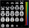
The SPECT imaging analysis exhibited decreased rCBF in the bilateral inferior frontal gyrus (p<0.001), bilateral temporal pole (p<0.001), and right precentral gyrus (p<0.001) in the AD patients with SL group compared with the AD patients without SL group (Table 2, Fig. 1). It also revealed increased rCBF in the right precuneus (p<0.001), right occipital pole (p=0.001), and left middle occipital gyrus (p=0.002) in the AD with SL group compared with the AD without SL group (Table 2, Fig. 1).
DISCUSSION
In this study, we investigated the changes in cerebral perfusion among AD patients who experience sleep disturbance using SPECT. The results of this study demonstrated that cerebral perfusion decreased in the temporal and frontal lobes and increased in the precuneus and occipital lobe in the AD patients who suffer from SL compared with AD patients who do not.
In line with the results of this study, functional deficits of the frontal and temporal cortices have been observed in previous neuroimaging studies of healthy individuals with compromised sleep quality. A study using positron emission tomography (PET) found decreases in metabolism in the frontal lobe including the inferior frontal gyrus and temporal regions after sleep deprivation.25 Similarly, other PET studies displayed a reduction of glucose uptake in the prefrontal cortex26 and changes in the rCBF pattern in the prefrontal and temporal areas during a verb generation task following sleep deprivation.27 Moreover, the electroencephalography (EEG) recordings of individuals with sleep deprivation exhibited increases in delta and theta activities in the frontal areas compared with the parietal and occipital areas, which indicated their vulnerability to the effects of sleep loss and their role in homeostatic function.28 Additionally, sleep problems were associated with impaired performance on tasks involving the frontal lobe including executive attention, working memory, and higher cognitive functions.29
Increased perfusion in the occipital areas in AD patients with SL was also found in this study. These findings are supported by previous PET studies that demonstrated increases in regional metabolism in the occipital cortex in individuals with sleep deprivation.2526 Since it has been suggested that the occipital areas are less susceptible to sleep loss than the frontal areas,30 decreased activity in the frontal lobe and increased activity in the occipital lobe may be due to the compensatory mechanisms of the occipital areas for frontal lobe deficits.25
On the other hand, our findings were not consistent with those from a previous SPECT study that demonstrated increased perfusion in the middle frontal gyrus in AD patients with SL.15 The inconsistencies may be explained in part by several differences between the studies including the study design, sample size, selection criteria, and patient samples.
The current study has limitations that merit mention. First, objective sleep measures such as a polysomnography were not used for group assignment in the study. Moreover, due to the cross-sectional design of the study, it is not possible to establish causal relationships.
In summary, the decreased perfusion in the frontal and temporal areas and increased rCBF in the parietal and occipital regions were evidenced in AD patients who suffer from sleep loss. The findings of this study suggest that functional alterations in these brain areas may be the underlying neural correlates of sleep disturbance in AD patients. Future studies with larger sample sizes and longitudinal designs are needed to confirm and further elucidate the effects of sleep disturbance on cerebral perfusion in AD patients.


 XML Download