

 PDF
PDF ePub
ePub Citation
 Print
Print


Transient global amnesia (TGA) is defined by a sudden onset of anterograde and retrograde amnesia that lasts up to 24 h. The clinical feature of this syndrome has been discussed in recent studies. Several etiological factors, such as epileptic phenomena, migraine-related mechanisms, focal ischemia, and venous flow abnormalities, have been implicated in its pathophysiology.1
A 68-year-old male visited our emergency room with temporary memory loss. He was taking medications for hypertension and benign prostate hypertrophy. His guardian described that the patient complained of a sudden onset of lightheadedness and mild headache while watching television the night prior to admission. After waking up the next day, he could not remember the events of the previous night and asked repetitive questions despite being provided with correct answers. Upon admission, he could not recall details of the night before the admission. He had no recent history of head trauma or seizure. Neurologic examination showed no focal neurological signs.
The patient scored 21 points on the Korean version of Mini-Mental Status Examination and 2 points on Global Deterioration Scale. These findings were indicative of mild cognitive impairment, considering his age and level of education (9 years).
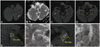
The patient was diagnosed with clinically typical TGA. Diffusion-weighted images obtained 12 h from the onset of symptoms revealed lesions with high signal intensity in the left cerebellum and localized lesions in the bilateral temporal lobe. Corresponding lesions showed decreased values in apparent diffusion coefficient maps (Fig. 1).
Typical findings of diffusion-weighted imaging in more than one-third of patients with transient complete amnesia include unilateral, 2–3 mm diffusion-limiting lesions in the cornu ammonis 1 area of the hippocampus. Diffusion-weighted MRI of the brain performed between 24 h and 72 h after the onset of symptoms revealed a high detection rate. However, these findings may not be detected on images obtained earlier.12
Therefore, TGA is attributed to an unspecified ischemic injury or delayed neuronal injury unlike a typical ischemic stroke. However, typical cerebral infarction was observed in the patient in question, suggesting that TGA may be associated with various mechanisms of onset.
 XML Download