

 PDF
PDF ePub
ePub Citation
 Print
Print


INTRODUCTION
The term ‘wandering’ is defined as seemingly aimless or disoriented ambulating behavior of demented persons with dimensions of pattern,1234 frequency,156 boundary transgressions,78 and deficits in wayfinding.5910 In addition, ‘wandering’ is frequently used as a broad term embracing diverse behaviors. It is often regarded as a kind of agitated behavior.2
Although a recent study has reported that 18.7% of patients with drug-naïve Alzheimer's disease (AD) are wanderers,11 the estimated prevalence of wandering differs across studies, ranging from 17.4% to 63%.1213 This rate can be as high as 100% if dementia patients with independent gait are selected.3 Another study has assessed the prevalence of dementia-related behaviors in a large and multi-ethnic sample of community-dwelling patients with moderate to severe dementia and reported that wandering is the most prevalent dementia related behavior regardless of ethnicity.14 These variations are partly due to differences in definitions used, characteristics of study setting, and different time periods covered. Regardless of the suggested definition, this behavior has a significant effect on the caregiver and the patient. However, little is known about the anatomical substrate according to specific wandering patterns in patients with AD. This might be due to difficulty in defining the specific pattern of wandering and in differentiating broad symptoms mimicking wanderings.
Wandering has received scant scientific attention. How such behavioral abnormalities are correlated with brain function is unclear. Little is known about its anatomic substrate. A major reason for limited progress in understanding biological cause of wandering is that wandering pattern is not simple. It has not been well characterized either. Despite these complex wandering patterns, encompassing various wandering pattern into “wandering” and correlating this phenomenon with specific brain area have resulted in conflicting findings.
The objective of this study was to determine specific patterns of wandering in patients with drug-naïve AD and explore anatomic substrate of these patterns by performing positron emission tomography with 18F fluorodeoxyglucose positron emission tomography (FDG PET). Differences between specific wandering patterns of AD patients with wandering (wanderer) and those without wandering (non-wanderer) were then determined.
METHODS
Participants
A total of 780 patients with dementia were screened from March 2012 to July 2017 at the Veteran Health Service Medical Center (Seoul, Korea) and Hyoja Geriatric Hospital (Yongin, Korea). Among these 780 patients with dementia, 342 patients with probable AD were selected as study subjects. They were not medicated before visiting the hospital. All study subjects met National Institute of Neurological and Communicative Disorders and Stroke-Alzheimer's Disease and Related Disorders Association (NINCDS-ADRDA) criteria for probable AD.15 Patients who were taking psychoactive drugs, including antidepressants, antipsychotics, anticonvulsants, benzodiazepines, and cholinesterase inhibitors, were excluded from this study. All study subjects underwent a complete medical history, physical and neurological evaluations, neuropsychological tests, routine laboratory tests, and a brain magnetic resonance imaging or computed tomography (CT) scan. To assess global dementia severity, the Korean version of the Mini-Mental State Examination (K-MMSE)16 and Clinical Dementia Rating (CDR) scale17 were used. Barthel index18 for activities of daily living (ADL) and Geriatric Depression Scale (GDS)19 for depression were adopted.
Screening tools used for diagnosing wandering were Caregiver-Administered Neuropsychiatric Inventory (CGA-NPI) “motor disturbance” item. Once this item was checked, structured interview was then performed for each subject. Using this interview, specific patterns of wanderings were clinically defined by neurologist independent of this study. The wandering behavior was measured using Korean-translated Revised Algase Wandering Scale–Nursing Home Version (KRAWS-NH).20 Based on an earlier version of the Algase Wandering Scale (AWS),21 the KRAWS-NH is a 59-item caregiver questionnaire consisting of six subscales: persistent walking, specific patterns, spatial disorientation, escape behaviour, attention shifting, and negative outcomes. Items were scored from 1 (never or unable) to 5 (always), with higher scores indicating more wandering. The scale took 10 minutes to complete.
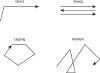
According to a previous study,6 we defined wandering subtypes (pacing, random, and lapping) at first instance by clinical judgement of a neurologist independent of this study (Fig. 1). We calculated average pacing score, random score, and lapping score for each patient. Average pacing score was derived from the sum of RAWS-NH items 2 and 20 divided by 2. Average random score was derived from the sum of RAWS-NH items 12, 15, 35, 37, and 41 divided by 5. The average lapping score was calculated from the sum of RAWS-NH items 22, 29, 32, 39, 52, and 57 divided by 6. The specific pattern of wandering type was operationally classified if the specific wandering score was at least twice the other specific wandering score and in agreement with clinical assessment. The remaining patients whose specific wandering scores were less than twice of other wandering scores or not in agreement with clinical judgement were classified as mixed type. They were excluded from this study. This study was approved by the Institutional Review Board of the Veteran Health Service Medical Center, Seoul, Korea (2016-05-020).
FDG PET
For 18F FDG PET scans, all study subjects were on nil per os for at least 4 hours and their pre-injection blood glucose levels were confirmed to be below 180 mg/dL. Images were acquired at approximately 45 minutes after fluorodeoxyglucose (FDG) injection (185–222 MBq) using a Discovery STE PET/CT scanner (GE Healthcare, Milwaukee, WI, USA). All studies were done in 3-dimensional acquisition mode. A 16-slice CT scan was performed for tissue correction of attenuation prior to FDG PET scan. These imaging data were analyzed using SPM 8 (Wellcome Trust Centre for Neuroimaging, University College London, London, UK) and implemented using MATLAB software (MathWorks Inc., Natick, MA, USA). Images were reconstructed using standard PET protocol, spatially normalized, and smoothed. Smoothing was performed using a 16-mm full-width-at-half-maximum isotropic Gaussian kernel.
Differences in glucose metabolism according to specific wandering patterns compared to non-wanderers were estimated on a voxel-by-voxel basis. The resultant set of t-values constituted SPM (t) map. The t-statistic image was acquired at a >3.93, corresponding to a uncorrected p value <0.05 in conjunction with a cluster filter of 50 voxels. For visualization and statistically significant hypometabolic anatomic localization, Talairach Coordinates system in MRIcro (Centre for Advanced Brain Imaging, Atlanta, GA, USA; http://www.cabiatl.com/mricro/mricro/index.html) was used.
Statistical analysis
Baseline demographic features and cognitive function of study participants (wanderers and non-wanderers) were assessed using Student's two-tailed t-test for continuous variables and χ2 test for categorical variables. Comparison of demographic features and FDG PET patterns among specific wandering patterns were assessed using one-way analysis of variance with subsequent Scheffe post hoc test. Values are expressed as means and standard deviations. Statistical analyses were performed with SPSS version 18.0 (SPSS Inc., Chicago, IL, USA).
RESULTS
Among 342 study subjects, 80 (23.4%) were classified as wanderers. Demographic characteristics of wanderers versus non-wanderers are shown in Table 1. K-MMSE and Barthel scores were significantly lower while CDR, GDS, and CGA-NPI scores were significantly higher in wanderers than those in non-wanderers. Gender, age, onset age, symptom duration, or education was not significantly different between wanderers and non-wanderers.
Table 1
Demographic and clinical features of AD patients with or without wandering

Among 80 wanderers, 62 showed distinct wandering patterns. Pacing was the most common wandering pattern, following by random pattern and lapping pattern. Patients with lapping pattern showed significantly lower K-MMSE score and Barthel index but higher CDR, and CGA-NPI scores than those with other wandering patterns. GDS scores were higher in patients with pacing pattern than those in patients with other wandering patterns (Table 2).
Table 2
Demographic and clinical features of wanderers with specific wandering types

K-MMSE: Korean version of the Mini-Mental State Examination, CDR: Clinical Dementia Rating, GDS: Geriatric Depression Scale, CGA-NPI: Caregiver-Administered Neuropsychiatric Inventory, P: pacing, R: random, L: lapping.
*Statistical significance was found between Lapping and other patterns of wandering; †Statistical significance was found among patterns of wandering.
In FDG PET, significant lower FDG uptakes in both middle cingulum and left putamen cluster were found in patients with pacing pattern compared to those of non-wanderers. The right precuneus and supplementary motor area (SMA) in patients with random pattern, both calcarine sulci, right middle cingulum, and right post central gyrus in patients with lapping pattern had significantly lower FDG uptakes than non-wanderers (Table 3).
Table 3
Location and peak of significant difference in regional cerebral hypometabolism compared with normal controls (p<0.05, FDR-corrected)

DISCUSSION
Up to date, no study has reported the anatomic substrate according to specific wandering pattern in patients with AD. In the absence of clearly known biological substrates, the cause of wandering has been mainly studied in other fields such as psychosocial and person-environment interaction. For example, environmental and need-driven factors can be main contributory factors to wandering risk. Wandering can occur when internal discomfort, especially when coupled with external demands (e.g., a noisy environment), exceeds the individual's threshold.22 However, there is a profound limitation in understanding wandering by these non-biological approaches because these patients clearly have neurological disorders with dementia symptoms.
Biological studies are mainly focused on the impairment of specific brain functions, especially in spatial memory, visuospatial processes, and executive functions.232425 Tetewsky and Duffy23 have shown that wandering accounts for visuospatial problems while McShane et al.24 have suggested that spatial memory problem is related to wandering in dementia. Other studies have reported that impairments in optic flow perception and interpretation can lead to spatial navigation failure as the cause of wandering in some patients with AD.2326 Execution problem from thoughts to actions involved in decision-making, planning, and monitoring has been suggested as another cause of wandering.27 Our previous study has also shown that wandering is associated with frontal and right parietal functions.11 Like other complex repetitive behaviors, wandering may also result from or be facilitated by aberrant distracted attention and executive functions, resulting in disinhibited motor programs.28 However, spatial representation deficits or spatial memory problems in AD cannot sufficiently explain wandering because these patients can reach a destination without having complete knowledge about it.29 Wayfinding difficulties might be due to their poorly structured overall decision plans rather than spatial memory problems.
In a single photon emission CT study, wanderers have more severely reduced regional cerebral blood flow in the left temporoparietal region than non-wanderers among patients with AD.30 Based on positron emission tomography, wandering patients with AD show frontotemporal glucose utilization and decreased dopamine metabolism in the striatum.31 These studies strongly suggest that the pathophysiological mechanism of wandering involves parietal and frontal dysfunction (and possibly temporal), suggesting that functionally impaired spatial and executive neural circuit can lead to wandering.32 However, these conflicting cognitive and brain imaging studies for wandering could not clearly outline suggested anatomic substrates for wandering. The reason of their inconsistent results is mainly due to lumping heterogenous phenomenology into a single termed “wandering.” The term ‘wandering’ is frequently used as a broad term encompassing diverse behaviors. It is often considered as a kind of agitated behavior.2 To overcome this problem, we first meticulously extracted “wander” from other behavioral and psychological symptoms of dementia. Because the pattern of wandering is very diverse, splitting this “wandering” into more homogenous sub-patterns might be important. Therefore, we operationally defined specific wandering pattern using both clinically visually determined wandering pattern by a neurologist and AWS specific items checked by caregivers. Three wandering patterns were operationally defined if both judgements were concordant. The concordant rate between clinically determined pattern and RAWS-NH was relatively high (77.5%).
Our study showed three distinct wandering patterns in patients with AD. These wandering patterns were related to their cognitive status. They were not greatly affected by other demographic characteristics. Among these wandering patterns, pacing (n=32, 51.6%) was the most frequently identified subtypes, followed by random (n=24, 38.7%) and lapping (n=6, 3.7%). These results are different from those of a previous study reporting that lapping is the most frequent type.6 Such discrepancy between the two studies might be due to inclusion bias. Our study subjects were recruited from hospital while study subjects recruited for the previous study were from a nursing home. Moreover, our study subjects were more biased for mild dementia. These differences might be important factors. Our study showed more cognitive dysfunctions in patients with lapping pattern, in agreement with results of the previous study.6 These clinical severity differences might account for less inclusion of lapping patterns in our study.
SPM analysis of FDG PET in wanderer showed distinct region hypometabolism according to specific wandering pattern. Significantly lower FDG uptakes were observed in both middle cingulum and left putamen in patients with pacing pattern compared to those in non-wanderers. Right precuneus and SMA in patients with random pattern and both calcarine sulci, right middle cingulum, and right postcentral gyrus in patients with lapping pattern had significantly lower FDG uptakes compared to those of non-wanderers. These findings suggest that specific wandering patterns are due to specific focal brain dysfunctions. How these specific areas are related to specific wandering pattern is currently unclear. The putamen is interconnected with many structures. It works in conjunction with those structures to control many types of motor skills. It also plays a role in the selection of movement (e.g., Tourette Syndrome) and automatic performance of previously learned movements.33 Cingulum is very important to brain structure connectivity. This area is also associated with automatic movements. These hypometabolic brain areas in patients with pacing may result in basic problem for movement including automaticity. Considering that pacing might be the simplest and easiest travel pattern, patients with dysfunctions in these areas might adopt this travel pattern.
The motor cortex contributes to steady state walking. During gait challenges, SMA is more active to account for continuous alteration necessary to navigate challenges.34 The SMA is known to be involved in the selection, planning, and coordination of voluntary movements as well as the suppression of stimulus-driven (although, purposeless) movements (i.e., grasping). This is supported by findings of profound activation of the SMA in the period prior to and around the start of locomotor tasks.35 The precuneus is a part of the superior parietal lobule in front of the occipital lobe. It is involved in episodic memory, visuospatial processing, reflections upon self, and aspects of consciousness.36 Especially, the precuneus is part of a neural network functionally specialized for the process of spatially guided behavior.37 This part acts in concert with lateral parietal areas to elaborate information about egocentric and allocentric spatial relations for body movement control (motor imagery) as well as higher-order processes such as voluntary attention shift and more abstract mental imagery tasks.38 We cautiously speculate that dysfunction in SMA may result in failure of suppressing stimulus-driven activity while dysfunction of precuneus may account for spatially guided behavior problems accompanying attention shift difficulty. These combined dysfunctions may manifest as random pattern of wandering.
The calcarine sulcus is located within the primary visual cortex of the occipital lobe. Visual feedback is important for most motor tasks.39 It allows for proper interpretation of the body in a certain environment. Visual feedback also plays an important role in the maintenance of balance.40 The primary somatosensory cortex is located in the postcentral gyrus. Lesions affecting this area can produce characteristic symptoms such as astereognosia, agraphesthesia, hemihypesthesia, and loss of vibration, proprioception, and fine touch. If it affects the non-dominant hemisphere, it can also produce hemineglect. Due to impaired visual feedback and interpretation from calcarine dysfunction and impaired sensory system from right post central gyrus, patients may go round to target destination with inefficient travel pattern like lapping.
SPM analysis was performed between AD patients with specific travel patterns (wanderers) and non-wanderers. Thus, commonly known hypometabolism in temporo-parietal or frontal cortical metabolism was not presented in this study. Considering commonly affected cortical area, interaction between these specific brain areas and commonly affected cortical area may result in specific wandering patterns. These explanations for specific wandering pattern is highly speculative without enough evidence. However, this study strongly suggests that wandering pattern mainly results from interaction between specific anatomic substrate dysfunction and commonly affected higher cortical dysfunction in patients with AD.
The present study has several limitations. First, although we used standardized wandering scale, valid methodology for defining specific wandering pattern is not established yet. Therefore, we operationally defined this. As a result, this study might include mistakenly classified wandering pattern or excluded the specific wandering pattern as mixed type. Second, the sample size was relatively small. Finally, the present study was a hospital-based study. It might not represent the real community.
Nonetheless, this study showed that wandering in patients with AD comprised of three distinct patterns, corresponding to low FDG uptake in specific brain areas. These findings can broaden our understanding of the biological cause of wandering pattern in AD patients. However, clinical implications of these findings are currently unclear. Therefore, further study is needed to explore such implications.
 XML Download