

 PDF
PDF ePub
ePub Citation
 Print
Print


INTRODUCTION
Primary progressive aphasia (PPA) is a clinical dementia syndrome in which language capabilities slowly and progressively become impaired.1 Three subtypes of PPA are recognized: nonfluent/agrammatic variant primary progressive aphasia (navPPA), semantic variant PPA, and logopenic variant PPA.2 Underlying pathologies of PPA are heterogeneous, including tauopathy, ubiquitinopathy (TAR-DNA binding protein 43), and Alzheimer's disease (AD) pathology.3456 The major neuropathology of sporadic navPPA is known to be tauopathy.789
Tau positron emission tomography (PET) imaging may allow better understanding of tau aggregation and deposition in various dementia syndromes including navPPA. Several radiotracers have been developed for in vivo visualization of tau pathology.10 18F-THK5351, a 18F-labed THK arylquinoline tracer, has a high binding affinity and selectivity for tau.11 However, off-target binding related to monoamine oxidase-B (MAO-B) has raised concerns about the specificity of 18F-THK5351 for tau recently.1213 The objective of this study was to analyze 18F-THK5351 PET scans14 of patients with clinically diagnosed navPPA.
METHODS
Participants
Thirty-one participants were recruited from a single tertiary hospital from April 2015 to April 2016. They underwent a clinical interview and standard neurologic examination as well as structural brain magnetic resonance imaging (MRI) as described below. Three patients who were clinically diagnosed with navPPA according to guidelines proposed by Gorno-Tempini et al.14 and 13 patients who were diagnosed with probable AD according to the National Institute of Neurological and Communicative Disorders and Stroke and the Alzheimer's Disease and Related Disorders Association15 criteria were included in this study. We included AD patients as a representative group to show the pattern of THK retention. None of the 3 patients diagnosed with navPPA had a family history of dementia or movement disorder. Fifteen normal control (NC) subjects with normal cognition without history of neurologic or psychiatric illness or abnormalities detected on neurologic examination were also included. All participants underwent a comprehensive neuropsychological evaluation using the Seoul Neuropsychological Screening Battery-II.16 Disease severity was assessed by the frontotemporal lobar degeneration (FTLD) modified Clinical Dementia Rating (CDR) containing 2 more domains (behavior and language) in addition to the 6 standard domains of the CDR scale.171819 In the NC group, the CDR score was zero, the Mini-Mental Status Examination (MMSE) score was above 25, and the overall neuropsychological test performance was 1.5 standard deviations (SDs) above age adjusted and education adjusted norms.
We excluded patients with structural, focal lesions on brain MRI such as stroke, intracranial hemorrhages, and evidence of traumatic brain injury, hydrocephalus, white matter hyperintensities associated with radiation, multiple sclerosis, or vasculitis. Potential secondary causes of dementia were excluded through laboratory tests such as complete blood count, vitamin B12 and folate levels, thyroid function test, metabolic profile, and syphilis serology. Apolipoprotein E (APOE) genotyping was also conducted as described previously.20 Our Institutional Review Board (IRB) approved the study protocol (IRB number: GDIRB2015-272). Informed consent was obtained from all participants.
Neuropsychological tests
All participants underwent neuropsychological evaluation using a battery that included assessments for the following cognitive domains: attention, language, praxis, visuoconstructive ability, elements of Gerstmann syndrome, visual and verbal memory, and frontal/executive function. Quantifiable tests among these evaluations included digit span test (forward and backward), the Rey-Osterrieth Complex Figure Test, the Korean version of the Boston Naming Test, the Seoul Verbal Learning Test, phonemic and semantic Controlled Oral Word Association Test, and a Stroop Test (color and word reading of 112 items during a 2-minute period). MMSE, CDR, CDR Sum of Boxes, FTLD modified CDR Sum of Boxes, and Geriatric Depression Scale results were also obtained. The Korean version of the Western Aphasia Battery (K-WAB) having subscales to assess spontaneous speech, auditory verbal comprehension, repetition, naming and word finding, reading, and writing was performed in 3 subjects diagnosed with navPPA.2122
Image acquisition and preprocessing
MRI
MRI of all participants was performed with a 3.0-T MRI scanner (Siemens with a Siemens matrix coil; Verio, Orem, Utah, USA) including a 3-dimensional (3D) magnetization prepared rapid gradient echo (MPRAGE) sequence. Imaging parameters used for 3D MPRAGE were as follows: repetition time=1900 m/s, echo time=2.93 m/s, flip angle=8°, pixel bandwidth=170 Hz/pixel, total acquisition time=4 minutes 10 seconds, and the iso-voxel resolution was 1.0 mm. Other clinical MRI sequences including fluid attenuated inversion recovery sequence were also acquired.
PET
All PET scans were acquired with a Siemens Biograph 6 Truepoint PET/computed tomography (CT) scanner (Siemens, Knoxville, TN, USA) with a list mode emission acquisition. Participants underwent a 20-minute emission scan at 90 minutes after intravenous injection of 185 MBq of 18F-flutemetamol (FLUTE). Within 3 months of undergoing the initial FLUTE PET scan, all participants underwent a 20-minute emission scan starting 50 minutes after intravenous injection of 185 MBq of THK. THK was synthesized and radiolabeled at Gachon University Neuroscience Research Institute. A low-dose CT was performed for attenuation correction prior to all scans. Images were reconstructed in a 256×256×109 matrix with a voxel size of 1.33×1.33×1.5 mm using a 2D Ordered Subset Expectation Maximization algorithm (8 iterations and 16 subsets). They were corrected for physical effects including radiation attenuation and scatter.
Image processing and analysis
THK retention was expressed as a standardized uptake value ratio (SUVR) using cerebellar gray matter as reference region. FLUTE retention was also expressed as SUVR using pons as the reference region.23 For both regional and voxel-wise group analyses of THK retention, individual sets of PET and MRI images were processed using PMOD 3.7 (PMOD Technologies Ltd., Zurich, Switzerland) as follows. Individual PET data were co-registered to 3D MRI images and corrected for grey matter atrophy and white matter spillover using MR-based partial volume correction (PVC). For the correction, we applied the geometric transfer matrix (GTM) method2425 to region of interest (ROI) data and the Müller-Gärtner (MG) method26 to voxel data. These were implemented in PMOD 3.7. ROI and tissue segments with GTM and MG, respectively, were obtained from individual MR images during the PMOD-based image processing step. In PVC-corrected images, voxels with low gray-matter probability (p<0.2) were masked out to prevent noise amplification. Individual 3D MRI slices were then normalized to the Montreal Neurological Institute template. Their transformation matrix was applied to MRI co-registered PVC PET images.
A voxel-wise analysis was also performed to compare regional pattern of THK retention using SPM12 (Statistical Parametric Mapping; Wellcome Trust Centre for Neuroimaging, London, UK). Before comparison, normalized PVC SUVR images were smoothed with an 8 mm Gaussian kernel to account for individual anatomical differences and improve signal to noise ratio.
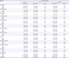
To compare THK retention in each group quantitatively, we defined 14 ROIs, including the precentral cortex, inferior frontal cortex(triangular part of inferior frontal gyrus, opercular part of inferior frontal gyrus, and orbital part of left inferior frontal gyrus), Broca's area (triangular part of inferior frontal gyrus and opercular part of inferior frontal gyrus), lateral temporal cortex (superior, middle, and inferior temporal cortex), mesial temporal cortex (hippocampus, parahippocampal gyrus, and amygdala), fusiform gyrus, superior parietal cortex, inferior parietal cortex (inferior parietal, supramarginal gyrus, and angular gyrus), insula cortex, anterior and posterior cingulate gyrus (posterior cingulate gyrus and precuneus), occipital cortex (superior, middle, inferior occipital gyrus, calcarine sulcus, and cuneus), basal ganglia (putamen, caudate nucleus, and pallidum), and brainstem. By using mean and SD of regional SUVR values of 15 NCs, regional Z-scores of SUVR values were calculated.
RESULTS
Demographic and clinical characteristics
Characteristics of navPPA patients are shown in Table 1. Those of NC and AD groups are presented in Supplementary Table 1. The first navPPA case was an 82-year-old man with 16 years of education. He presented with 3-year history of slow effortful speech. He showed slow speech and used short sentences. Neuropsychiatric evaluation revealed dysfunctions in verbal memory, phonemic/semantic fluency, and frontal/executive domains. Results of his K-WAB were consistent with transcortical motor aphasia. He was found to have diffuse brain atrophy. However, predominant atrophy was seen on the left frontal area on brain MRI. High THK retention was also observed in the left frontal cortex (Fig. 1). FLUTE PET scan was negative for amyloid pathology.
Table 1
Characteristics of patients with navPPA

Fig. 1
18F-THK5351 PET and MRI (T1-MPRAGE) scans of navPPA patients.
PET: positron emission tomography, MRI: magnetic resonance imaging, MPRAGE: magnetization prepared rapid gradient echo, navPPA: nonfluent/agrammatic variant primary progressive aphasia.
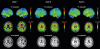
The second case was a 73-year-old man with 6 years of education who complained of difficulties with speech production that started 1.5 years prior to his initial visit. Neuropsychiatric evaluation indicated dysfunctions in verbal memory, phonemic/semantic fluency, and frontal/executive domains. Results of his K-WAB were compatible with transcortical motor aphasia. FLUTE PET scan was negative for amyloid pathology. MRI showed bilateral frontal atrophy (left > right). High THK retention was observed in the left frontal region (Fig. 1).
Case 3 was a 76-year-old man with 6 years of education who developed slow effortful speech from 1 year prior to enrolling in this study. Neuropsychiatric evaluation revealed dysfunction in verbal fluency. Results of his K-WAB were consistent with anomic aphasia. Atrophy on the left frontal area was seen in MRI. High THK retention was observed in the left frontal region (Fig. 1). FLUTE PET scan was negative for amyloid pathology.
ROI-based analysis of THK retention in navPPA, NC, and AD groups
Statistical results of ROI analysis after PVC are summarized in Table 2. With cut-off Z-score of 2.5, typical AD patients showed greater binding than NC in widespread cortical regions except bilateral mesial temporal, right precentral, and left anterior cingulate cortices. Patients with navPPA had higher levels of THK retention in the Broca's area, bilateral inferior frontal lobes, bilateral precentral gyri, and bilateral basal ganglia (Table 2). Details of ROI analysis results of 3 patients with navPPA are presented in Supplementary Table 2.
Table 2
Regional binding values of 18F-THK5351 PET in NC, AD, and navPPA patients
Voxel-based analysis of THK retention in PPA, NC, and AD groups
NavPPA vs. NC
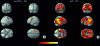
Patients with navPPA showed higher levels of THK retention in the bilateral frontal lobe (mainly left side) than NC (Fig. 2A).
Fig. 2
Statistical parametric mapping analysis of THK SUVR images after PVC. Colored areas represent brain regions corresponding to increased THK retention from voxel-wise statistical analyses in those with navPPA (A) and AD (B) compared to NC (p<0.001, uncorrected for multiple comparisons after adjusting for age, gender, and years of education, a cluster >100).
SUVR: standardized uptake value ratio, PVC: partial volume correction, navPPA: nonfluent/agrammatic variant primary progressive aphasia, AD: Alzheimer's disease, NC: normal control, T: T value.
AD vs. NC
Patients with AD had higher levels of THK retention in widespread cortical regions (including the frontal, parietal, occipital, anterior/posterior cingulate, and mesial/lateral temporal cortices) than NC while relatively sparing the primary sensorimotor cortices bilaterally (Fig. 2B).
DISCUSSION
In our study, THK retention in navPPA patients was mainly distributed at the frontal region (left > right). This regional pattern of 18F-THK5351 binding is well correlated with the functional-radiological distribution of navPPA. To account for potential confounding effects of marked cortical atrophy, all images were corrected for partial volume effects and white matter spillover in this study to confirm higher cortical THK retention.
The pathology of navPPA is heterogeneous. However, many neuropathologic studies have shown that the most common pathologic subtype of navPPA is tauopathy.3727 Many clinically diagnosed navPPA cases are known to be often associated with corticobasal degeneration (CBD) or progressive supranuclear palsy (PSP) pathology.28 Recent in vivo studies have suggested that 18F-THK5351 PET scan might be suitable for the assessment of tau deposition in 4-repeat tauopathy disease spectrum including CBD29 and PSP.3031 Along with these studies, our study suggests that tau PET imaging could be useful not only in AD, but also in non-AD tauopathies.
We observed that navPPA patients also showed increased THK binding in the bilateral basal ganglia. Basal ganglia atrophy and extrapyramidal sign are commonly found in navPPA patients.323334 In many navPPA patients, there are clinical and pathological overlaps with CBD or PSP.28 Recent reports have shown greater THK binding in the basal ganglia in PSP and CBD patients.2931 However, possibility of off-target signal should also be considered because off-target signal in the MAO-B rich basal ganglia has been reported.12
There is a possibility that THK retention in our navPPA patients might reflect neuroinflammation or neurodegeneration rather than tau pathology itself. A recent study has shown a possibility that the interpretation of 18F-THK5351 PET images could be confounded by the high MAO-B availability across the entire brain because 18F-THK5351 might bind to MAO-B and tau paired helical filaments with similar affinity.13 MAO-B has been proposed as a biomarker for astrocytosis in various conditions associated with neuroinflammation and neurodegeneration.35
Although 18F-THK5351 has limited utility for selective detection of tau pathology, 18F-THK5351 could be a supportive tool for diagnosis of navPPA because this tracer reflects the combination of tau pathology with associated neuroinflammatory changes and distribution is well matched with functional-radiological distribution of navPPA. An important advantage of tau PET might be its potential for early diagnosis of neurodegenerative disease due to its greater sensitivity and clarity. In some of our cases with early stage of navPPA, it was not easy to differentiate them with other types of PPA before obtaining tau PET imaging.
This study has some limitations. This was a study examining only a small number of patients. Thus, validation in a large population is needed in the future. The major caveat of our study was the lack of pathologic confirmation. Autopsy data are needed to resolve this problem.
 XML Download