

 PDF
PDF ePub
ePub Citation
 Print
Print


Small molecular weight gadolinium chelate is a positive contrast agent (CA) for magnetic resonance imaging (MRI), used to diagnose many tissue and vascular diseases that may not be detected in unenhanced MR images (1). However, over the last decade, safety issues have arisen regarding the use of gadolinium CAs, because some Gd3+- based CAs have been associated with nephrogenic systemic fibrosis (NSF), that can arise in patients with impaired renal function (23). Furthermore, recent reports revealed another possible safety issue when long-term retention of Gd3+-based CAs, was found in the brains of patients with normal renal function (4).
There is a consensus in the scientific literature that NSF and brain retention of Gd3+-based CAs are strongly associated with the ligand structure of Gd3+-based CAs. Several studies have shown that Gd3+-based CAs with linear ligand structures were associated with a much higher incidence for NSF and brain retention of CAs, than Gd3+-based CAs with macrocyclic ligand structures (35). The underlying principle behind the difference between the two types of ligand structures is that the Gd3+-based CAs with linear ligand structure are less resistant to the release of free gadolinium than the Gd3+-based CAs with macrocyclic ligand structure (6). Due to safety concerns, among all commercialized Gd3+-based CAs with linear ligand structures, 4 extracellular fluid (ECF) CAs were recommended for withdrawal from the market in Europe (7). However, the liver-specific Gd3+-based CAs with linear ligand structures, were not recommended for market withdrawal because there are no functional Gd3+-based CAs with macrocyclic ligand structures.
Recently, we synthesized a highly stable multifunctional Gd3+-based CA with a macrocyclic ligand structure (HNP-2006) (8). In the current study, we report the in vivo biodistribution profile of this new multifunctional Gd3+-based CA. We also demonstrated that this new multifunctional Gd3+-based CA, with macrocyclic ligand structure holds promise for several clinical imaging applications.
In vivo MRI biodistribution experiments were performed using a 1.5T MRI system (GE Healthcare, Milwaukee, WI, USA). The experimental protocol was approved by the animal research committee of Kyungpook National University (KNU). Homemade birdcage-type radiofrequency (RF) coil, which was a receiver-only type with an inner diameter of 50 mm, was used for small animals. The imaging parameters for spin echo sequence were as follows: repetition time (TR) = 300 ms; echo time (TE) = 13 ms; field of view (FOV) = 8 mm; 192 × 128 matrix size; 1.2 mm slice thickness; number of excitation (NEX) = 8. Total scan time was 2 minutes, 37 seconds. The imaging parameters for spin echo sequence were as follows: TR = 300 ms; TE = 13 ms; FOV = 8 mm; 192 × 160 matrix size; 1.2 mm slice thickness; NEX = 8. Total scan time was 2 minutes, 37 seconds. Six-week old male mice (weighing 25–27 g), obtained from the Institute of Cancer Research (ICR) were used. Animals were anesthetized by 1.5% isoflurane in oxygen. For the tumor model, the mice were placed in a stereotaxic apparatus. After scalp incision to expose bregma, the skull was drilled at an exact position of 1 mm posterior and 2 mm lateral from the bregma for stereotaxic injections. C6 glioma cells were prepared (1.0 × 10⁵) in DMEM, and injected for 5 minutes at a rate of 1 µL/min, and depth of 2 mm into the hole. The injection needle was kept in this position to prevent cell leaks for 5 minutes, then slowly removed from the brain. The MR study was performed 2 weeks after C6 glioma implantation. MR images were acquired before and after tail vein injection of multifunctional Gd3+-based CA with macrocyclic ligand structure (HNP-2006). The CA injection dose was 0.1 mmol Gd/kg for MR images. Anatomical locations of liver, bile duct, heart, and kidney were identified in post-contrast enhanced MR images. For quantitative measurements, the signal intensities in specific regions of interest (ROI) were measured using Advantage Window software (GE Medical, USA). The contrast-to-noise ratio (CNR) was calculated using equation [1], where SNR is the signal-to-noise ratio.
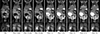
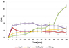
Figure 1 shows the coronal T1-weighted images of 6-week-old male ICR mice before and after HNP-2006 injection. Figures 1 and 2 demonstrated that several organs showed significant contrast enhancements after CA injection. The strong heart enhancement demonstrated that HNP-2006 can be used for possible cardiovascular application. The most characteristic features of HNP-2006 as a CA are that the liver and gallbladder showed significant signal enhancement. Contrast enhancement of the liver and gallbladder established HNP-2006 as the first Gd3+-based CAs with macrocyclic ligand structure, with possible hepatobiliary applications. Currently, two Gd3+-based CAs with linear ligand structures, Gd-BOPTA (Multihance) and Gd-EOB-DTPA (Primovist), were reported to show enhancement of the liver and gallbladder. The in vivo biodistribution patterns of Gd-BOPTA and Gd-EOB-DTPA were thought to result from the fact that these agents exhibit lipophilic properties, and thus are excreted through the hepatocytes and biliary tract. In the hepatocyte, these agents are known to be excreted through organic anion-transporting polypeptide-mediated protein channels (9). Therefore, HNP-2006 seems to use a similar organic anion-transporting polypeptide-mediated protein channel for hepatocyte uptake. Furthermore, the strong contrast enhancement in kidney and bladder confirmed that HNP-2006 was excreted by glomerular filtration, in addition to hepatobiliary excretion. The in vivo biodistribution pattern of HNP-2006 through kidney and bladder is very similar with that of commercialized macrocyclic Gd3+-based ECF agents, such as Gd-DOTA (Dotarem®) and Gd-HP-DO3A (Prohance®). After injection, these CAs were excreted by the kidneys and administration of these CAs resulted in the same diagnostic imaging and in vivo biodistribution.
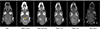
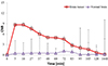
Figure 3 shows the coronal T1-weighted images of brain tumors in an animal model before and after HNP-2006 injection. The tumor, which was not visible in the pre-contrast image, was clearly depicted in the post-contrast image. Figure 4 shows the significant difference in CNR between tumor and normal brain tissue. Time series data suggested that wash out patterns differentiated the active tumor margin from the central necrosis-like region. Therefore, tumor images using HNP-2006 confirmed that HNP-2006 can effectively be used for enhanced imaging.
To our knowledge, HNP-2006 is the first macrocyclic Gd3+-based multifunctional CA. As mentioned above, several macrocyclic Gd3+-based ECF CAs have already been commercialized. However, the macrocyclic Gd3+-based functional CA is not commercially available. For example, the Gd3+-based liver specific CAs, Gd-BOPTA (Multihance) and Gd-EOB-DTPA (Primovist), have linear ligand structures, and thus showed relatively low kinetic stability compared with HNP-2006 (8). The low stability of the linear type liver-specific CAs may increase the chance of release of free Gd3+ions, compared to the macrocyclic HNP-2006. This instability of linear type functional CAs may be implicated in the higher incidence of in vivo toxicities, such as NSF and brain accumulation, compared to HNP-2006 with macrocyclic ligand structure.
In conclusion, the in vivo biodistribution profile of a new multifunctional Gd3+-based CA (HNP-2006) demonstrated significant contrast enhancement in many different organs. Furthermore, contrast enhanced tumor imaging using HNP-2006 confirmed that this new macrocyclic CA can be used for detecting tumors in the central nervous system. Therefore, this new multifunctional Gd3+-based CAs with macrocyclic ligand structure, is a promising contrast agent for use in whole-body clinical applications.
 XML Download