

 PDF
PDF ePub
ePub Citation
 Print
Print


Abstract
Purpose:
This study conducts a comparative evaluation of the skin dose in CyberKnife (CK) and Helical Tomotherapy (HT) to predict the accurate dose of radiation and minimize skin burns in head-and-neck stereotactic body radiotherapy.
Materials and Methods:
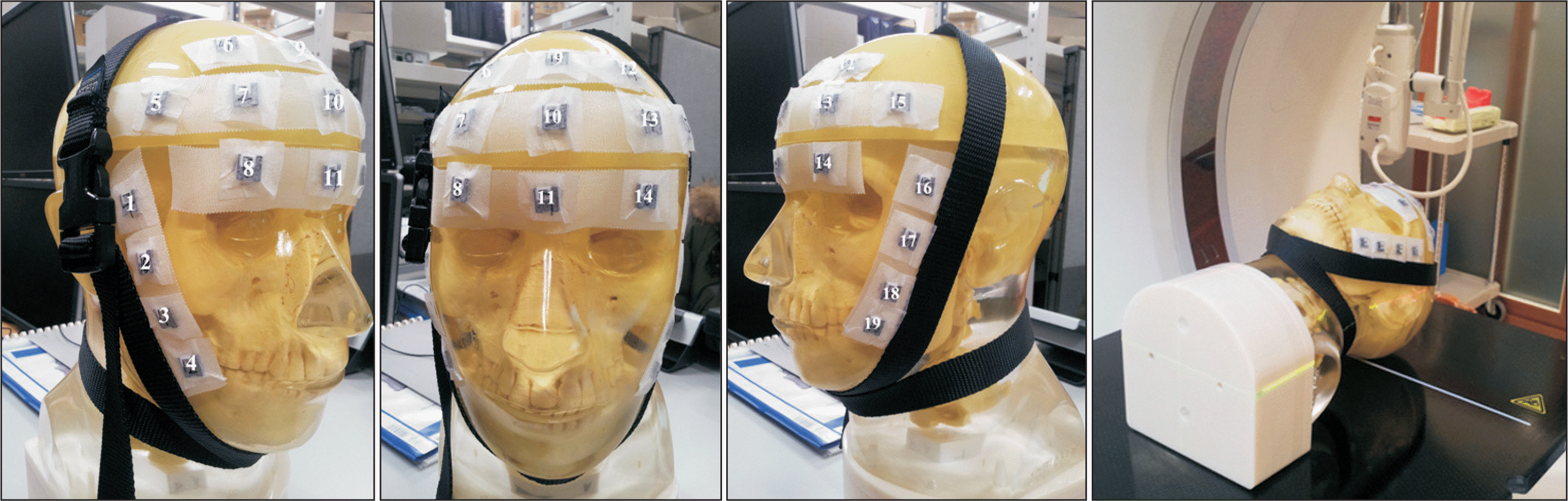
Arbitrarily-defined planning target volume (PTV) close to the skin was drawn on the planning computed tomography acquired from a head-and-neck phantom with 19 optically stimulated luminescent dosimeters (OSLDs) attached to the surface (3 OSLDs were positioned at the skin close to PTV and 16 OSLDs were near sideburns and forehead, away from PTV). The calculation doses were obtained from the MultiPlan 5.1.2 treatment planning system using raytracing (RT), finite size pencil beam (FSPB), and Monte Carlo (MC) algorithms for CK. For HT, the skin dose was estimated via convolution superposition (CS) algorithm from the Tomotherapy planning station 5.0.2.5. The prescribed dose was 8 Gy for 95% coverage of the PTV.
Results and Conclusions:
The mean differences between calculation and measurement values were −1.2±3.1%, 2.5±7.9%, −2.8±3.8%, −6.6±8.8%, and −1.4±1.8% in CS, RT, RT with contour correction (CC), FSPB, and MC, respectively. FSPB showed a dose error comparable to RT. CS and RT with CC led to a small error as compared to FSPB and RT. Considering OSLDs close to PTV, MC minimized the uncertainty of skin dose as compared to other algorithms.
Go to : 
REFERENCES
1.Guadagnolo BA., Zagars GK., Araujo D., Ravi V., Shellenberg-er TD., Sturgis EM. Outcomes after definitive treatment for cutaneous angiosarcoma of the face and scalp. Head Neck. 2011. 33(5):661–67.

2.Mellenberg DE., Schoeppel SL. Total scalp treatment of mycosis fungoides: the 4× 4 technique. Int J Radiat Oncol Biol Phys. 1993. 27(4):953–58.
3.Stelzer KJ., Griffin TW. A randomized prospective trial of radiation therapy for AIDS-associated Kaposi's sarcoma. Int J Radiat Oncol Biol Phys. 1993. 27(5):1057–61.
4.Katayama S., Haefner MF., Mohr A, et al. Accelerated tomotherapy delivery with TomoEdge technique. J Appl Clin Med Phys. 2015. 16(2):33–42.
5.Sterzing F., Uhl M., Hauswald H, et al. Dynamic jaws and dynamic couch in helical tomotherapy. Int J Radiat Oncol Biol Phys. 2010. 76(4):1266–73.
6.Lee E., Park K., Kim JS., Kim YB., Lee H. Practical Implementation of Patient-Specific Quality Assurance for Small and Multiple Brain Tumors in CyberKnife with Fixed Collimators. Prog Med Phys. 2018. 29(2):53–8.
7.Dieterich S., Cavedon C., Chuang CF, et al. Report of AAPM TG 135: quality assurance for robotic radiosurgery. Med Phys. 2011. 38(6):2914–36.
8.Yoon J., Park K., Kim JS., Kim YB., Lee H. Acceptance Testing and Commissioning of Robotic Intensity-Modulated Radiation Therapy M6 System Equipped with InCiseTM 2 Multileaf Collimator. Prog Med Phys. 2018. 29(1):8–15.
9.Jursinic PA. Changes in optically stimulated luminescent dosimeter (OSLD) dosimetric characteristics with accumulated dose. Med Phys. 2010. 37(1):132–40.
10.Jursinic PA. Characterization of optically stimulated luminescent dosimeters, OSLDs, for clinical dosimetric measurements. Med Phys. 2007. 34(12):4594–604.
11.Kim J., Park K., Yoon J, et al. Feasibility Study of a Custom-made Film for End-to-End Quality Assurance Test of Robotic Intensity Modulated Radiation Therapy System. Prog Med Phys. 2016. 27(4):189–95.
12.Sterpin E., Salvat F., Olivera G., Vynckier S. Monte Carlo evaluation of the convolution/superposition algorithm of Hi–ArtTM tomotherapy in heterogeneous phantoms and clinical cases. Med Phys. 2009. 36(5):1566–75.
13.Okoye CC., Patel RB., Hasan S, et al. Comparison of ray tracing and Monte Carlo calculation algorithms for thoracic spine lesions treated with CyberKnife-based stereotactic body radiation therapy. Technol Cancer Res Treat. 2016. 15(1):196–202.
14.Yoon J., Lee E., Park K., Kim JS., Kim YB., Lee H. Patient-Specific Quality Assurance in a Multileaf Collimator-Based CyberKnife System Using the Planar Ion Chamber Array. Prog Med Phys. 2018. 29(2):59–65.
15.Mcguinness C., Descovich M., Barani I. CyberKnife image-guided hypofractionated stereotactic radiotherapy. Image-Guided Hypofractionated Stereotactic Radiosurgery: A Practical Approach to Guide Treatment of Brain and Spine Tumors: CRC Press, Taylor and Francis Group. 2016. 49.
16.Kang KM., Jeong BK., Choi HS, et al. Combination effects of tissue heterogeneity and geometric targeting error in stereotactic body radiotherapy for lung cancer using CyberKnife. J Appl Clin Med Phys. 2015. 16(5):193–204.
Go to :
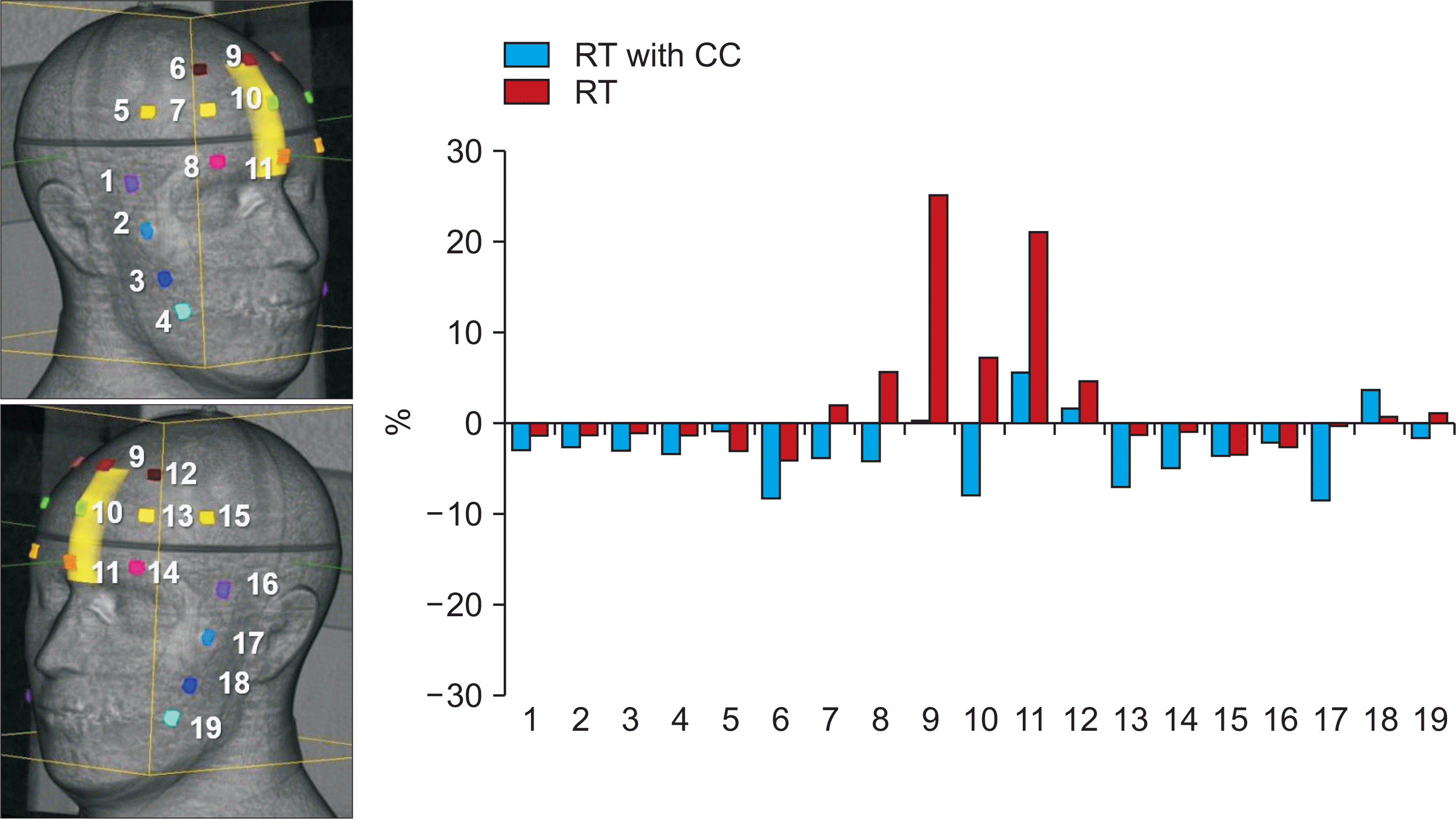 | Fig. 2Comparison of the dose difference between calculated and measured values in 19 OSLDs using RT with and without CC. |
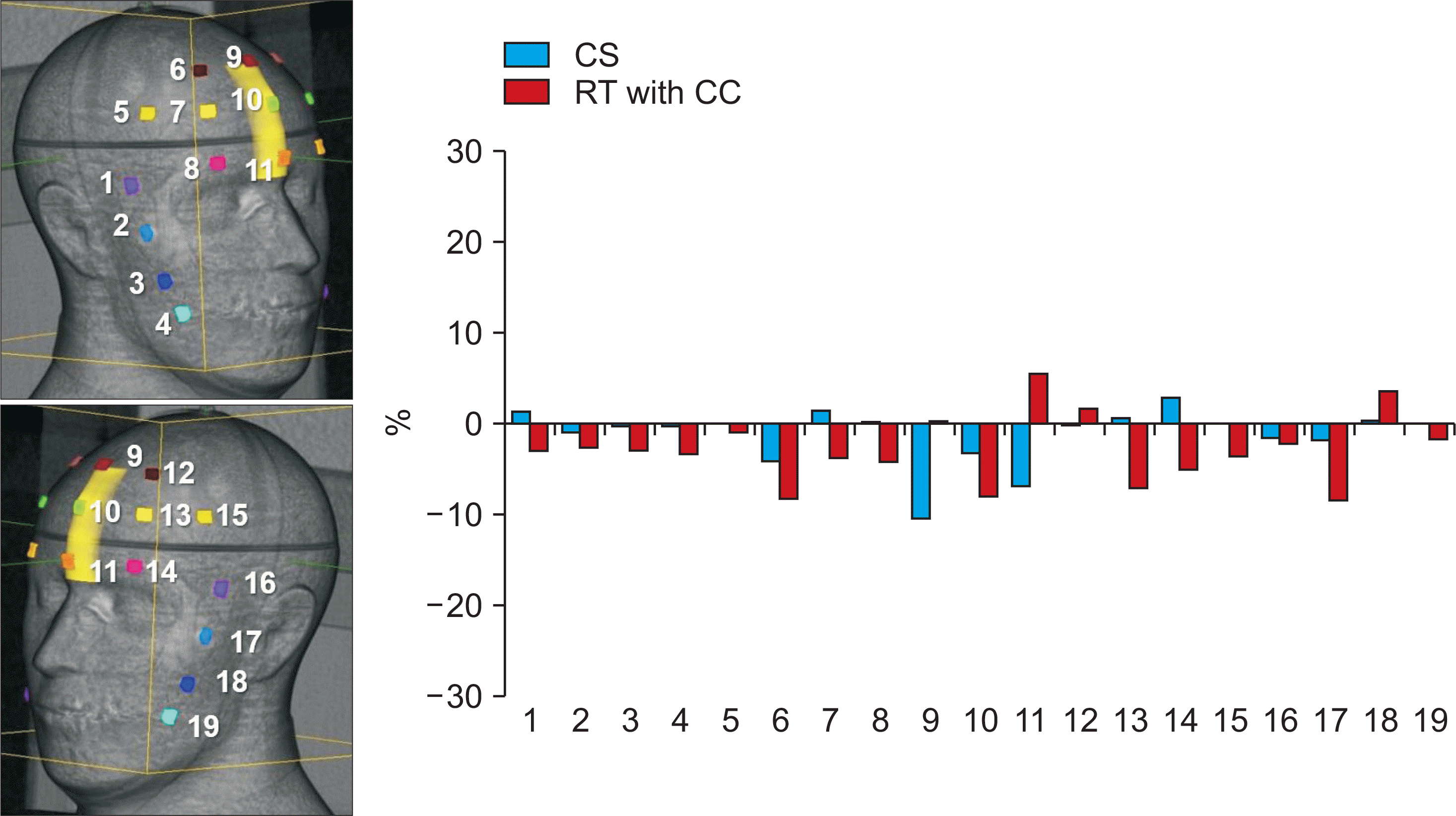 | Fig. 3Comparison of the dose difference between calculated and measured values in 19 OSLDs using CS and RT with CC. |
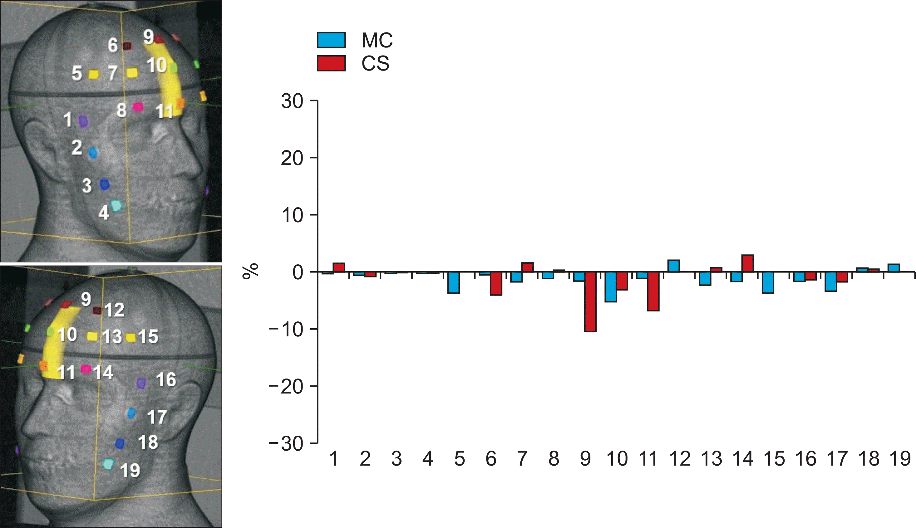 | Fig. 4Comparison of the dose difference between calculated and measured values in 19 OSLDs using MC and CS. |
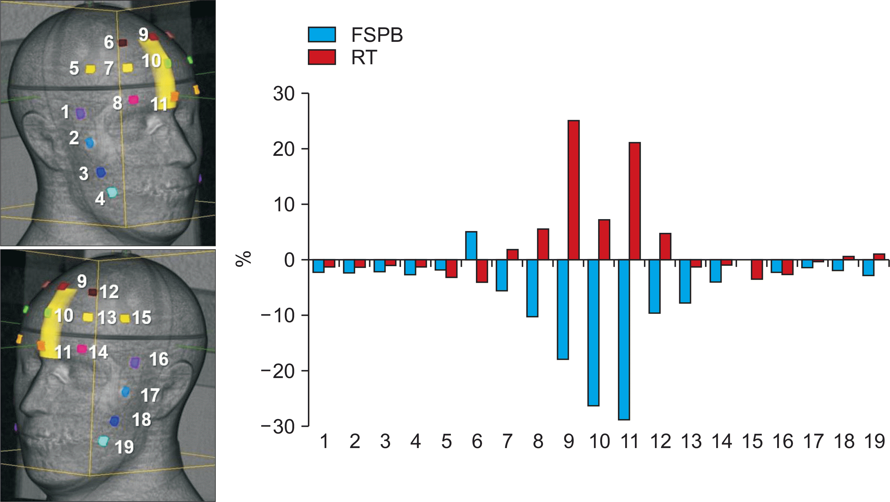 | Fig. 5Comparison of the dose difference between calculated and measured values in 19 OSLDs using FSPB and RT. |
Table 1.
Comparison of dose calculated by five dose calculation algorithms and measured dose.
 XML Download