

 PDF
PDF Citation
 Print
Print


INTRODUCTION
Crossed aphasia (CA) refers to language disturbance caused by right-hemisphere brain lesion in right-handed individuals. CA is rarely observed, with incidence estimated to be between 0.38% and 3% [1]. Mariën et al. [2] conducted a thorough analysis of previous CA literature and proposed a diagnostic algorithm for CA which is still widely used today. The diagnostic criteria include the following: 1) aphasia; 2) right-hemisphere lesion; 3) right-handedness without family history of left-handedness; 4) morphological integrity of left-hemisphere; and 5) absence of brain damage or seizures in childhood.
Although growing numbers of cases have been reported last decade, pathophysiology of CA is not yet completely understood. It has been proposed that several brain lesions are responsible for CA as well [2], and the lentiform nucleus has been suggested as an important neural substrate in recent study by using lesion mapping [1].
The corpus callosum is a massive fiber bundle that interconnects bilateral cerebral hemispheres, taking a crucial part in integrating sensory and motor functions. There have been increasing reports of studies that white matter tracts are essential components in neural mechanisms of language process [3] in which the importance of corpus callosum in aphasia has been suggested [4]. An infarction of the corpus callosum is uncommon due to the relatively rich blood supply [5] which makes CA caused by a corpus callosum infarction much scarcer, with the first case being reported in 2012 [6]. The patient presented with speech difficulty with left-handed apraxia after a right corpus callosum infarction. We describe another rare case of CA caused by right corpus callosum infarction.
CASE REPORT
A 74-year-old female patient had experienced her first infarction of right thalamus 7 months prior to second infarction (Fig. 1A). She had no sequela after that, with intact orientation, speech and motor function. She could walk independently on even level and had no difficulties with activities of daily living. Then after 7 months, she suddenly developed loss of spontaneous speech and dizziness, at which diffusion weighted magnetic resonance imaging (DW-MRI) revealed a newly developed right corpus callosum infarction (Fig. 1B). She was referred to our hospital for rehabilitation after 1 month of the second infarction. Her past medical history included hypertension and diabetes, and she had been on 200 mg of cilostazol and 100 mg of aspirin since first infarction. She was strongly right-handed with Edinburgh Handedness Inventory Laterality Quotient + 100 and had no family history of left-handedness nor previous brain injuries or seizures.
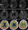 | Fig. 1Structural and functional brain imaging of the brain. (A) The brain MRI after first infarction shows increased signal intensity (arrows) at right thalamus. (B) The brain MRI after second infarction shows increased signal intensity (arrows) at right corpus callosum. (C) The brain PET-CT image demonstrates decreased metabolism (arrows) at left frontal, temporal cortex and right corpus callosum.MRI, magnetic resonance imaging; PET-CT, positron emission tomography-computed tomography.
|
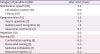
In the initial evaluation, her mental status was alert but showed disorientation to time and place. A cranial nerve function test did not show abnormal findings with no signs of extraocular movement limitations or facial palsy. Her muscle power was relatively well preserved with Medical Research Council scale grading 5 in the right side and 4 in the left side. Independent gait with normal pattern was observed on even level. Unilateral spatial neglect, apraxia, and agnosia were not seen. She could follow simple commands but had decreased spontaneous speech with barely a single-word answer to questions. The aphasia quotient of Korean version of the Western Aphasia Battery was 2.5 after 2 weeks of onset done at her previous hospital, indicating severe global aphasia (Table 1).
Table 1
K-WAB after 2 weeks of onset
![]()
Positron emission tomography-computed tomography (PET-CT) after 2 months of the second infarction showed hypometabolism of the right corpus callosum and mild, diffuse hypometabolism of the left frontal and temporal cortex (Fig. 1C).
DISCUSSION
Corpus callosum is positioned between bilateral cerebral hemispheres. It is supplied by four vessels, the anterior communicating artery, the pericallosal artery, the posterior choroidal artery, and the posterior cerebral artery [7]. Due to its dual rich blood supply, even with arterial stenosis on one side, blood flow is compensated by anastomosis, which makes corpus callosum infarction very rare. Clinical manifestation of corpus callosum infarction is usually non-specific because it is often combined with infarctions at other locations [8]. Li et al. [5] did retrospective analysis of 59 cases of corpus callosum infarction and clinical symptoms varied from movement disorders and cognitive impairment to language disturbance. Association between language function and corpus callosum is not clearly understood. Speech difficulties including stuttering [9] or dysarthria [10] have been reported in several studies, but aphasia after corpus callosum infarction is rare [4].
Definite mechanism underlying CA is obscure as well [6]. Several hypotheses have been proposed; 1) congenital or acquired left hemisphere dysfunction causing lateralization shift [11], 2) bilateral representation of linguistic functions [12], 3) absence of right-shift gene that gives relative advantage toward left hemisphere [13], and 4) diaschisis [14].
The development of functional brain imaging such as single-photon emission computed tomography (SPECT) or PET-CT has introduced concept of diaschisis in CA, in which language functions in left hemisphere could be functionally depressed by functional connection to right hemisphere lesion. Several studies have confirmed metabolic depression of left hemisphere in CA patients [1516].
The first report to describe CA after right corpus callosum infarction [6] has also suggested transcallosal diaschisis as possible cause of aphasia. Our patient had severe global aphasia, but was not accompanied by typical callosal disconnection syndrome such as neglect, apraxia or alien hand syndrome. Akinetic mutism has also been reported to be possibly accompanied by a similar lesion [17], but our patient showed isolated aphasia. An MRI showed anatomically intact left hemisphere but decreased metabolism at left frontal and temporal cortex in PET-CT, implying interhemispheric diaschisis as possible mechanism.
In conclusion, isolated manifestation of CA without disconnection syndrome succeeding right corpus callosum infarction is presented in our report.
 XML Download