

 PDF
PDF Citation
 Print
Print


INTRODUCTION
CASE REPORT
Fig. 1
T2-weighted brain MRI showing encephalomalacic changes in both cerebellar hemispheres. The left middle cerebellar peduncle (arrow, A), left pons (arrow, B), left anterior (arrow, D), and right lateral medulla (arrowhead, D).
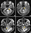
Table 1
The respiratory rehabilitation protocols for the patient and their expected effects
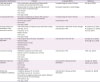
1. Chest wall ROM exercise: before starting active respiratory rehabilitation, 5-minute warm-up stretching of intercostalis, pectoralis, sternocleidomastoid muscles was done. Because the patient's dynamic sitting balance was poor, this stretching exercise was done with manual assist of a physiotherapist. 5 minutes per session, once a day, 5 days a week [6].
2. Air stacking exercise: the patient was encouraged to inhale as much air as she can, and then therapist infused additional air two times by bagging (M.O.W. Silicone resuscitators MR010; MOW Medical, Wonju, Korea) via tracheostomy. The patient holds the breath for 2 seconds and then started to exhale. 10 times per session, once a day, 5 days a week. This exercise was done in a supine position on the first week, and then changed to a sitting position [7].
3. NMES on abdominal muscles: active electrodes were placed on the bilateral rectus abdominis muscles (Microstim; MEDEL, Hamburg, Germany). The patient was encouraged to exhale during the stimulation. 50 mA of intensity, 50 Hz of frequency, for 15 minutes, once a day, 5 days a week, in a sitting position [789].
4. Upper extremity ergometer: arm crank exercise was performed at 60% of the peak work rate. Intensity was progressed maintaining symptom scores at a moderate level of 3 on the modified Borg scale. For 15 minutes, 3 times a week (Isokinetic upper body ergometer, PRO1000 seated upper body; SCIFIT, Tulsa, OK, USA) [1011].
5. Locomotor training: wheelchair propulsion with both upper extremities, started at 1.0 km/h, increased up to 1.5 km/h. For 10 minutes, twice a day, started with 3 days a week and increased to 5 days a week [12].
6. High-frequency chest wall oscillator: vest pressure setting 6 (The Vest; Hill-Rom, Chicago, IL, USA), 12 Hz of frequency, for 15 minutes, once a day, 5 days a week, in a sitting position [1314].
7. MIE: insufflation and exsufflation pressures of +30/−30 cmH2O were applied via tracheostomy (Cough & Suction, CNS-100; Sungdo MC, Siheung, Korea), and changed to oronasal mask after the removal of the tracheostomy. Five positive-to-negative pressure cycles per session, followed by 20 seconds of normal breathing. Total 5 session per day, 5 days a week, in a sitting position [15].
Fig. 2
Changes in spirometry before (left) and after (right) 4 weeks of a respiratory rehabilitation program.
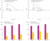
Table 2
Changes in pulmonary function test parameters after 4 weeks of respiratory rehabilitation

 XML Download