

 PDF
PDF ePub
ePub Citation
 Print
Print


INTRODUCTION
Splenosis refers to heterotopic autotransplantation of the splenic tissue occurring after splenic trauma or splenectomy (12). This condition is usually incidentally found without clinical symptoms. Mostly it is found in the intraperitoneal cavity but extraperitoneal locations have also been rarely reported as follows: intrathoracic, pericardium or subcutaneous tissue (3). In the literature, about 20 cases of splenosis involving the liver and their radiologic characteristics have been reported to date (45).
We discuss a case in which a patient with liver cirrhosis (LC) and previous splenectomy due to trauma is diagnosed with intrahepatic splenosis. We describe the imaging findings of intrahepatic splenosis on ultrasonography (US), CT and MRI. The final diagnosis was confirmed by the radionuclide imaging.
CASE REPORT
A 43-year-old man was on regular visit to the outpatient clinic after acute myocardial infarction. He was a heavy alcoholic and the level of liver enzyme was elevated on screening blood tests. Initial laboratory results were as follows; albumin 3.4 g/dL (3.8–4.7), glutamic oxaloacetic transaminase 46 IU (11–37), alkaline phosphatase 226 IU (45–130). Although the Alpha-fetoprotein was within normal range, but the protein induced by absence of vitamin K (PIVKA II) was elevated as 20677 mAU/mL (9.1–26.8). Under diagnosis of alcoholic LC, the clinician examined him with US as an initial imaging workup.
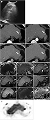
US showed typical characteristics of LC and a homogeneous hypoechoic lesion measuring 3.3 × 4.0 cm in dimension, located in the subcapsular region of the left hepatic lobe (Fig. 1A).
Multiphase contrast-enhanced CT scan was performed for further evaluation (Fig. 1B). The lesion appeared as a low-attenuated area compared to the surrounding liver parenchyma on pre-contrast image. After contrast administration, the lesion showed strong enhancement on the arterial and portal phases and mild washout on delayed phase.
On MRI (Fig. 1C), the lesion showed homogeneous low signal intensity (SI) on T1-weighted image (T1WI) and high SI on T2WI. After intravenous administration of hepatocyte-specific contrast agent (Gadoxetic acid disodium, Primovist; Bayer-Schering, Berlin, Germany), the lesion showed strong enhancement on arterial phase, washout on 3-minute delayed image and low SI on 20-minute delayed image leading to high suspicion for hepatocellular carcinoma (HCC). The lesion seemed to be intrahepatic location surrounded by thin hepatic parenchyma.
However, the diagnosis of intrahepatic splenosis was more favored because he underwent splenectomy 20 years ago due to traffic accident. The patient was referred to the department of nuclear medicine to perform radionuclide scintigraphy for confirmative diagnosis. Heat damaged red blood cell (RBC) scintigraphy was performed with 2 mCi (37 MBq) 99m technetium (Tc)-labelled autologous RBCs and the uptake was observed at the corresponding area (Fig. 1D).
DISCUSSION
The term splenosis was first described in 1939 defined as auto-transplantation of the splenic tissue to other parts of body following splenic injury (4). Splenosis is believed to occur in up to 67% of patients who suffered from splenic trauma or splenectomy, but the true incidence is unknown because it is usually asymptomatic and incidentally found during imaging workup or surgery for an unrelated disease or at autopsy (2).
Splenosis rarely causes symptoms. Some lesions, however, can twist a long pedicle attached to the splenic nodule, causing acute abdominal pain. There also have been reports of spontaneous rupture causing intra-abdominal hemorrhage or gastrointestinal bleeding related to mucosal erosion as complications of splenosis (46).
Hepatic splenosis is generally thought to occur with spillage and implantation of damaged splenic pulp into subcapsular area of liver. Alternatively, another proposed mechanism is the migration of the splenic pulp or erythrocyte progenitor cells through portal vein and proliferation and differentiation of splenic erythrocytic progenitor cells induction by local hypoxia of the liver (17).
Choi et al. (2) reviewed 10 cases of hepatic splenosis. In 5 cases, the splenoses were between the diaphragm and segment II of liver as same as in our case. They proposed that the space between diaphragm and segment II of the liver is located close to the spleen so that it can be easily exposed during splenectomy. The chronic compression of the diaphragm might induce splenic tissue to invade into the liver parenchyma.
In previous reports (1), splenosis showed hypo- or isoechogenicity on US. The lesions mostly appeared as low attenuated area distinguished from the surrounding liver parenchyma on the pre-contrast CT scan, heterogeneously enhanced on the arterial phase and showed washout on the delayed phase. On MRI, the diagnosis was suggested by low SI on T1WI, high SI on T2WI, strong arterial enhancement and delayed washout after intravenous hepatocyte-specific contrast agent administration. Although such imaging findings are compatible with the characteristics of splenic tissue, these are indistinguishable from hepatic tumors, such as HCC, hepatic adenoma or focal nodular hyperplasia.
In our case, the lesion showed intrahepatic location surrounded by thin hepatic parenchyma on CT and MRI. Moreover, the clinical presentation, regarding hypervascular mass in cirrhotic liver and marked elevation of serum PIVKA II level, led to the primary suspicion for a HCC. But the possibility of hepatic splenosis was more favored based on the patient's history of previous splenectomy and the dynamic characteristics on CT and MRI. Unlike in a typical HCC, the mass did not show definite washout on portal phases on both CT and MRI. We recommended the radionuclide scintigraphy using heat damaged 99m Tc-labeled RBCs for the confirmative diagnosis of intrahepatic splenosis because it was described as the most efficient and accurate diagnostic method on the previous literature (157).
After the confirmative diagnosis, the clinician supposed that the marked elevation of the level of PIVKA II was probably related to longstanding administration of oral warfarin due to the patient's past history of acute myocardial infarction and thrombus in the left ventricle of heart. On follow-up blood tests, the level of PIVKA II was normalized.
In conclusion, hepatic splenosis should be considered if the subcapsular mass is found in patients with a history of splenectomy. The imaging findings of US, CT and MRI can reveal characteristics of splenic tissue but still insufficient for diagnosis of splenosis. Thus, if the hepatic splenosis is suspected, the radiologist should recommend the most specific imaging technique, radionuclide scintigraphy, for the confirmative diagnosis. Unnecessary liver resection or invasive percutaneous liver biopsy in cases misdiagnosed as HCC should be avoided.
 XML Download