

 PDF
PDF ePub
ePub Citation
 Print
Print


Postmortem human brain tissues are critical for the advancement of neuroscience. In basic science dealing with neurochemistry and signaling pathways as well as translational research pursuing therapeutic goals, the final step is often ver-ification through human brain tissues. Obtainment and storage of human brain tissues with reliable medical records and clinical data are invaluable for neuroscience. Antemortem workup using relevant biomarkers and imaging techniques can result in clinical diagnosis, even to the level of specific disorder subtypes. Indeed, brain autopsy remains the gold standard for confirmative diagnosis of neurodegenerative disorders.1 Adequate categorization of postmortem brain tissue according to the final pathologic diagnosis expedites proper use of limited human materials.
In 2016, we established the national neuropathology reference and diagnostic laboratory for Alzheimer's disease (AD). This project aimed to standardize a neuropathology-based diagnosis of dementia by establishing a country-wide brain tissue bank network. Herein, we reviewed brain autopsy procedures performed in brain banks within and outside the co-untry, and also designed an optimal brain autopsy procedure to be performed for brain bank in South Korea. This proposed guideline covers the overall processes of brain autopsy, diagnostic processes, and quality control of stored tissues. The ethical, legal, and procedural issues associated with disbursement of postmortem human brain tissue are not considered, because these issues are beyond the scope of this article. We rather focused on the methodology for management and pa-thologic workup of autopsied human brains, and the section below provides recommendations about the acquisition of human brain tissue, dissection and processing of the tissue, his-tological evaluation to reach a pathologic diagnosis, and quality control of the tissue.
Dissection and preparation of frozen tissue
A brain donated to the brain bank should be processed as soon as possible after death in order to minimize autolytic effects during the postmortem interval. Degenerative processes in the brain are believed to begin at death, but a large body of evidence indicates that the effects of autolysis within 24 hours following death are smaller than antemortem hypoxic events.234 After removal of the brain, it should be examined, photographed, and weighed. Surface pH should be measured with a pH meter if available. Next, the brain should be separated into two hemispheres. In general, the dominant hemisphere (the left hemisphere for a right-handed person) should be fixed for thorough neuropathologic evaluation, while the contralateral hemisphere should be left unfixed, dissected into slabs, and immediately snap-frozen. However, if a unilateral lesion or more severe lesion with asymmetric changes is identified on gross examination, the side involved should be reserved for pathologic workup and fixed in formalin.2
Before the two hemispheres are separated, the brainstem and cerebellum need to be removed from the cerebrum. To se-parate the brainstem, the mammillary body should be pressed softly with a cutting blade, and the midbrain is detached from the cerebral hemisphere transversely through the crus cerebri up to the dorsal aspect of the superior colliculus.2 When the red nuclei and substantia nigra are clearly visible, the cutting level is appropriate. The cerebellum should be separated from the brainstem by sequential sectioning through the superior, middle, and inferior cerebellar peduncles. The brain stem should then be cut sagittally into two parts 2-mm to the right of the midline. The bigger left side should be fixed in formalin and blocked for pathologic examination. The smaller right side should be left unfixed and immediately snap-frozen for storage at −80℃. The cerebellar hemispheres should be separated sagittally through the vermis. The right cerebellar hemisphere should be dissected parasagittally into three slabs and snap-frozen. The left hemisphere should be reserved and fixed in formalin. The left cerebral hemisphere, left cerebellar hemisphere, and left-side brainstem should be fixed in 10–20% neutral buffered formalin (pH 7.4) for 2–4 weeks before tissue blocks are prepared.
The unfixed cerebral hemisphere should be dissected immediately into slabs following the same steps as described for the fixed hemisphere. First, the hemisphere should be cut coronally into two parts at the level of the mammillary bodies. The sectioning should be performed gently with one stroke per sagittal cut along an axis perpendicular to a ventrodorsal line that connects the temporal pole and occipital pole.2 The anterior part should then be dissected into 1-cm-thick slabs forwardly and numbered “S, minus, and sequential number” from the mammillary bodies throughout the frontal pole; for example, S-1, S-2, S-3, and so on. The posterior portion should also be dissected into 1-cm-thick slabs backwardly and numbered “S, plus, and sequential number” from S0, S+1, S+2, and so on. Unfixed slabs should be placed on metal trays covered with powdered dry ice with their anterior surface facing down. They should then be wrapped in aluminum foil, individually put into labeled airtight storage bags, and stored in a −80℃ freezer until investigational use. Frozen brain parenchyma aliquots when fresh or from stored frozen tissue on request for disbursement later are optional.2
Block preparation following standard section list
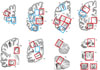
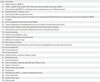
After sufficient fixation, the left hemisphere should be dissected into 1-cm-thick slabs, and blocks are prepared according to the proposed standard set of brain regions (Table 1, Fig. 1). By sharing the standard set of brain blocks, information on stored brains can easily be accessed by researchers through the data network hub. Two groups of block sites are listed in Table 1. Core blocks are crucial for pathologic confirmation of neurodegenerative diseases. Additional blocks are optional and should be based on the academic interests of pathologists or researchers. In these guidelines, we defined core blocks and additional blocks using previously published data. The recent National Institute on Aging-Alzheimer's Association (NIA-AA) revision of criteria for the pathologic diagnosis of AD proposed a minimum set of 13 histologic sections for evaluation of major neurodegenerative diseases.56 In addition to the minimum set of 13 regions, brain section lists were obtained from the brain donation and neuropathology manual of the Alzheimer's Disease Neuroimaging Initiative-Neuropathology Core (ADNI-NPC) at Washington University (kindly provided by Dr. Cairns), the neuropathology section list from the University of California San Francisco (kindly provided by Dr. Seeley), and the neuropathology core manual from Northwestern Memorial Hospital (kindly provided by Dr. Bigio).
Consistently identifying the central sulcus from the dissected slabs is important when blocking the motor cortex and sensory cortex. The precentral gyrus should be inked as needed. The blocks should be post-fixed for an additional 2–4 days and then processed in an automatic tissue processor. The tissue processing program should be adjusted to increase the time in ethanol tanks in order to allow sufficient dehydration of brain tissues.
Gross examination
A careful gross examination can provide a good amount of useful information for pathologic diagnosis. Vascular pathology, including stenosis or atherosclerosis, can be estimated semiquantitatively. Regional atrophy should be assessed using a semi-quantitative method involving a four-tier system of none, mild, moderate, and severe. Atrophy of the hippocampus, caudate, subthalamic nucleus, brainstem, cerebellum, and neocortices must be documented. Pallor of the substantia nigra and locus ceoruleus should be recorded. Unbalanced pallor in the substantia nigra and locus ceoruleus can help a conjectural diagnosis; AD is favored with greater pallor in the locus ceoruleus, and the opposite is seen in frontotemporal lobar degeneration (FTLD).1 Furthermore, the degree of degeneration in the cerebellar dentate nucleus and color changes of basal ganglia should be recorded.
Microscopic workup using immunohistochemical and special stains
The three most frequently encountered neurodegenerative disorders are AD, dementia with Lewy bodies (DLB) or Lewy body disease (LBD), and FTLD.1 Elements of pathologic diagnosis of neurodegenerative diseases in our guidelines are based on recent guidelines released by the NIA-AA.5 The guidelines summarize remarkable advances made since publication of the NIA/Reagan Institute of the AA Consensus Recommendations for the Postmortem Diagnosis of AD or NIA-Reagan Criteria in 1997.7
Alzheimer's disease neuropathologic change (ADNC) should be evaluated with an “ABC” score along with Aβ (β-amyloid) plaque score based on Thal phases, Braak neurofibrillary tangle (NFT) stage, and Consortium to Establish a Registry for Alzheimer's Disease (CERAD) neuritic plaque score.56 Immunohistochemistry (IHC)-based analysis of Aβ should be used to assess Thal phases based on progressive Aβ deposition.8 IHC staining for phospho-tau can be used to assess NFTs.8910 The scope of tau pathology observed in AD includes pretangles, neurophil threads in neuronal processes, and dystrophic neuritis in neuritic plaques, as well as NFTs in cell bodies.10 Bielschowsky or Gallyas silver or thioflavin-S fluorescent special staining can be performed for differential detection of neuritic plaques from Aβ deposits.1112 By combining ABC scores, ADNC can be transformed into one of four levels: not, low, intermediate, or high.6
LBD is a neuropathological term that encompasses the two clinical entities of Parkinson disease and DLB.13 Currently, LBD is classified as follows: brainstem-predominant, limbic (transitional), neocortical (diffuse), or amygdala-predominant.514 Assessment of LBD pathology includes identification of Lewy bodies on H&E staining, mainly in neurons of the brainstem sections. IHC staining for α-synuclein is the preferred method because of its high sensitivity for revealing Lewy body pathology, including Lewy neurites and variable neuronal perikaryal inclusions that comprise the continuum of immunoreactive pathology leading to Lewy body formation.13 α-synuclein IHC staining should be performed on amygdala and anterior cingulate sections. If α-synuclein pathology is identified in the anterior cingulate, additional stains can be performed on neocortical sections to determine the stage of LBD pathology.115
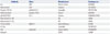
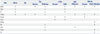
To explore FTLD pathology, tau, ubiquitin/p62, and transactive response DNA-binding protein 43 (TDP-43) staining of hippocampal and neocortex sections should be performed. To specifically examine FTLD-tau, such as corticobasal degeneration, progressive supranuclear palsy, and Pick disease, tau immunostaining should be performed on the neocortex, basal ganglia, brainstem, and dentate gyrus to determine the pathognominic inclusions of each entity.1 IHC for 3-repeat and 4-repeat tau can be beneficial in some cases. To diagnose FTLD-TDP, FTLD-fused in sarcoma (FUS), or FTLD-ubiquitin-proteasome system (UPS), a set of immunostains of corresponding antibodies should be performed on the hippocampus, cerebellum, neocortex, and deep nuclei. For diagnostic details of a wider pathologic spectrum, please refer to previously published articles.1617 To evaluate amyotrophic lateral sclerosis, additional TDP-43 staining can be performed on spinal cord sections and medulla with hypoglossal and vagus nuclei as well as the motor cortex. Commonly used antibodies and manufacturer information are listed in Table 2. Basic stain sets for screening and an extended set for specific diagnosis are summarized in Table 3.
Neurodegenerative disorders are frequently accompanied by other forms that can contribute to cognitive impairment of the affected individuals.18 Nevertheless, comparative estimation for weighing of co-existing pathologic changes in terms of contribution to cognitive impairment is rarely proposed except for AD and LBD.14 It is demanding to gauge the extent to which each disease course might have contributed to cognitive dysfunction.5 Nonetheless, all observed pathologic changes should be described with regard to disorder type and severity.
As a progression of the molecular genetic study in the neurodegenerative diseses, DNA and RNA analysis from the autopsy brain sections should be recommended.192021
Cerebrovascular diseases that cause vascular brain injury are mainly atherosclerosis, arteriolosclerosis, and cerebral amyloid angiopathy. Vascular brain injury manifests as hemorrhages or infarcts. Infarcts are classified according to dimensions as territorial infarcts, lacular infarcts, and microinfarcts.5 All infarcts and hemorrhages observed should be documented by description of location, size, and chronicity, as a comorbidity of other degenerative illnesses, or as an isolated lesion.
In conclusion, a nationwide brain bank system is being established in South Korea. Coordination of brain donation, processing, storage, and research steps will result in a synergistic chain that might help alleviate the burden of devastating neurodegenerative diseases.2 For efficient utilization of resources and to establish a functional and efficient system, we herein proposed tentative standard operating protocols for data systemization. We hope that our proposed guidelines will lead to constructive debate and commentary that will facilitate the development of an advanced brain bank system in Korea. We anticipate that updated protocols and procedures will be developed and shared with the medical community in the future.
 XML Download