Abstract
Summary of Literature Review
AR is a new technology that simulates interactions with real-world surroundings using computer graphics, and it is a field that has recently been highlighted as part of the fourth industrial revolution.
Go to : 
REFERENCES
1. Shuhaiber JH. Augmented reality in surgery. Arch Surg. 2004 Feb; 139(2):170–4. DOI: 10.1001/archsurg. 139.2.170.

2. Graham M, Zook M, Boulton A. Augmented reality in urban places: contested content and the duplicity of code. Transactions of the Institute of British Geographers. 2013 Jul; 38(3):464–79. DOI: 10.1111/j.1475-5661.2012.00539. x.

3. Yoon JW, Chen RE, Kim EJ, et al. Augmented reality for the surgeon: Systematic review. Int J Med Robot. 2018 Aug; 14(4):E1914. DOI: 10.1002/rcs.1914.

4. Ma L, Fan Z, Ning G, et al. 3D Visualization and Augmented Reality for Orthopedics. Adv Exp Med Biol. 2018; 1093:193–205. 10.1007/978-981-13-1396-7_16.

5. Fida B, Cutolo F, di Franco G, et al. Augmented reality in open surgery. Updates Surg. 2018 Sep; 70(3):389–400. DOI: 10.1007/s13304-018-0567-8.

6. Weiss CR, Marker DR, Fischer GS, et al. Augmented reality visualization using Image-Overlay for MR-guided inter-ventions: system description, feasibility, and initial evaluation in a spine phantom. AJR Am J Roentgenol. 2011 Mar; 196(3):W305–7. DOI: 10.2214/AJR.10.5038.

7. Terander AE, Burstrom G, Nachabe R, et al. Pedicle Screw Placement Using Augmented Reality Surgical Navigation with Intraoperative 3D Imaging: A First In-Human Prospective Cohort Study. Spine (Phila Pa 1976). 2019 Apr; 44(7):517–25. DOI: 10.1097/BRS. 0000000000002876.
8. Elmi-Terander A, Nachabe R, Skulason H, et al. Feasibility and Accuracy of Thoracolumbar Minimally Invasive Pedicle Screw Placement With Augmented Reality Navigation Technology. Spine (Phila Pa 1976). 2018 Jul; 43(14):1018–23. DOI: 10.1097/BRS.0000000000002502.

9. Elmi-Terander A, Skulason H, Soderman M, et al. Surgi-cal Navigation Technology Based on Augmented Reality and Integrated 3D Intraoperative Imaging: A Spine Cadaveric Feasibility and Accuracy Study. Spine (Phila Pa 1976). 2016 Nov; 41(21):E1303–11. DOI: 10.1097/BRS.0000000000001830.
10. Umebayashi D, Yamamoto Y, Nakajima Y, et al. Aug-mented Reality Visualization-guided Microscopic Spine Surgery: Transvertebral Anterior Cervical Foraminotomy and Posterior Foraminotomy. J Am Acad Orthop Surg Glob Res Rev. 2018 Apr; 2(4):e008. DOI: 10.5435/JAAOSGlobal-D-17-00008.

11. Cho HS, Park YK, Gupta S, et al. Augmented reality in bone tumour resection: An experimental study. Bone Joint Res. 2017 Mar; 6(3):137–43. DOI: 10.1302/2046-3758.63.BJR-2016-0289.R1.
12. Hodges SD, Eck JC, Newton D. Analysis of CT-based navigation system for pedicle screw placement. Orthopedics. 2012 Aug; 35(8):e1221–4. DOI: 10.3928/01477447-20120725-23.

Go to : 
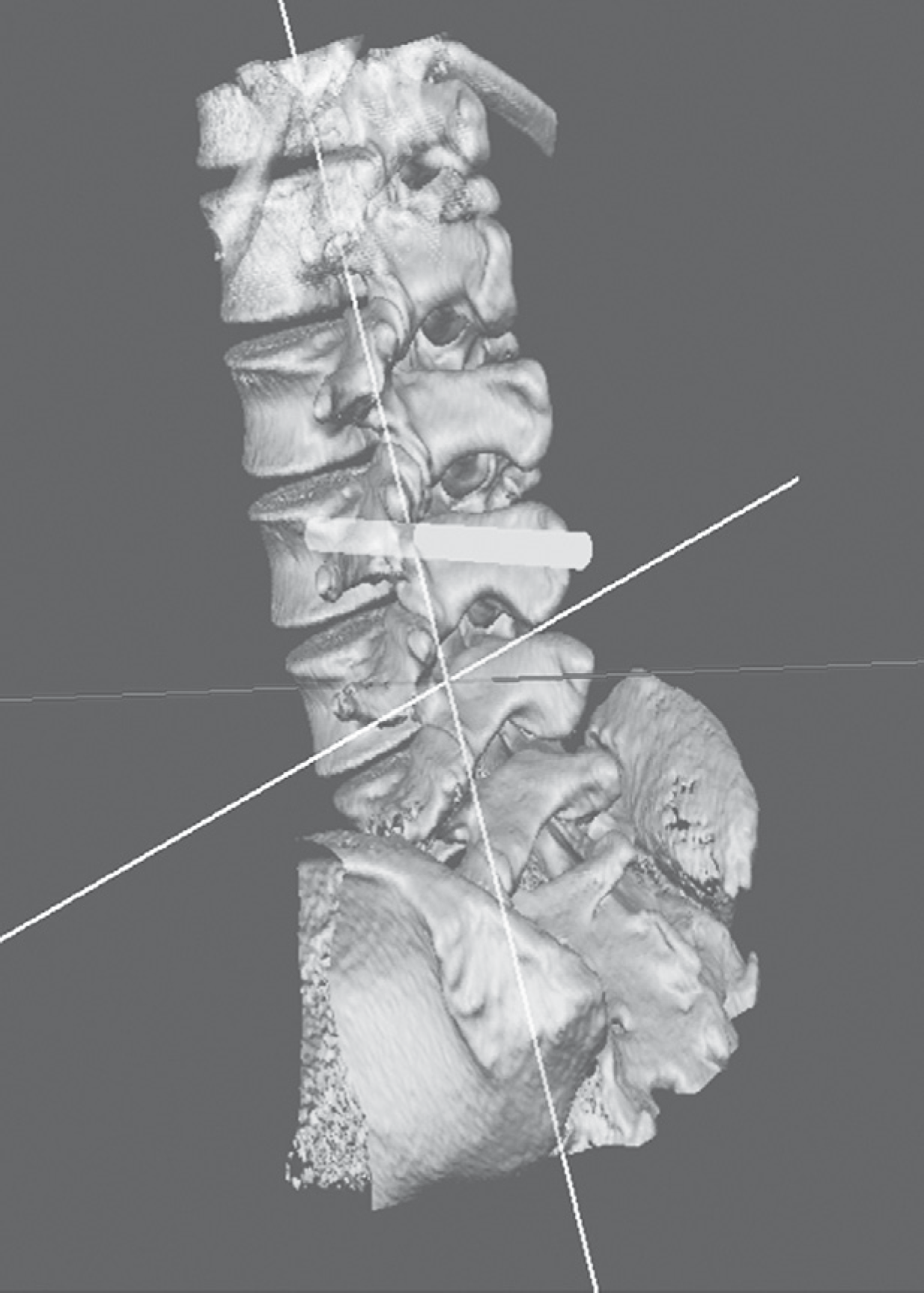 | Fig. 2.Three-dimensional reconstructed model of the spine, simulated by pedicle screw simulator software. |
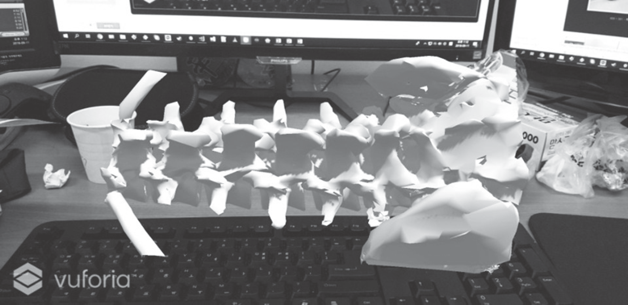 | Fig. 3.Three-dimensional reconstructed model of the spine, which can be seen using a head-mounted device. |
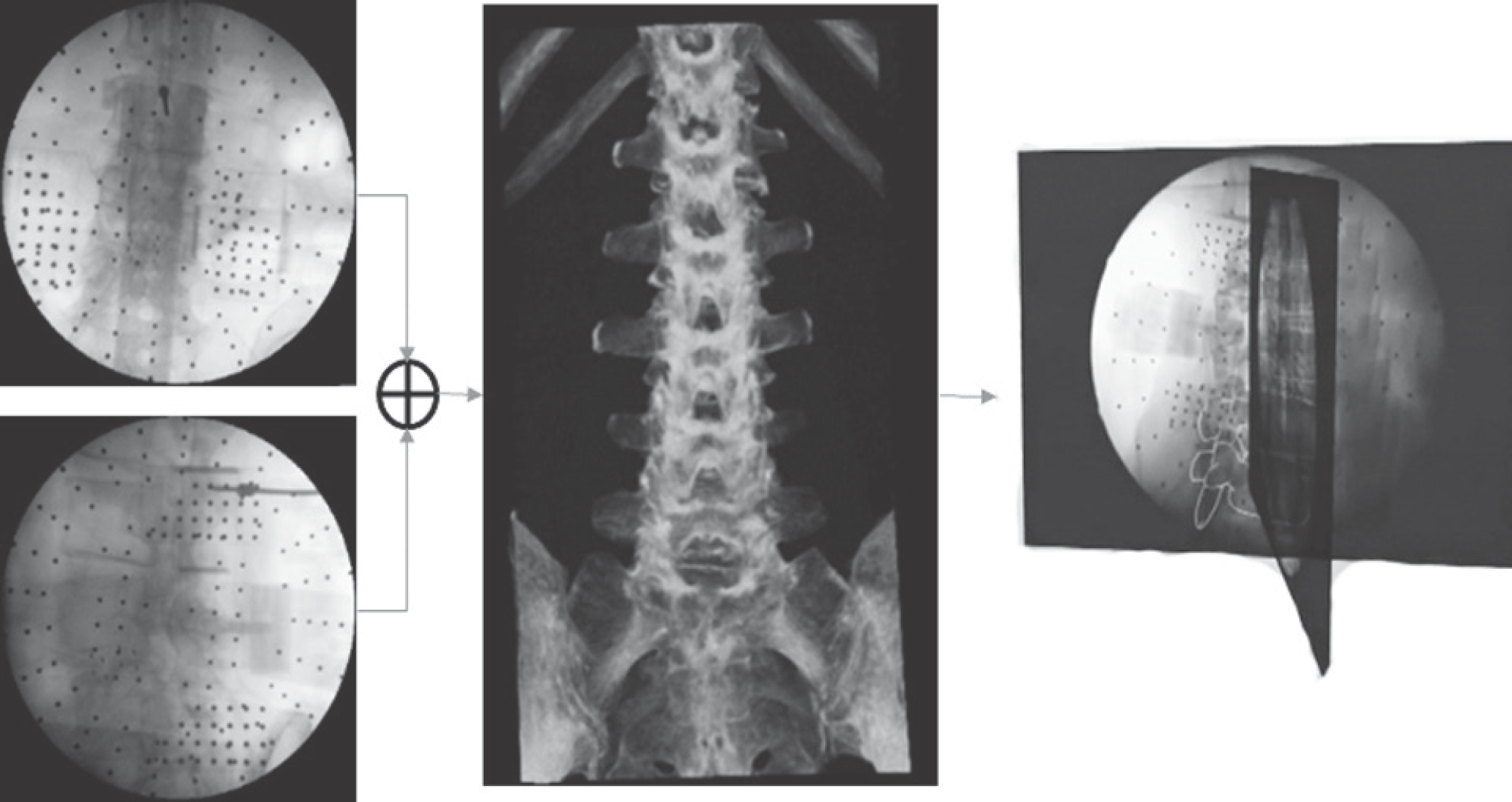 | Fig. 5.Schematic illustration of computed tomography (CT)-fluoroscopy registration. After creating a virtual C-arm image from CT, it is compared with the actual anteroposterior and lateral C-arm images to perform registration. |
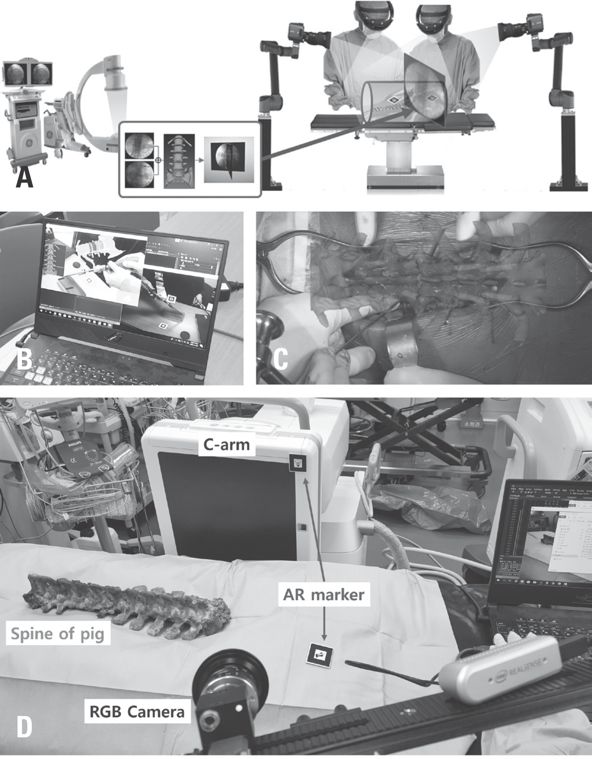 | Fig. 6.
(A) Schematic illustration of augmented reality (AR)-based spine surgery. After attaching the AR marker (DRB) to the patient's vertebrae, and completing the computed tomography (CT)-fluoroscopy or paired-point matching, the motion of the vertebrae can be tracked by only tracking the position of the DRB with the RGB camera. After the matching process, the 3-dimensional (3D) reconstructed vertebrae from CT images and surgical instruments can be overlaid on the actual patient's vertebrae. (B) Demonstration of the 3D reconstructed model on a notebook display. (C) Example of an augmented spine 3D reconstruction model during spine surgery. (D) The experimental setup consisted of vertebrae, a RGB cam-era, the C-arm, and the AR marker. |


 PDF
PDF ePub
ePub Citation
 Print
Print


 XML Download